Colorectal Cancer Information

Colorectal Cancer

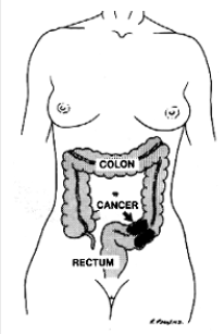
Colorectal cancer is the second most common cancer in the United States, striking 140,000 people annually and causing 60,000 deaths. That’s a staggering figure when you consider the disease is potentially curable if diagnosed in the early stages.

Who is at risk?
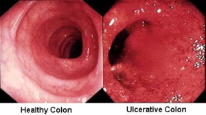
Though colorectal cancer may occur at any age, more than 90% of the patients are over age 40, at which point the risk doubles every ten years. In addition to age, other high risk factors include a family history of colorectal cancer and polyps and a personal history of ulcerative colitis, colon polyps or cancer of other organs, especially of the breast or uterus.

How does it start?
It is generally agreed that nearly all colon and rectal cancer begins in benign polyps. These pre-malignant growths occur on the bowel wall and may eventually increase in size and become cancer. Removal of benign polyps is one aspect of preventive medicine that really works!

What are the symptoms?
The most common symptoms are rectal bleeding and changes in bowel habits, such as constipation or diarrhea. (These symptoms are also common in other diseases so it is important you receive a thorough examination should you experience them.) Abdominal pain and weight loss are usually late symptoms indicating possible extensive disease.

Unfortunately, many polyps and early cancers fail to produce symptoms. Therefore, it is important that your routine physical includes colorectal cancer detection procedures once you reach age 50. There are several methods for detection of colorectal cancer.

These include digital rectal examination, a chemical test of the stool for blood, flexible sigmoidoscopy and colonoscopy (lighted tubular instruments used to inspect the lower bowel) and barium enema. Be sure to discuss these options with your surgeon to determine which procedure is best for you. Individuals who have a first-degree relative (parent or sibling) with colon cancer or polyps should start their colon cancer screening at the age of 40.

How is colorectal cancer treated?
Colorectal cancer requires surgery in nearly all cases for complete cure. Radiation and chemotherapy are sometimes used in addition to surgery. Between 80-90% are restored to normal health if the cancer is detected and treated in the earliest stages. The cure rate drops to 50% or less when diagnosed in the later stages. Thanks to modern technology, less than 5% of all colorectal cancer patients require a colostomy, the surgical construction of an artificial excretory opening from the colon.

Can colon cancer be prevented?
Colon cancer is preventable. The most important step towards preventing colon cancer is getting a screening test. Any abnormal screening test should be followed by a colonoscopy. Some individuals prefer to start with colonoscopy as a screening test.

Colonoscopy provides a detailed examination of the bowel. Polyps can be identified and can often be removed during colonoscopy.

Though not definitely proven, there is some evidence that diet may play a significant role in preventing colorectal cancer. As far as we know, a high fiber, low fat diet is the only dietary measure that might help prevent colorectal cancer.

Finally, pay attention to changes in your bowel habits. Any new changes such as persistent constipation, diarrhea, or blood in the stool should be discussed with your physician.

Can hemorrhoids lead to colon cancer?
No, but hemorrhoids may produce symptoms similar to colon polyps or cancer. Should you experience these symptoms, you should have them examined and evaluated by a physician, preferably by a colon and rectal surgeon. Colorectal Cancer Surgery-Follow Up Evaluation

Colorectal Cancer Surgery-Follow Up Evaluation

Why should there be a postoperative follow-up program?
Surgery is the most effective treatment for colorectal cancer. Even when all visible cancer has been removed, it is possible for cancer cells to be present in other areas of the body. These cancer deposits, when very small, are undetectable at the time of surgery, but they can begin to grow at a later time. The chance of recurrence depends on the characteristics of the original cancer and the effectiveness of chemotherapy, if needed, or other follow up treatment. Patients with recurrent cancers – if diagnosed early – may benefit, or be cured, by further surgery or other treatment.

Another good reason for postoperative follow up is to look for new colon or rectal polyps. Approximately one in five patients who has had colon cancer will develop a new polyp at a later time in life. It is important to detect and remove these polyps before they become cancerous.

How long will my follow-up program last?
Most recurrent cancers are detected within the first two years after surgery. Therefore, follow up is most frequent during this period of time. After five years, nearly all cancers that are going to recur will have done so. Follow up after five years is primarily to detect new polyps, and can, therefore, be less frequent but advisable for life.

What might I expect at my follow-up visit?
Your doctor will examine you approximately every two or three months for the first two years, and discuss your progress. A CEA blood test can be done, as a method of trying to detect recurrence of cancer. Because this test is not totally reliable, other follow up examinations may be advised. These examinations may include flexible sigmoidoscopy (an examination of the rectum and lower colon with a flexible, lighted instrument), colonoscopy (examination of the entire colon with a long flexible instrument), chest x-rays, and sometimes CT scans or ultrasound tests.

What about my family?
Close relatives of patients with colon and rectal cancer (parents, brothers, sisters, children) are at increased risk for the disease. Because of this, periodic examination of the lining of the colon, using a colonoscope to detect small polyps, is advised. If polyps are promptly detected and removed, cancers cannot develop. Other factors which increase the risk of developing polyps or cancer include cancer occurring at an early age, and a personal history of breast or female genital cancer.

Colon Cancer Treatment Guidelines

Download PDF

Practice Parameters for the Management of Colon Cancer

George J. Chang, M.D. • Andreas M. Kaiser, M.D. • Steven Mills, M.D.
Janice F. Rafferty, M.D. • W. Donald Buie, M.D., on behalf of the Standards Practice Task Force of the American Society of Colon and Rectal Surgeons

The American Society of Colon and Rectal Surgeons is dedicated to ensuring high quality patient care by advancing the science, prevention, and management of disorders and diseases of the colon, rectum, and anus. The Standards Committee is composed of Society members who are chosen because they have demonstrated expertise in the specialty of colon and rectal surgery. This Committee was created to lead international efforts in defining quality care for conditions related to the colon, rectum, and anus. This is accompanied by developing Clinical Practice Guidelines based on the best available evidence. These guidelines are inclusive, and not prescriptive. Their purpose is to provide information on which to base decisions, rather than dictate a specific form of treatment. These guidelines are intended for the use of all practitioners, health care workers, and patients who desire information about the management of the conditions addressed by the topics covered in these guidelines. It should be recognized that these guidelines should not be deemed inclusive of all proper methods of care or exclusive of methods of care reasonably directed to obtaining the same results. The ultimate judgment regarding the propriety of any specific procedure must be made by the physician in light of all of the circumstances presented by the individual patient.

STATEMENT OF THE PROBLEM
Colon cancer affects approximately 107,000 new patients in the United States each year and is the third leading cause of cancer death among men and women. Despite significant improvements in prevention and treatment of colon cancer over the past several years, in 2010 approximately 30,000 deaths are estimated to have been due to colon cancer.1 Most patients will present with localized disease amenable to curative surgical resection, but approximately 20% of patients will still present with distant metastases. The treatment of patients with colon cancer is largely guided by stage at presentation, emphasizing the importance of a comprehensive strategy of diagnosis, evaluation, and treatment.

The scope of this parameter will be to address the issues related to the evaluation and treatment of patients who have been diagnosed with colon cancer. Issues pertinent to colon cancer screening and surveillance after colon cancer treatment as well as rectal cancer will be addressed in separate documents.

METHODS
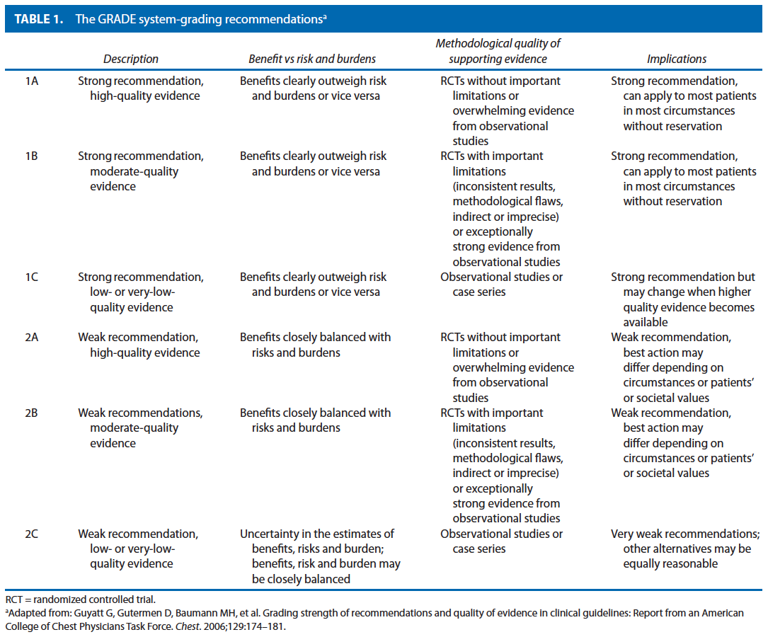
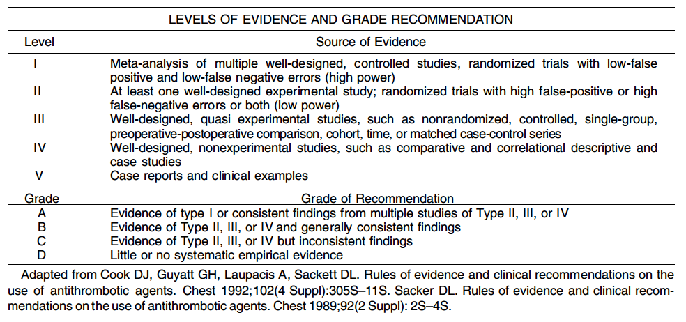
This parameter is based on the previous parameter published in 2004. An organized search of MEDLINE, PubMed, Embase, and the Cochrane Database of Collected Reviews was performed through February 2010. Key word combinations included colonic or colorectal neoplasms, adenocarcinoma, chemotherapy, colonoscopy, staging, lymph node, neoplasm metastasis, peritoneal neoplasm, surgical procedures, and recurrence. Directed searches of the embedded references from the primary articles were also performed in selected circumstances. All English language articles and studies of adults were reviewed by the primary authors. In selected instances where a full article was not yet available, reports of conference proceedings were reviewed. Recommendations were formulated by the primary authors and reviewed by the entire Standards Committee. The final grade of recommendation was performed by using the Grades of Recommendation, Assessment, Development, and Evaluation (GRADE) system (Table 1) and reviewed by the entire Standards Committee.

EVALUATION AND RISK ASSESSMENT
A thorough disease history should be obtained eliciting diseasespecific symptoms, associated symptoms, and family history. Routine laboratory values, including CEA levels, should also be evaluated, as indicated. Grade of Recommendation: 1C

Patients with colon cancer are often asymptomatic. Some have symptoms of change in bowel habits, blood in their stool, anemia, or are found to be fecal occult blood positive. Less often, a patient may have pain or obstructive symptoms or symptoms of metastatic disease. A complete history, including family history and colon cancerspecific history can guide the surgeon to suspect hereditary cancer syndromes, look for associated pathology or metastatic disease, and initiate additional workup such as mutational analysis of the tumor. Patients meeting clinical criteria for or having a family history of an increased susceptibility to colorectal cancer should be referred for genetic counseling for formal evaluation.

Routine laboratory examinations including complete blood cell count, liver function tests, and chemistry panel should be performed, based upon patient comorbidities, as indicated for preparation for general anesthesia.2 Because anemia can be common in patients with colon cancer, a complete blood count including platelets is recommended before surgical intervention. Carcinoembryonic antigen levels should be assessed before elective surgery for colon cancer for the establishment of baseline values and during the surveillance period to monitor for signs of recurrence.3 Although higher levels of CEA have been correlated with poorer prognosis, the data are insufficient to justify the use of a high preoperative CEA as an indication for adjuvant therapy.4,5 A confirmed rise in the postoperative CEA during surveillance should prompt further investigation for recurrent disease.6 At present there is insufficient evidence to support the routine use of other tumor markers such as CA199 in the routine evaluation of patients with colon cancer.4

When possible, all patients with presumed or proven colon cancer should undergo a full colonic evaluation with histologic assessment of the colonic lesion before treatment. Grade of Recommendation: 1C

Although the majority of patients are diagnosed with colon cancer during a full colonoscopy, new screening recommendations have recently been published that give alternatives to endoluminal examination.7–10 An increasing number of patients may be diagnosed by these newer methods and may be referred for surgical therapy without having previously undergone complete endoluminal examination with histologic tissue diagnosis. In cases without documented complete intracolonic evaluation, full endoluminal examination with biopsy is advocated. Whenever possible, the histologic diagnosis of colon cancer should be confirmed before elective surgical resection because nonneoplastic processes such as diverticulitis or IBD may be associated with the endoscopic appearance of colon cancer.

The risk for synchronous carcinomas or adenomas within the colon may be as high as 10% in the general population. Preoperative evaluation and diagnosis allow the surgeon to diagnose and potentially treat other colonic polyps, or, in the case of a synchronous cancer, choose the correct extent of colonic resection. The identification of synchronous cancers may also lead to workup for underlying predisposing risk factors such as inheritable colorectal cancer syndromes. In addition, endoscopic marking of the lesion location (tattoo) could be performed, especially in cases where laparoscopic resection is planned.

Some patients undergo colonoscopy, but the examination cannot be completed. In the absence of a clinically complete obstruction or perforation, a radiological study should be obtained to complete the colonic evaluation. These include contrast enemas (eg, barium enema) or preferably CT colonography or PET/CT colonography.11–13 In circumstances where the examination could not be completed but the patient meets indications for adjuvant chemotherapy, the completion colonoscopy should be performed soon after completion of chemotherapy.

Preoperative radiologicalo staging should be routinely performed. Grade of Recommendation: 1B

Preoperative radiographic staging including a CT scan of the chest, abdomen, and pelvis should be routinely performed before the elective surgical resection of colon cancer. This permits the detection and evaluation of the extent of synchronous metastases that may require a change in the treatment strategy, eg, chemotherapy rather than surgery first or potential simultaneous resection of both the primary tumor and the metastatic sites. The preoperative CT scan findings may also result in the operative plan being altered based on identification of the tumor location, adjacent organ or abdominal wall involvement, in addition to the presence of metastatic disease. In the event that a patient has been referred with CT imaging only of the abdomen and pelvis, at minimum, a preoperative chest xray should be obtained and a CT of the chest should be performed postoperatively. In patients with hypersensitivity to the iodine contrast dye, or in the appropriate clinical setting to work up indeterminate lesions on CT, an 18FDGPET fused CT scan or noncontrast chest CT with an MRI of the abdomen and pelvis may be considered. In some situations, a preoperative or intraoperative ultrasound may provide additional information.

The importance of preoperative imaging is also supported by practice guidelines of a number of national and international organizations, including the American Society of Clinical Oncology, the National Comprehensive Cancer Network, and the Association of Coloproctology of Great Britain and Ireland, the European Society of Medical Oncology, and others.3,14,15

STAGING OF COLON CANCER
Colon cancer staging should be performed according to the American Joint Committee on Cancer (AJCC)/TNM system and include an assessment of the completeness of surgical resection designated by the residual tumor code “R.” Grade of Recommendation: 1B

The tumor depth, nodal metastasis, and distant metastasis have been shown to be predictors of prognosis in colon cancer. These characteristics are described by the Union for International Cancer Control/AJCC TNM staging system, initially described in 2002, but that recently has been updated with the 7th edition of the AJCC Cancer Staging Manual16 and is presented in Table 1. The new system is based on observed survival outcomes within the US populationbased Surveillance Epidemiology and End Results cancer registry.17 Further stratification of tumor deposits and types of metastases to reflect the continuously evolving treatment of patients with advanced disease has been incorporated into the new edition of the staging system.18 In addition, prognostic calculators and nomograms have been proposed and may be considered in the future.19,20

In addition to TNM staging, the histologic grade of the tumor as well as the completeness of the resection should be assessed. Histologic grade has also been shown to be an important predictor of outcome and is an important consideration for treatment recommendations. The absence or presence of residual tumor following resection is designated by the letter R in accordance with the AJCC prognostic factors, as indicated below, and where possible should be indicated in the operative report:

• R0—complete tumor resection with all margins histologically negative
• R1—incomplete tumor resection with microscopic surgical resection margin involvement (margins grossly uninvolved)
• R2—incomplete tumor resection with gross residual tumor that was not resected (primary tumor, regional nodes, macroscopic margin involvement).21

SURGICAL TREATMENT OF THE PRIMARY TUMOR
The primary treatment for localized resectable colon cancer is colectomy with en bloc removal of all associated regional lymph nodes and involved adjacent structures. A thorough exploration should be performed at the time of resection. The value of the “no touch” technique in which the vascular supply to and from the tumor are divided before manipulating the tumor has remained controversial, and definite benefit has not been demonstrated.22,23 However a principle of gentle handling of the tumor during operation should be observed to avoid the risk for tumor spillage or perforation, in particular, in the management of locally advanced tumors or those with associated abscess.

a. A thorough surgical exploration should be performed and the findings documented in the operative report. Grade of Recommendation: 1C

The surgical exploration includes a visual or palpatory assessment of the peritoneal cavity and the abdominal organs to detect or rule out synchronous lesions, more advanced malignant disease (carcinomatosis, adjacent organ involvement, occult metastasis), or coexisting pathology (eg, adhesions, hernia, cholelithiasis, cirrhosis, etc.).14,21

b. The extent of resection of the colon should correspond to the lymphovascular drainage of the site of the colon cancer. The lymphadenectomy should be complete and en bloc with the bowel segment. Grade of Recommendation: 1A

The extent of a curative resection for colon cancer depends on (1) the site of the primary lesion and (2) the lymphovascular drainage of the cancer site. The length of bowel resected is governed by the blood supply to that segment. In the absence of synchronous pathology, an anatomic colon resection for cancer should achieve at least a 5cm negative margin on either side of the tumor. A colotomy and local excision of a colon cancer is not an adequate surgical technique for curative resection. It is associated with a risk of tumor spillage into the peritoneal cavity, and the lack of a lymphadenectomy increases the risk of tumor progression.3,14,21

The mesentery to the tumorbearing segment of bowel should be removed to the origin of the named primary feeding vessel(s). This resection should be performed en bloc with preservation of the integrity of the colonic mesentery.24,25 In the absence of clinical involvement, a more radical resection above the primary feeding vessel has not been associated with improved survival (eg, superior mesenteric versus ileocolic lymph node dissection for ascending colon cancer). The complete surgical removal of the regional lymph nodes within the mesocolon allows for a curative resection and accurate pathologic staging of the disease. When suspected to be involved, the most apical lymph nodes should be marked on the specimen as their metastatic involvement is a negative prognostic indicator. Because the total number of lymph nodes evaluated at the time of resection has been associated with survival, the lymph node examination should be as complete as possible.26,27 It is recommended that at least 12 lymph nodes be evaluated to assign N0 stage, and the examination of fewer than 12 lymph nodes is a highrisk feature for stage II colon cancer.3 In the event that fewer than 12 lymph nodes are reported on the pathology report, the surgeon is encouraged to request processing and reporting of the specimen in accordance to the guidelines set forth by the College of American Pathologists.28,29

c. Clinically positive lymph nodes located outside the standard field of resection identified at the time of resection and suspected to contain metastatic disease should be biopsied or removed at the time of primary resection. Grade of Recommendation: 2B

If residual tumorbearing lymph nodes remain following sampling, the resection will be considered incomplete. If no residual tumorbearing lymph nodes remain, the resection may be considered complete.21 High ligation is defined by an extended lymphadenectomy beyond the primary lymph node distribution (eg, ligation and resection of inferior mesenteric vessels and lymph nodes at the aorta, rather than superior rectal vessels and lymph nodes at their origin for a distal sigmoid carcinoma). Standard ligation is performed at the origin of the primary feeding vessel and should include all associated lymph nodes. In the absence of clinical evidence for metastasis to the extended lymph node distribution, high ligation beyond the origin of the primary feeding vessel has not been shown to improve survival.30 However, in the context of modern systemic chemotherapy, resection of isolated metastases to retroperitoneal lymph nodes may be considered in the context of a multidisciplinary setting.

d. Resection of involved adjacent organs should be en bloc. Grade of Recommendation: 1B

Local tumor control is achieved by complete resection of the tumor en bloc with contiguously involved structures. It may not be possible to distinguish between inflammatory and malignant adhesions, and peritumoral adhesions have been shown to harbor malignant cells in more than 40% of cases. Therefore, peritumoral adhesions should not be divided, and the adherent structure should be excised en bloc.3,14,21 Available diagnostic modalities (eg, CT scan or MRI scan) should be used to facilitate the identification of adjacent organ involvement before surgical exploration, so that adequate preparation and assembly of a multidisciplinary team may be performed. Local tumor control is achieved by complete resection of the tumor en bloc with contiguously involved structures.31–33 Tumor debulking in the setting of resectable adjacent organ involvement should not be performed.

Synchronous colon cancers can be treated by 2 separate resections or subtotal colectomy. Grade of Evidence: 1B

In recent years, the incidence of synchronous primary colorectal cancers is estimated to be 2% to 5% of all patients presenting with primary colorectal cancer, although the true incidence is probably related to underlying screening intensity.9,34,35 Patients with synchronous tumors should be evaluated for an associated genetic colorectal cancer syndrome and their treatment appropriately tailored. Synchronous pathology (cancer or endoscopically unresectable polyps) may be safely managed by an extended resection incorporating both lesions or 2 separate resections.36

When associated with underlying colonic disease (eg, chronic ulcerative colitis or hereditary nonpolyposis colorectal cancer syndrome), the extent of resection should consider treatment of the underlying disorder. For example, carcinoma arising in the setting of chronic ulcerative colitis should be treated with a proctocolectomy, whereas carcinoma arising in the setting of hereditary nonpolyposis colorectal cancer may be treated by either tumordirected segmental resection or by a more extensive resection tailored to the underlying risk of the patient37,38

Sentinel lymph node (SLN) mapping for colon cancer does not replace standard lymphadenectomy. Grade of Recommendation: 1B

Despite continued interest in SLN mapping and ultrastaging for colon cancer, to date, the results of SLN mapping for staging remain discordant and are not sufficiently accurate for identifying lymph node metastases, with a particular concern for the high rate of falsenegative nodal staging.39–43 A potential benefit of SLN techniques could be to support the pathologist in identifying lymph nodes ex vivo and hence decrease the risk for understaging the tumor. Furthermore, the clinical significance of micrometastatic disease within the SLN (as identified by hematoxylin and eosin staining or immunohistochemical and APC gene polymerase chain reaction analysis) remains undefined.44,45 Immunohistochemical ultrastaging currently does not appear to be clinically relevant, but polymerase chain reaction amplification of tumorrelated genes may show promise.

Laparoscopic and open colectomy achieve equivalent oncological outcomes for localized colon cancer. The use of the laparoscopic approach should be based on the surgeon’s documented experience in laparoscopic surgery as well as on patient and tumorspecific factors. Grade of Recommendation: 1A

The laparoscopic procedure should achieve the same goals as the open approach; a conversion to a laparotomy approach is otherwise recommended. A number of large multiinstitutional randomized trials with experienced participating surgeons in the United States and internationally have demonstrated equivalent overall and recurrencefree survival rates after laparoscopic in comparison with open surgical resection of localized colon cancer, excluding tumors within the rectum or transverse colon. One randomized trial did demonstrate a survival benefit with laparoscopic surgery, but other studies have not reproduced this observation.46 The short and longterm oncological outcomes have also been shown to be equivalent between patients treated with an open or laparoscopic approach.43,47–52 This practice parameter advocates the American Society of Colon and Rectal Surgeons position statement regarding credentialing of surgeons to perform laparoscopic colectomy for cancer.53

Treatment of the malignant polyp is determined by the morphology and histology of the polyp. Grade of Recommendation: 1B

Adenomatous polyps of the colon can be classified to be pedunculated or sessile. A malignant adenomatous polyp is defined as one in which cancer is invading through the muscularis mucosae into the submucosa (T1). It is estimated that 2% to 5% of adenomatous polyps will be associated with invasive malignancy. A number of classification systems have been developed to describe malignant polyps on the basis of which guidance for clinical management can be developed (Haggitt, Sm, Japanese Society for Cancer of the Colon and Rectum, and the Paris endoscopic classification of superficial GI neoplastic lesions).54–56

Endoscopic management can be sufficient for selected malignant polyps, if they are pedunculated or have protruding morphology and are lowrisk lesions with no adverse histologic features. Lowrisk polyps are those that are completely removed, preferably after submucosal elevation, without specimen fragmentation, are not poorly differentiated, and show no lymphatic or vascular invasion, or extension of the tumor to the margin of stalk resection. For lesions meeting lowrisk criteria, endoscopic resection followed by observation is appropriate. Those with adverse histologic features are at a higher risk for nodal metastases and formal oncological resection should be performed after weighing the risk of surgery against the risk of tumor progression.7,8,57

PROPHYLACTIC ONCOLOGICAL RESECTION OF EXTRAINTESTINAL ORGANS
Oophorectomy is advised for grossly abnormal ovaries or contiguous extension of the colon cancer, but routine prophylactic oophorectomy is not necessary. Grade of Recommendation: 1C

The ovaries are the site for colon cancer metastasis in fewer than 15% of patients, but colon cancer metastases to the ovaries can reach a considerable size (Krukenberg tumor). At this time, there are insufficient data to support routine prophylactic oophorectomy at the time of colectomy; however, oophorectomy should be performed during resection of the primary tumor with curative intent in patients suspected or known to have ovarian involvement, either by direct extension or metastasis.58 If 1 ovary is involved with metastatic disease, a bilateral oophorectomy should be performed. Limited data exist regarding prophylactic oophorectomy in women with colon cancer without other risk factors for ovarian pathology such as hereditary nonpolyposis colorectal cancer or BRCA.59 Routine prophylactic oophorectomy of normalappearing ovaries has not been associated with improved survival; however, there are insufficient data to recommend strongly for or against it.54 Oophorectomy may be considered in postmenopausal women after preoperative consultation, or in women at risk for ovarian cancer.

MANAGEMENT OF SYNCHRONOUS STAGE IV DISEASE
Approximately 15% to 20% of patients with have liver or lung metastases at the time of initial presentation with colon cancer. Among these approximately 20% to 25% of patients will have potentially resectable disease.60 The treatment of patients presenting with synchronous stage IV disease should be individualized and guided by a multidisciplinary team of diseasesite surgeons (colorectal surgeon, hepatic and/or thoracic surgeon) and medical oncologist.61,62 Patients may be classified as initially having resectable or potentially resectable disease, and unresectable disease with respect to both their primary tumor site and metastases.

Resectable Stage IV Disease
The treatment of patients with resectable stage IV colon cancer should be individualized based on comprehensive multidisciplinary evaluation. Grade of Recommendation: 1B

The treatment of patients with resectable metastatic colon cancer should be individualized and determined by multidisciplinary consensus. When the metastatic disease is potentially resectable, resection of the primary tumor should be complete and radical consistent with oncological principals of resection for localized disease as previously outlined in this document. In general, medically fit patients with resectable hepatic and/or pulmonary metastases will benefit from curative resection of the metastases.63 The sequence of chemotherapy, resection of the primary tumor, and resection of metastasis should be individualized and determined by multidisciplinary consensus.61,62 The disease of some patients may be converted to resectable after systemic chemotherapy.64 Neoadjuvant approaches to systemic chemotherapy before surgical resection may assist in identifying patients who are candidates for surgical resection.65,66 Patient survival is improved by the addition of systemic chemotherapy to surgical resection.60,67

Unresectable Stage IV Disease
Palliative intervention or resection of the symptomatic primary tumor should be considered, but routine resection of the asymptomatic primary tumor is not recommended. Grade of Recommendation: 1B

Patients with unresectable metastatic disease should be treated with systemic chemotherapy with palliative intent. With sequential therapies, the current median survival among patients with unresectable metastatic colon cancer is currently greater than 24 months and may be as long as 34 months.68,69 Previous studies have evaluated the role of primary resection in patients with stage IV disease and demonstrated an association with improved survival.70 However, these observational studies are limited by a significant influence of selection bias and outdated chemotherapy regimens. More recent prospective data support only selective primary tumor resection for treatment of symptoms, and patients who are asymptomatic from their primary tumor may therefore be closely followed with serial endoscopic evaluation for obstruction.71 Routine resection of the asymptomatic primary tumor is not recommended.72,73

TUMORRELATED EMERGENCIES
Tumor complications (bleeding, perforation, and obstruction) are serious and potentially lifethreatening conditions of locally advanced tumors. The goals of treatment for these conditions are to 1) avert the immediate negative impact of the complication (eg, death, sepsis), 2) achieve the best possible tumor control, and 3) ensure timely recovery to permit initiation of appropriate adjuvant or systemic treatment.

Bleeding
Surgical resection to stop severe blood loss from localized colon cancer should follow the same oncological principles as in elective resection. Grade of Recommendation: 2C

Acute massive lower GI bleeding from a colon cancer is a rare complication, whereas chronic blood loss is very common. Immediate management includes resuscitation of the patient and potential selective embolization, but surgical resection is the most effective and definitive approach. Preoperative or intraoperative efforts to localize the site of bleeding may be pursued in the clinically stable patient.74 In the very uncommon instance in which the site of bleeding cannot be determined either before operation or intraoperatively, but a colonic tumor is suspected, a subtotal colectomy adhering to oncological principles to each segment of the colon may be considered.

Perforation
Perforation is a lifethreatening complication. After resuscitation of the patient, surgical resection to address both the perforation and the tumor should be performed, if at all possible. Grade of Recommendation: 1B

The overall prognosis of colon perforation due to a colon cancer is significantly worse (associated with advanced tumor disease or sepsis) than perforation from other causes, but it is influenced by factors such as whether the perforation occurs at the tumor proximal to an obstructing tumor in an uninvolved segment of the bowel.75,76 When perforation of uninvolved colon proximal to an obstructing tumor has occurred, whenever possible, resection of the tumor following the oncological principles outlined above should be performed in addition to resection of the perforated segment. In most instances, an ostomy will provide effective fecal diversion and allow for patient recovery until the acute peritonitis has resolved. If the perforation occurs at the site of the tumor but is contained by adjacent structures, resection should ideally incorporate the adjacent structures en bloc. In cases of free perforation with peritonitis, the involved segment should be resected and proximal fecal diversion constructed. A primary anastomosis (with/without proximal diversion) may be considered in selected patients with minimal contamination, healthy tissue quality, and clinical stability.77

Obstruction
The management of patients with an obstructing cancer should be individualized but may include a definitive surgical resection with primary anastomosis. Grade of Recommendation: 1B

Options for the treatment of obstructing tumors depend on the site of obstruction and the presence of proximal colonic distention with fecal load. Options for treatment may include resection with or without anastomosis (eg, Hartmann resection), resection of the distended bowel (eg, subtotal/total colectomy), or temporary relief of obstruction and fecal load (eg, preoperative stenting as a bridge to resection).78,79 The prognosis among patients with obstructing cancers may be worse than among those without obstruction because of the inherently more advanced nature of their disease. However, this does not preclude the potential for curative resection.80

For tumors of the right or transverse colon, a tumordirected resection removes the distended colon segment, and an enterocolonic anastomosis can generally be safely achieved. Performance of an anastomosis in this setting depends on the patient’s general condition at the time of resection and the absence of other factors that indicate the need for a stoma to be created. During curative resection, the principles of oncological resection, including radical lymphadenectomy, should be observed.

A variety of surgical options exists for patients who present with a leftsided colon obstruction from cancer. Appropriate surgical approaches include resection with end colostomy and Hartmann pouch, resection with primary anastomosis, and subtotal colectomy with ileorectal anastomosis. In selected patients, successful preoperative stenting may allow for colonic decompression, metabolic and nutritional recovery, and adequate workup (operability, colonic evaluation) to optimize subsequent elective resection.81 Patients should be carefully selected, however, because a randomized trial of palliative stenting versus surgery was prematurely closed owing to an unexpectedly high rate of perforations in the stented group.82 The selection of the surgical approach should consider the patient’s general condition at the time of resection as well as the quality of the proximal bowel. The morbidity and mortality of a segmental resection, following intraoperative colonic irrigation, among patients with left colonic malignant obstruction has been compared with subtotal colectomy and has not been shown to differ in a randomized trial.83 More recent studies have demonstrated that colonic irrigation may not be mandatory before primary bowel anastomosis in this setting.84,85

Management of Locoregional Recurrence
The treatment of patients with locoregionally recurrent colon cancer should be multidisciplinary, and curative resection should adhere to the principles of primary resection. Grade of Recommendation: 1C

The risk for locoregional recurrence as the first and only site of recurrence following curative resection of localized colon cancer is low, approximately 2% to 3%.86,87 Salvage surgical resection is possible in up to approximately 30% of patients.88 Multimodality treatment, where indicated, provides the patients with the greatest potential for cure with a 5year survival estimate of 27% to 37%.89,90 Factors predictive of prolonged survival following surgical salvage include the completeness of resection (R0), early stage of initial disease, no associated distant disease, unifocal site of recurrence, and lack of retroperitoneal involvement.

Management of Peritoneal Carcinomatosis
The treatment of patients with peritoneal carcinomatosis should be multidisciplinary and individualized and may include surgical cytoreduction. The role of intraperitoneal chemotherapy remains insufficiently defined.

Grade of Recommendation: 2C
Peritoneal carcinomatosis will occur in an estimated 10% to 15% of patients with colorectal cancer. Newer and more effective systemic chemotherapeutic agents and targeted biological therapies have improved survival outcomes in patients with metastatic colorectal cancer. However, there remains a paucity of experimental evidence to guide the treatment of patients with carcinomatosis. The most welldescribed approach includes the combination of cytoreductive surgery in conjunction with perioperative intraperitoneal chemotherapy with or without hyperthermia.91,92 However, most studies report small singlecenter experiences with predominantly 5fluorouracil (5FU)onlybased systemic therapy, and the morbidity associated with cytoreductive surgery with multivisceral resection and hyperthermic intraperitoneal chemotherapy is as high as 60% to 80%.93 As of yet, unanswered questions include the definition of optimal cytoreduction, the impact of systemic therapy by using modern agents, and the role for intraperitoneal chemotherapy.94,95 Registration of patients in clinical treatment protocols is strongly encouraged.

Palliative Procedures
In patients with extensive incurable extent of tumor burden, palliative surgical interventions should be individualized based on the presence of symptoms. Grade of Recommendation: 1B

Patients who present with widely metastatic colon cancer are usually not candidates for surgical cure. Other patients may not be candidates for radical, curative resection because of systemic comorbidities. In these situations, a multidisciplinary approach to potential palliation should be recommended. The goals of palliation should be relief of symptoms caused by the cancer and maintenance of quality of life. In asymptomatic patients, prophylactic resection of the primary tumor is generally not necessary.71,72,96 Patients with asymptomatic primary lesions in the setting of distant metastasis should be referred for systemic chemotherapy unless initial resection of the primary tumor is determined to be the first stage of the multidisciplinary curative treatment plan. Palliative surgical interventions for obstruction of the GI tract or intractable bleeding caused by colon cancer include resection, endoluminal stent therapy, ablative procedures, internal bypass, or creation of a diverting stoma.81 The avoidance of resection in asymptomatic patients allows the patient to more rapidly initiate systemic chemotherapy, averts the risk for surgical morbidity, and results in improved outcomes. While observing patients with intact primary tumors, serial endoscopic evaluation should be performed to detect evidence for progressive disease and permit interventions to avoid acute obstruction. An individual patient’s overall life expectancy should also be considered in the decision for the type of palliative intervention (eg, resection or stent).

Recommendations Regarding Documentation
The surgical report for colorectal cancer should include information regarding the diagnostic workup, intraoperative findings, and technical details of the procedure. Grade of Recommendation: 1C

The ideal surgical report should clearly communicate the workup, intraoperative findings, and technical details of the procedure. The report should include a description of preoperative treatments and relevant workup and findings on exploration, including the presence of synchronous metastases or gross involvement of mesenteric lymph nodes, tumor site, and adjacent organ involvement. The report should also describe treatment details, including type of incision, extent of bowel and mesenteric resection, anastomotic technique, en bloc resection of contiguously involved organs, and an intraoperative assessment of the completeness of resection including margin status.14,21,28

Adjuvant Therapy
Decisions regarding adjuvant treatment following curatively resected colon cancer should be based on the clinical findings at resection, including stage of disease and patient comorbidities. The choice of the adjuvant chemotherapy regimen should be made jointly by the patient and the physician. Radiation therapy plays a minimal role in the adjuvant treatment of colon cancer.

Adjuvant chemotherapy should be recommended for patients with stage III colon cancer. Grade of Recommendation: 1A

A number of large multiinstitutional US and international randomized clinical trials have demonstrated the survival benefit with adjuvant chemotherapy. Pooled data from randomized trials demonstrates a 30% reduction in the risk for recurrence and a 26% reduction in the risk for death with fluoropyrimidinebased therapy administered for 6 months.97–99 More recently, the addition of oxaliplatin to fluoropyrimidine (eg, 5FU) chemotherapy has been shown to effect an additional approximately 20% reduction in relative risk for recurrence or death corresponding to an approximately 5% absolute survival benefit at 5 years with combination 5FU and leucovorin (LV) with oxaliplatin in comparison with 5FU alone.100,101 Therefore, the firstline adjuvant chemotherapy regimen should include a fluoropyrimidine (5FU/LV or capecitabine) and oxaliplatin. However, grade 3 peripheral sensory neuropathy occurs in approximately 12% of patients who receive oxaliplatin, which may make it unsuitable for some patients.102 The addition of irinotecan in combination with 5FU was studied in several phase III randomized controlled trials in the United States and internationally and was demonstrated to yield no survival benefit when compared to 5FU/LV alone.103,104 At present, there is no role for the addition of irinotecan in the adjuvant setting after resection of localized colon cancer.

The role of the biological agents, such as the vascular endothelial growth factor inhibitor bevacizumab or the epidermal growth factor receptor inhibitor cetuximab and panitumumab along with other targeted agents, has been the subject of recent investigation. Unfortunately, 3 separate phase III multiinstitutional trials have failed to demonstrate added benefit with the addition of either bevacizumab (NSABP C08, AVANT) or epidermal growth factor receptor inhibitors (NCCTG N0147) to FOLFOX alone105–107 At present, there is no evidence to support the routine addition of biological agents in the adjuvant setting.

Adjuvant chemotherapy may be considered for patients with highrisk stage II colon cancer. Grade of Recommendation: 2B

There are conflicting data regarding the role of adjuvant chemotherapy in stage II colon cancer. Most of the previous randomized trials of adjuvant therapy for colon cancer have enrolled both stage II and stage III patients, and some have demonstrated a small difference corresponding to a potential absolute improvement in overall survival of approximately 2% to 3% with 5FU/LV and 3% to 4% with FOLFOX.101,108–110 However, the proportion of patients with stage II cancers were approximately 20% to 25% in these trials, and definitive conclusions, given such a small effect, have not been possible. However, a subgroup of highrisk patients with nodenegative colon cancer may benefit. In the United Kingdom, a study of 5FU adjuvant chemotherapy for stage II colon cancer was associated with a 20% relative risk reduction for recurrence associated with adjuvant therapy. However, a significant proportion of the patients had fewer than 12 lymph nodes examined, and the potential impact of understaging in this study is unknown.111 The benefit of adjuvant chemotherapy in stage II patients has not been definitively shown, and patients with stage II colon cancer should be encouraged to participate in adjuvant therapy clinical trials. Patients with stage II colon cancer are considered to be at high risk in the presence of T4 stage, perforation, peritumoral lymphovascular or neural involvement, or poorly differentiated histology. In addition, those patients in whom fewer than 12 lymph nodes were evaluated may also be considered to be at high risk, and adjuvant chemotherapy may be recommended.3

More recently, new molecular prognostic markers have emerged and are under investigation. Tumors that are microsatellite instabilityhigh (MSIH) appear to have improved prognosis and limited benefit from 5FUbased chemotherapy. Conversely, loss of heterozygosity at chromosome 18q (DCC) has been associated with poor prognosis, although the true value of this factor has not yet been fully elucidated.4,112 Although recently developed and commercially available genomic profiling tools have demonstrated prognostic information in patients with stage II colon cancers, their utility for determining treatment response could not be established, and there is no clear role for their use in treatment stratification.113,114

APPENDIX A: CONTRIBUTING MEMBERS OF THE ASCRS STANDARDS COMMITTEE
Committee Members: W. Donald Buie, M.D., Chair, Janice Rafferty, M.D., Cochair, Jose Guillem, M.D., Council Representative, Robin Boushey, M.D., George Chang, M.D., Daniel Feingold, M.D., Philip Fleshner, M.D., Jill Genua, M.D., Kerry Hammond, M.D., William Harb, M.D., Samantha Hendren, M.D., Daniel Herzig, M.D., Andreas Kaiser, M.D., David Larson, M.D., Sang Lee, M.D., James McCormick, D.O., Genevieve MeltonMeaux, M.D., Steven Mills, M.D., John Monson, M.D., Harvey Moore III, M.D., W. Brian Perry, M.D., P. Terry Phang, M.D., David Rivadeneira, M.D., Howard Ross, M.D., Scott Steele, M.D., Scott Strong, M.D., Charles Ternent, M.D., Madhulika Varma, M.D., Martin Weiser, M.D., Kirsten Wilkins, M.D.
REFERENCES
1. Jemal A, Siegel R, Xu J, Ward E. Cancer statistics, 2010. CA Cancer J Clin. 2010;60:277–300.
2. Practice advisory for preanesthesia evaluation: a report by the American Society of Anesthesiologists Task Force on Preanesthesia Evaluation. Anesthesiology. 2002;96:485–496.
3. Engstrom PF, Arnoletti JP, Benson AB III, et al. NCCN clinical practice guidelines in oncology: colon cancer. J Natl Compr Canc Netw. 2009;7:778–831.
4. Locker GY, Hamilton S, Harris J, et al. ASCO 2006 update of recommendations for the use of tumor markers in gastrointestinal cancer. J Clin Oncol. 2006;24:5313–5327.
5. Harrison LE, Guillem JG, Paty P, Cohen AM. Preoperative carcinoembryonic antigen predicts outcomes in nodenegative colon cancer patients: a multivariate analysis of 572 patients. J Am Coll Surg. 1997;185:55–59.
6. Wiratkapun S, Kraemer M, SeowChoen F, Ho YH, Eu KW. High preoperative serum carcinoembryonic antigen predicts metastatic recurrence in potentially curative colonic cancer: results of a fiveyear study. Dis Colon Rectum. 2001;44:231–235.
7. McFarland EG, Fletcher JG, Pickhardt P, et al. ACR Colon Cancer Committee white paper: status of CT colonography 2009. J Am Coll Radiol. 2009;6:756–772.e4.
8. Regge D, Laudi C, Galatola G, et al. Diagnostic accuracy of computed tomographic colonography for the detection of advanced neoplasia in individuals at increased risk of colorectal cancer. JAMA. 2009;301:2453–2461.
9. Johnson CD, Chen MH, Toledano AY, et al. Accuracy of CT colonography for detection of large adenomas and cancers. N Engl J Med. 2008;359:1207–1217.
10. Levin B, Lieberman DA, McFarland B, et al. Screening and surveillance for the early detection of colorectal cancer and adenomatous polyps, 2008: a joint guideline from the American Cancer Society, the US MultiSociety Task Force on Colorectal Cancer, and the American College of Radiology. CA Cancer J Clin. 2008;58:130–160.
11. Macari M, Berman P, Dicker M, Milano A, Megibow AJ. Usefulness of CT colonography in patients with incomplete colonoscopy. AJR Am J Roentgenol. 1999;173:561–564.
12. Yucel C, LevToaff AS, Moussa N, Durrani H. CT colonography for incomplete or contraindicated optical colonoscopy in older patients. AJR Am J Roentgenol. 2008;190:145–150.
13. VeitHaibach P, Kuehle CA, Beyer T, et al. Diagnostic accuracy of colorectal cancer staging with wholebody PET/CT colonography. JAMA. 2006;296:2590–2600.
14. Association of Coloproctology of Great Britain and Ireland. Guidelines for the Management of Colorectal Cancer. 3rd ed. London, United Kingdom: The Association of Coloproctology of Great Britain and Ireland; 2007.
15. Van Cutsem E, Oliveira J. Primary colon cancer: ESMO clinical recommendations for diagnosis, adjuvant treatment and followup. Ann Oncol. 2009;20(suppl 4):49–50.
16. Edge SB, Byrd DR, Compton CC, Fritz AG, Greene FL, Trotti A, eds. AJCC Cancer Staging Manual. 7th ed. New York, NY: Springer; 2010.
17. Gunderson LL, Jessup JM, Sargent DJ, Greene FL, Stewart AK. Revised TN categorization for colon cancer based on national survival outcomes data. J Clin Oncol. 2010;28:264–271.
18. Puppa G, Ueno H, Kayahara M, et al. Tumor deposits are encountered in advanced colorectal cancer and other adenocarcinomas: an expanded classification with implications for colorectal cancer staging system including a unifying concept of intransit metastases. Mod Pathol. 2009;22:410–415.
19. Chang GJ, Hu CY, Eng C, Skibber JM, RodriguezBigas MA. Practical application of a calculator for conditional survival in colon cancer. J Clin Oncol. 2009;27:5938–5943.
20. Weiser MR, Landmann RG, Kattan MW, et al. Individualized prediction of colon cancer recurrence using a nomogram. J Clin Oncol. 2008;26:380–385.
21. Nelson H, Petrelli N, Carlin A, et al. Guidelines 2000 for colon and rectal cancer surgery. J Natl Cancer Inst. 2001;93:583–596.
22. Hayashi N, Egami H, Kai M, Kurusu Y, Takano S, Ogawa M. Notouch isolation technique reduces intraoperative shedding of tumor cells into the portal vein during resection of colorectal cancer. Surgery. 1999;125:369–374.
23. Slanetz CA Jr, Grimson R. Effect of high and intermediate ligation on survival and recurrence rates following curative resection of colorectal cancer. Dis Colon Rectum. 1997;40:1205–1219.
24. West NP, Hohenberger W, Weber K, Perrakis A, Finan PJ, Quirke P. Complete mesocolic excision with central vascular ligation produces an oncologically superior specimen compared with standard surgery for carcinoma of the colon. J Clin Oncol. 2010;28:272–278.
25. West NP, Morris EJ, Rotimi O, Cairns A, Finan PJ, Quirke P. Pathology grading of colon cancer surgical resection and its association with survival: a retrospective observational study. Lancet Oncol. 2008;9:857–865.
26. Chang GJ, RodriguezBigas MA, Skibber JM, Moyer VA. Lymph node evaluation and survival after curative resection of colon cancer: systematic review. J Natl Cancer Inst. 2007;99:433–441.
27. Wong SL, Ji H, Hollenbeck BK, Morris AM, Baser O, Birkmeyer JD. Hospital lymph node examination rates and survival after resection for colon cancer. JAMA. 2007;298:2149–2154.
28. Compton CC. Key issues in reporting common cancer specimens: problems in pathologic staging of colon cancer. Arch Pathol Lab Med. 2006;130:318–324.
29. Washington MK, Berlin J, Branton P, et al. Protocol for the examination of specimens from patients with primary carcinoma of the colon and rectum. Arch Pathol Lab Med. 2009;133:1539–1551.
30. Kawamura YJ, Umetani N, Sunami E, Watanabe T, Masaki T, Muto T. Effect of high ligation on the longterm result of patients with operable colon cancer, particularly those with limited nodal involvement. Eur J Surg. 2000;166:803–807.
31. Gebhardt C, Meyer W, Ruckriegel S, Meier U. Multivisceral resection of advanced colorectal carcinoma. Langenbecks Arch Surg. 1999;384:194–199.
32. Govindarajan A, Fraser N, Cranford V, et al. Predictors of multivisceral resection in patients with locally advanced colorectal cancer. Ann Surg Oncol. 2008;15:1923–1930.
33. Poeze M, Houbiers JG, van de Velde CJ, Wobbes T, von Meyenfeldt MF. Radical resection of locally advanced colorectal cancer. Br J Surg. 1995;82:1386–1390.
34. Takeuchi H, Toda T, Nagasaki S, et al. Synchronous multiple colorectal adenocarcinomas. J Surg Oncol. 1997;64:304–307.
35. Kim MS, Park YJ. Detection and treatment of synchronous lesions in colorectal cancer: the clinical implication of perioperative colonoscopy. World J Gastroenterol. 2007;13:4108–4111.
36. Whelan RL, Wong WD, Goldberg SM, Rothenberger DA. Synchronous bowel anastomoses. Dis Colon Rectum. 1989;32:365–368.
37. Church J, Simmang C. Practice parameters for the treatment of patients with dominantly inherited colorectal cancer (familial adenomatous polyposis and hereditary nonpolyposis colorectal cancer). Dis Colon Rectum. 2003;46:1001–1012.
38. Guillem JG, Wood WC, Moley JF, et al. ASCO/SSO review of current role of riskreducing surgery in common hereditary cancer syndromes. J Clin Oncol. 2006;24:4642–4660.
39. Lim SJ, Feig BW, Wang H, et al. Sentinel lymph node evaluation does not improve staging accuracy in colon cancer. Ann Surg Oncol. 2008;15:46–51.
40. Bertagnolli M, Miedema B, Redston M, et al. Sentinel node staging of resectable colon cancer: results of a multicenter study. Ann Surg. 2004;240:624–630.
41. Bembenek AE, Rosenberg R, Wagler E, et al. Sentinel lymph node biopsy in colon cancer: a prospective multicenter trial. Ann Surg. 2007;245:858–863.
42. Stojadinovic A, Nissan A, Protic M, et al. Prospective randomized study comparing sentinel lymph node evaluation with standard pathologic evaluation for the staging of colon carcinoma: results from the United States Military Cancer Institute Clinical Trials Group Study GI01. Ann Surg. 2007;245:846–857.
43. Des Guetz G, Uzzan B, Nicolas P, et al. Is sentinel lymph node mapping in colorectal cancer a future prognostic factor? A metaanalysis. World J Surg. 2007;31:1304–1312.
44. Bilchik AJ, Hoon DS, Saha S, et al. Prognostic impact of micrometastases in colon cancer: interim results of a prospective multicenter trial. Ann Surg. 2007;246:568–577.
45. Redston M, Compton CC, Miedema BW, et al. Analysis of micrometastatic disease in sentinel lymph nodes from resectable colon cancer: results of Cancer and Leukemia Group B Trial 80001. J Clin Oncol. 2006;24:878–883.
46. Lacy AM, Delgado S, Castells A, et al. The longterm results of a randomized clinical trial of laparoscopyassisted versus open surgery for colon cancer. Ann Surg. 2008;248:1–7.
47. A comparison of laparoscopically assisted and open colectomy for colon cancer. The clinical outcomes of Surgical Therapy Study Group. N Engl J Med. 2004;350:2050–2059.
48. Bonjer HJ, Hop WC, Nelson H, et al. Laparoscopically assisted vs open colectomy for colon cancer: a metaanalysis. Arch Surg. 2007;142:298–303.
49. Buunen M, Veldkamp R, Hop WC, et al. Survival after laparoscopic surgery versus open surgery for colon cancer: longterm outcome of a randomised clinical trial. Lancet Oncol. 2009;10:44–52.
50. Liang JT, Shieh MJ, Chen CN, Cheng YM, Chang KJ, Wang SM. Prospective evaluation of laparoscopyassisted colectomy versus laparotomy with resection for management of complex polyps of the sigmoid colon. World J Surg. 2002;26:377–383.
51. Jayne DG, Guillou PJ, Thorpe H, et al. Randomized trial of laparoscopicassisted resection of colorectal carcinoma: 3year results of the UK MRC CLASICC Trial Group. J Clin Oncol. 2007;25:3061–3068.
52. Kuhry E, Schwenk WF, Gaupset R, Romild U, Bonjer HJ. Longterm results of laparoscopic colorectal cancer resection. Cochrane Database Syst Rev. 2008:CD003432.
53. American Society of Colon and Rectal Surgeons Position Statement. Laparoscopic Colectomy for Curable Cancer. 2004. Available at: http://www.fascrs.org/physicians/position_statements/laparoscopic_colectomy/. Accessed March 15, 2012.
54. YoungFadok TM, Wolff BG, Nivatvongs S, Metzger PP, Ilstrup DM. Prophylactic oophorectomy in colorectal carcinoma: preliminary results of a randomized, prospective trial. Dis Colon Rectum. 1998;41:277–285.
55. Kudo S. Endoscopic mucosal resection of flat and depressed types of early colorectal cancer. Endoscopy. 1993;25:455–461.
56. Repici A, Pellicano R, Strangio G, Danese S, Fagoonee S, Malesci
A. Endoscopic mucosal resection for early colorectal neoplasia: pathologic basis, procedures, and outcomes. Dis Colon Rectum. 2009;52:1502–1515.
57. Cooper HS. Pathologic issues in the treatment of endoscopically removed malignant colorectal polyps. J Natl Compr Canc Netw. 2007;5:991–996.
58. Sielezneff I, Salle E, Antoine K, Thirion X, Brunet C, Sastre B. Simultaneous bilateral oophorectomy does not improve prognosis of postmenopausal women undergoing colorectal resection for cancer. Dis Colon Rectum. 1997;40:1299–1302.
59. Banerjee S, Kapur S, Moran BJ. The role of prophylactic oophorectomy in women undergoing surgery for colorectal cancer. Colorectal Dis. 2005;7:214–217.
60. Nordlinger B, Sorbye H, Glimelius B, et al. Perioperative chemotherapy with FOLFOX4 and surgery versus surgery alone for resectable liver metastases from colorectal cancer (EORTC Intergroup trial 40983): a randomised controlled trial. Lancet. 2008;371:1007–1016.
61. Fahy BN, D’Angelica M, DeMatteo RP, Blumgart LH, Weiser MR, Ostrovnaya I, Gonen M, Jarnagin WR. Synchronous hepatic metastases from colon cancer: changing treatment strategies and results of surgical intervention. Ann Surg Oncol. 2009;16:361–370.
62. Hillingso JG, WilleJorgensen P. Staged or simultaneous resection of synchronous liver metastases from colorectal cancer: a systematic review. Colorectal Dis. 2009;11:3–10.
63. Kopetz S, Chang GJ, Overman MJ, et al. Improved survival in metastatic colorectal cancer is associated with adoption of hepatic resection and improved chemotherapy. J Clin Oncol. 2009;27:3677–3683.
64. Adam R, Wicherts DA, de Haas RJ, et al. Patients with initially unresectable colorectal liver metastases: is there a possibility of cure? J Clin Oncol. 2009;27:1829–1835.
65. Folprecht G, Grothey A, Alberts S, Raab HR, Kohne CH. Neoadjuvant treatment of unresectable colorectal liver metastases: correlation between tumour response and resection rates. Ann Oncol. 2005;16:1311–1319.
66. Gruenberger B, Tamandl D, Schueller J, Scheithauer W, Zielinski C, Herbst F, Gruenberger T. Bevacizumab, capecitabine, and oxaliplatin as neoadjuvant therapy for patients with potentially curable metastatic colorectal cancer. J Clin Oncol. 2008;26:1830–1835.
67. Portier G, Elias D, Bouche O, et al. Multicenter randomized trial of adjuvant fluorouracil and folinic acid compared with surgery alone after resection of colorectal liver metastases: FFCD ACHBTH AURC 9002 trial. J Clin Oncol. 2006;24:4976–4982.
68. Bokemeyer C, Bondarenko I, Makhson A, et al. Fluorouracil, leucovorin, and oxaliplatin with and without cetuximab in the firstline treatment of metastatic colorectal cancer. J Clin Oncol. 2009;27:663–671.
69. Grothey A, Sugrue MM, Purdie DM, et al. Bevacizumab beyond first progression is associated with prolonged overall survival in metastatic colorectal cancer: results from a large observational cohort study (BRiTE). J Clin Oncol. 2008;26:5326–5334.
70. Cook AD, Single R, McCahill LE. Surgical resection of primary tumors in patients who present with stage IV colorectal cancer: an analysis of surveillance, epidemiology, and end results data, 1988 to 2000. Ann Surg Oncol. 2005;12:637–645.
71. McCahill LE, Yothers GA, Sharif S, et al. A phase II trial of
5 fluorouracil, leucovorin, and oxaliplatin (mFOLFOX6) chemotherapy plus bevacizumab for patients with unresectable stage IV colon cancer and a synchronous asymptomatic primary tumor: results of NSBP C10 [abstract 3527]. J Clin Oncol. 2010;28(suppl):15s.
72. Poultsides GA, Servais EL, Saltz LB, et al. Outcome of primary tumor in patients with synchronous stage IV colorectal cancer receiving combination chemotherapy without surgery as initial treatment. J Clin Oncol. 2009;27:3379–3384.
73. Gunderson LL, Jessup JM, Sargent DJ, Greene FL, Stewart
A. Revised tumor and node categorization for rectal cancer based on surveillance, epidemiology, and end results and rectal pooled analysis outcomes. J Clin Oncol. 2010;28:256–263.
74. Barnert J, Messmann H. Management of lower gastrointestinal tract bleeding. Best Pract Res Clin Gastroenterol. 2008;22:295–312.
75. Abdelrazeq AS, Scott N, Thorn C, et al. The impact of spontaneous tumour perforation on outcome following colon cancer surgery. Colorectal Dis. 2008;10:775–780.
76. McArdle CS, McMillan DC, Hole DJ. The impact of blood loss, obstruction and perforation on survival in patients undergoing curative resection for colon cancer. Br J Surg. 2006;93:483–488.
77. Smithers BM, Theile DE, Cohen JR, Evans EB, Davis NC. Emergency right hemicolectomy in colon carcinoma: a prospective study. Aust N Z J Surg. 1986;56:749–752.
78. Govindarajan A, Naimark D, Coburn NG, Smith AJ, Law CH. Use of colonic stents in emergent malignant left colonic obstruction: a Markov chain Monte Carlo decision analysis. Dis Colon Rectum. 2007;50:1811–1824.
79. Brehant O, Fuks D, Bartoli E, Yzet T, Verhaeghe P, Regimbeau JM. Elective (planned) colectomy in patients with colorectal obstruction after placement of a selfexpanding metallic stent as a bridge to surgery: the results of a prospective study. Colorectal Dis. 2009;11:178–183.
80. De Salvo GL, Gava C, Pucciarelli S, Lise M. Curative surgery for obstruction from primary left colorectal carcinoma: primary or staged resection? Cochrane Database Syst Rev. 2004:CD002101.
81. Watt AM, Faragher IG, Griffin TT, Rieger NA, Maddern GJ. Selfexpanding metallic stents for relieving malignant colorectal obstruction: a systematic review. Ann Surg. 2007;246:24–30.
82. van Hooft JE, Fockens P, Marinelli AW, Bossuyt PM, Bemelman WA. Premature closure of the Dutch Stentin I study. Lancet. 2006;368:1573–1574.
83. Singlestage treatment for malignant leftsided colonic obstruction: a prospective randomized clinical trial comparing subtotal colectomy with segmental resection following intraoperative irrigation. The SCOTIA Study Group. Subtotal Colectomy versus Ontable Irrigation and Anastomosis. Br J Surg. 1995;82:1622–1627.
84. Lee YM, Law WL, Chu KW, Poon RT. Emergency surgery for obstructing colorectal cancers: a comparison between rightsided and leftsided lesions. J Am Coll Surg. 2001;192:719–725.
85. Lim JF, Tang CL, SeowChoen F, Heah SM. Prospective, randomized trial comparing intraoperative colonic irrigation with manual decompression only for obstructed leftsided colorectal cancer. Dis Colon Rectum. 2005;48:205–209.
86. Tsikitis VL, Malireddy K, Green EA, et al. Postoperative surveillance recommendations for early stage colon cancer based on results from the clinical outcomes of surgical therapy trial. J Clin Oncol. 2009;27:3671–3676.
87. Harris GJ, Church JM, Senagore AJ, et al. Factors affecting local recurrence of colonic adenocarcinoma. Dis Colon Rectum. 2002;45:1029–1034.
88. Delpero JR, Pol B, Le Treut P, et al. Surgical resection of locally recurrent colorectal adenocarcinoma. Br J Surg. 1998;85: 372–376.
89. Bowne WB, Lee B, Wong WD, et al. Operative salvage for locoregional recurrent colon cancer after curative resection: an analysis of 100 cases. Dis Colon Rectum. 2005;48:897–909.
90. Taylor WE, Donohue JH, Gunderson LL, et al. The Mayo Clinic experience with multimodality treatment of locally advanced or recurrent colon cancer. Ann Surg Oncol. 2002;9:177–185.
91. Glehen O, Kwiatkowski F, Sugarbaker PH, et al. Cytoreductive surgery combined with perioperative intraperitoneal chemotherapy for the management of peritoneal carcinomatosis from colorectal cancer: a multiinstitutional study. J Clin Oncol. 2004;22:3284–3292.
92. Elias D, Benizri E, Di Pietrantonio D, Menegon P, Malka D, Raynard B. Comparison of two kinds of intraperitoneal chemotherapy following complete cytoreductive surgery of colorectal peritoneal carcinomatosis. Ann Surg Oncol. 2007;14: 509–514.
93. Franko J, Gusani NJ, Holtzman MP, et al. Multivisceral resection does not affect morbidity and survival after cytoreductive surgery and chemoperfusion for carcinomatosis from colorectal cancer. Ann Surg Oncol. 2008;15:3065–3072.
94. Chang GJ, Lambert LA. Hidden opportunities in cytoreductive surgery for peritoneal carcinomatosis of colorectal origin. Ann Surg Oncol. 2008;15:2993–2995.
95. Bloemendaal AL, Verwaal VJ, van Ruth S, Boot H, Zoetmulder FA. Conventional surgery and systemic chemotherapy for peritoneal carcinomatosis of colorectal origin: a prospective study. Eur J Surg Oncol. 2005;31:1145–1151.
96. Scoggins CR, Meszoely IM, Blanke CD, Beauchamp RD, Leach SD. Nonoperative management of primary colorectal cancer in patients with stage IV disease. Ann Surg Oncol. 1999;6:651–657.
97. Haller DG, Catalano PJ, Macdonald JS, et al. Phase III study of fluorouracil, leucovorin, and levamisole in highrisk stage II and III colon cancer: final report of Intergroup 0089. J Clin Oncol. 2005;23:8671–8678.
98. Gill S, Loprinzi CL, Sargent DJ, et al. Pooled analysis of fluorouracilbased adjuvant therapy for stage II and III colon cancer: who benefits and by how much? J Clin Oncol. 2004;22:1797–1806.
99. Di Costanzo F, Sobrero A, Gasperoni S, et al. Adjuvant chemotherapy in the treatment of colon cancer: randomized multicenter trial of the Italian National Intergroup of Adjuvant Chemotherapy in Colon Cancer (INTACC). Ann Oncol. 2003;14:1365–1372.
100. Andre T, Boni C, Navarro M, et al. Improved overall survival with oxaliplatin, fluorouracil, and leucovorin as adjuvant treatment in stage II or III colon cancer in the MOSAIC trial. J Clin Oncol. 2009;27:3109–3116.
101. Kuebler JP, Wieand HS, O’Connell MJ, et al. Oxaliplatin combined with weekly bolus fluorouracil and leucovorin as surgical adjuvant chemotherapy for stage II and III colon cancer: results from NSABP C07. J Clin Oncol. 2007;25:2198–2204.
102. Land SR, Kopec JA, Cecchini RS, et al. Neurotoxicity from oxaliplatin combined with weekly bolus fluorouracil and leucovorin as surgical adjuvant chemotherapy for stage II and III colon cancer: NSABP C07. J Clin Oncol. 2007;25:2205–2211.
103. Van Cutsem E, Labianca R, Bodoky G, et al. Randomized phase III trial comparing biweekly infusional fluorouracil/leucovorin alone or with irinotecan in the adjuvant treatment of stage III colon cancer: PETACC3. J Clin Oncol. 2009;27:3117–3125.
104. Saltz LB, Niedzwiecki D, Hollis D, et al. Irinotecan fluorouracil plus leucovorin is not superior to fluorouracil plus leucovorin alone as adjuvant treatment for stage III colon cancer: results of CALGB 89803. J Clin Oncol. 2007;25:3456–3461.
105. Allegra CJ, Yothers G, O’Connell MJ, et al. Phase III trial assessing bevacizumab in stages II and III carcinoma of the colon: results of NSABP protocol C08. J Clin Oncol. 2011;29:11–16.
106. NCT00265811. Combination chemotherapy with or without cetuximab in treating patients with stage III colon cancer that was completely removed by surgery. Available at: http://clinicaltrials.gov/ct/show/NCT00265811.
107. de Gramont A, Chibaudel B, Bachet JB, et al. From chemotherapy to targeted therapy in adjuvant treatment for stage III colon cancer. Semin Oncol. 2011;38:521–532.
108. Andre T, Boni C, MounedjiBoudiaf L, et al. Oxaliplatin, fluorouracil, and leucovorin as adjuvant treatment for colon cancer. N Engl J Med. 2004;350:2343–2351.
109. Quasar Collaborative Group, Gray R, Barnwell J, McConkey C, et al. Adjuvant chemotherapy versus observation in patients with colorectal cancer: a randomised study. Lancet. 2007;370:2020–2029.
110. Wilkinson NW, Yothers G, Lopa S, Costantino JP, Petrelli NJ, Wolmark N. Longterm survival results of surgery alone versus surgery plus 5fluorouracil and leucovorin for stage II and stage III colon cancer: pooled analysis of NSABP C01 through
C 05. A baseline from which to compare modern adjuvant trials. Ann Surg Oncol. 2010;17:959–966.
111. Gray R, Barnwell J, McConkey C, Hills RK, Williams NS, Kerr DJ. Adjuvant chemotherapy versus observation in patients with colorectal cancer: a randomised study. Lancet. 2007;370:2020–2029.
112. Vicuna B, Benson AB III. Adjuvant therapy for stage II colon cancer: prognostic and predictive markers. J Natl Compr Canc Netw. 2007;5:927–936.
113. O’Connell MJ, Lavery I, Yothers G, et al. Relationship between tumor gene expression and recurrence in four independent studies of patients with stage II/III colon cancer treated with surgery alone or surgery plus adjuvant fluorouracil plus leucovorin. J Clin Oncol. 2010;28:3937–3944.
114. Haince JF, Houde M, Beaudry G, et al. Comparison of histopathology and RTqPCR amplification of guanylyl cyclase C for detection of colon cancer metastases in lymph nodes. J Clin Pathol. 2010;63:530–537.

Rectal Cancer Treatment Guidelines

Download PDF

Practice Parameters for the Management of Rectal Cancer (Revised)

Prepared by
The Standards Practice Task Force
The American Society of Colon and Rectal Surgeons
Joe J. Tjandra, M.D., John W. Kilkenny, M.D., W. Donald Buie, M.D.,
Neil Hyman, M.D., Clifford Simmang, M.D., Thomas Anthony, M.D.,
Charles Orsay, M.D., James Church, M.D., Daniel Otchy, M.D., Jeffrey Cohen, M.D.,
Ronald Place, M.D., Frederick Denstman, M.D., Jan Rakinic, M.D.,
Richard Moore, M.D., Mark Whiteford, M.D.

The American Society of Colon and Rectal Surgeons is dedicated to assuring highquality patient care by advancing the science, prevention, and management of disorders and diseases of the colon, rectum, and anus. The Standards Committee is composed of Society members who are chosen because they have demonstrated expertise in the specialty of colon and rectal surgery. This Committee was created to lead international efforts in defining quality care for conditions related to the colon, rectum, and anus. This is accompanied by developing Clinical Practice Guidelines based on the best available evidence. These guidelines are inclusive, and not prescriptive. Their purpose is to provide information on which decisions can be made, rather than dictate a specific form of treatment. These guidelines are intended for the use of all practitioners, health care workers, and patients who desire information about the management of the conditions addressed by the topics covered in these guidelines. It should be recognized that these guidelines should not be deemed inclusive of all proper methods of care or exclusive of methods of care reasonably directed to obtaining the same results. The ultimate judgment regarding the propriety of any specific procedure must be made by the physician in light of all of the circumstances presented by the individual patient.


STATEMENT OF THE PROBLEM
Colorectal adenocarcinoma is the second leading cause of cancer deaths in western countries. Rectal cancer comprises approximately 25 percent of the malignancies arising in the large bowel. The estimated occurrence of new rectal cancer cases in the United States was projected to be 40,570 during 2004.1

Anatomically, the rectum is the distal 18cm of the large bowel leading to the anal canal.2 Cancers of the intraperitoneal rectum behave like colon cancers with regard to recurrence patterns and prognosis.3 By contrast, the extraperitoneal rectum resides within the confines of the bony pelvis; it is this distal 10 to 12 cm that constitutes the rectum from the oncologic standpoint.

PREOPERATIVE ASSESSMENT
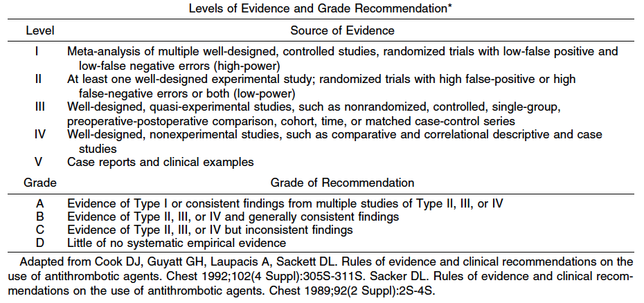
1. Patients should be evaluated for their medical fitness to undergo surgery. When an ostomy is a consideration, preoperative counseling with an enterostomal therapist should be offered when available. Level of Evidence: III; Grade of Recommendation: B.

Appraisal of operative risk, especially with respect to cardiopulmonary comorbidity, is an essential part of the preoperative process. History and physical examination are the cornerstones of diagnostic evaluation and may prompt further investigation and intervention to optimize operative risk. In selected cases, a nonsurgical approach to the lesion may be necessary. Several perioperative, riskassessment scoring systems have been published to help guide the surgeon.4–6 The need for ancillary laboratory tests is guided by history and physical examination.

Retrospective studies have indicated that patients who had access to enterostomal therapy counseling before surgery enjoyed a better quality of life postoperatively.7 Thus preoperative siting and counseling by an enterostomal therapist helps to improve outcomes in patients requiring a stoma.8

2. Clinical assessment should include a family history to identify patients with familial cancer syndromes and to evaluate familial risk. Level of Evidence: III; Grade of Recommendation: B.

A family medical history should be taken from patients with rectal cancer to identify close relatives with a cancer diagnosis. The clinician should look for patterns consistent with the genetic syndromes of hereditary nonpolyposis colorectal cancer, familial adenomatous polyposis, and familial colorectal cancer because this may affect surgical decisions.9

The colorectal cancer risk in family members increases with the number of affected members, the closeness of the relationship to the patient, and earlier age of onset.10,11 Medical information that patients provide about their relatives often is inaccurate.12–16 If a family medical history seems to be significant but proves difficult to confirm, it may be appropriate to seek expert help from a familial cancer clinic.

3. Digital rectal examination and rigid proctosigmoidoscopy are typically required for accurate tumor assessment. Level of Evidence: Class V; Grade of Recommendation: D.

Digital rectal examination enables detection and assessment of the size and degree of fixation of mid and low rectal tumors. Although digital assessment of the extent of local disease may be imprecise, it provides a rough estimate of the local staging of rectal cancer.17 Rigid proctosigmoidoscopy is usually performed in conjunction with the digital rectal examination. It usually allows the most precise assessment of tumor location and the distance of the lesions from the anal verge. These issues are critical in optimizing preoperative planning.

4. Full colonoscopy should be performed to exclude synchronous neoplasms. Barium enema may be used for those patients unable to undergo complete colonoscopy. Level of Evidence: III; Grade of Recommendation: B.

Colonoscopy is currently the most accurate tool for screening the colon and rectum for neoplasms.18 The sensitivity of colonoscopy for colon cancer is typically in the range of 95 percent.19–21 Colonoscopy allows biopsy and histologic confirmation of the diagnosis. It also allows for identification and endoscopic removal of synchronous polyps. A study by the U.S. National Polyp Study found that colonoscopy was significantly more accurate than doublecontrast barium enema in diagnosing colorectal polyps.18

5. CT scanning of the abdomen and pelvis and transrectal ultrasound (TRUS) or magnetic resonance imaging (MRI) should typically be performed in patients who are potentially surgical candidates. Level of Evidence: III; Grade of Recommendation: B.

Transrectal ultrasound has emerged as the diagnostic modality of choice for preoperative local staging of mid and distal rectal cancers.22 Abdominal and pelvic CT scans often provide highly useful information regarding the presence of distant metastases as well as adjacent organ invasion in advanced lesions. However, its role in local staging is limited.23,24 TRUS more accurately assesses bowel wall penetration and lymph node involvement.25 MRI, bolstered by the recent introduction of phased array coils, has improved spatial resolution. Overall MRI has similar accuracy to TRUS in tumor staging. MRI seems to be more accurate in assessing T3 and T4 lesions, whereas TRUS may be more accurate in defining earlierstage lesions (T1, T2).26,27 Nodal staging seems to be comparable between TRUS and MRI. MRI has the added advantage of a multiplanar and larger field of view of the mesorectal fascia and more accurately predicts the likelihood of obtaining a tumorfree circumferential resection margin.28,29 Because of technical reasons, TRUS is less useful for the evaluation of more proximal rectal cancers. Both modalities have interobserver issues and a demonstrable learning curve. TRUS is more accessible, portable, and less expensive.

6. Routine chest radiographs or chest CT scanning should usually be performed. Level of Evidence: III; Grade of Recommendation: B.

Rectal cancer is more likely than colon cancer to be associated with lung metastases without liver metastases. The finding of pulmonary metastases often will alter patient management decisions and therefore is warranted in most clinical situations. Abnormal findings on plain radiographs usually warrant chest CT scanning.30

7. Carcinoembryonic antigen level should usually be determined preoperatively. Level of Evidence: III; Grade of Recommendation: B.

Carcinoembryonic antigen (CEA) level is most useful when found to be elevated preoperatively and then normalizes after resection of the tumor. Subsequent elevations suggest recurrence or metastatic disease. Because of a lack of sensitivity and specificity, its utility as a screening test has never been demonstrated.31 Preoperative liver function tests may suggest metastatic disease, but are nonspecific and insensitive. Therefore, routine liver function tests are not warranted.32

TREATMENT CONSIDERATIONS
Surgery is the mainstay of treatment for rectal cancer. The risk of recurrence is dependent on the TNM stage (Table 1).33 Early stage cancer can be treated by surgical resection alone. More advanced lesions require adjuvant therapy to increase the probability of cure.34

The surgeon is a critical variable with respect to morbidity, sphincter preservation rate, and local recurrence.35–38 Phillips found that local recurrence ranged from <5 to 15 percent amongst different surgeons with no difference in case mix.39 In a Scottish study,40 the operative mortality and tenyear survival rate after “curative” surgery varied with the surgeon, ranging from 0 to 20 percent and 20 to 63 percent, respectively. Adequate training35,41 and surgical volume35,42,43 both seem to be important factors. These data emphasize the technical aspect of rectal cancer surgery and the need for a standardized surgical approach.

SURGICAL THERAPY
Resection Margin
A 2cm distal margin is adequate for most rectal cancers. Level of Evidence: Class III; Grade of Recommendation: B.

In smaller cancers of the low rectum without adverse histologic features, a 1cm distal margin is acceptable. Level of Evidence: Class III; Grade of Recommendation: B.

The principle objective of surgical treatment is to obtain clear surgical margins.44 The proximal resection margin is determined by blood supply considerations. Multiple studies have demonstrated that 81 to 95 percent of rectal cancers have intramural spread <1 cm from the primary lesion.45–49 Rectal carcinomas with intramural spread beyond 1 cm tend to be highgrade, nodepositive, or have distant metastases45–48 In the majority of cases, a distal surgical margin of 2 cm would remove all microscopic disease. In patients with advanced disease, more extensive microscopic intramural disease may be present, but the resection is typically palliative because of a high likelihood of occult distant metastases.46,50 For cancers of the distal rectum (<5 cm from the anal verge), the minimum acceptable length of the distal margin is 1 cm.51–54 Margins >1 cm should be obtained with larger tumors, especially those demonstrating adverse histologic features.55 The margins of resection should be measured in the fresh, pinned out specimen. The formalinfixed specimen may shrink up to 50 percent in length.45

Level of Proximal Vascular Ligation
Proximal lymphovascular ligation at the origin of the superior rectal artery is adequate for most rectal cancers. Level of Evidence: Class III; Grade of Recommendation: B.

Appropriate lymphadenectomy is based on the ligation of the major vascular trunks. There is no demonstrable survival advantage for a high ligation of the inferior mesenteric artery at its origin. Available evidence suggests that for colorectal cancer without clinically suspicious nodal disease, removal of lymphovascular vessels up to the origin of the primary feeding vessel is adequate.56–58 Thus for rectal cancer, this is at the origin of the superior rectal artery, just distal to the origin of the left colic artery.59 In patients with lymph nodes thought to be involved clinically, removal of all suspicious nodal disease up to the origin of inferior mesenteric artery is recommended.57 Suspicious periaortic nodes may be biopsied for staging purposes. High ligation of the inferior mesenteric vessels may be helpful to provide additional mobility of the left colon, as often is required for a low colorectal anastomosis or a colonic Jpouch construction.60

Circumferential Resection Margin
For distal rectal cancers, total mesorectal excision (TME) is recommended. For upper rectal cancers, a tumorspecific mesorectal resection is adequate. Level of Evidence: Class II; Grade of Recommendation: A.

The mesorectum is the fatty tissue that encompasses the rectum. It contains lymphovascular and neural elements. Surgical excision of the mesorectum is accomplished by sharp dissection in the plane between the fascia propria of the rectum and the presacral fascia. Radial clearance of mesorectal tissue enables the en bloc removal of the primary rectal cancer with any associated lymphatic, vascular, or perineural tumor deposits. Total mesorectal excision is associated with the lowest reported local recurrence rates.61–63

The importance of en bloc resection of an intact mesorectum is supported by pathologic studies that demonstrated tumor deposits in the mesorectum separate from the primary tumor.64,65 A similar local recurrence rate has been noted by others who practice wide anatomic resection in the mesorectal plane without routine total mesorectal excision.66,67 The degree of mesorectal involvement on pathologic examination correlates with recurrence and survival.65 Pathologic assessment of rectal cancer specimens suggests that distal mesorectal spread may occur up to 4 cm away from the primary tumor.68,69 Thus, a cancer in the distal rectum should be treated with a total mesorectal excision in most cases.70 Upper rectal cancers may be treated with a tumorspecific mesorectal resection.

Pathologic studies also have drawn attention to the circumferential margin and the importance of radial clearance. In a prospective study by Quirke et al.,71 when the resected specimen had negative lateral margins, cancer recurred locally in only 3 percent of cases compared with an 85 percent local recurrence rate if the lateral margins were involved with tumor. Pathologic studies of mesorectal specimens have confirmed these findings.72–75 In the presence of negative circumferential margins, specimens with an intact or nearly intact mesorectum are associated with a lower overall recurrence rate compared with an incomplete specimen.75

Circumferential margin involvement in the presence of an intact mesorectal specimen is a strong predictor for local recurrence and is independent of TNM classification. This finding is a marker for advanced or aggressive disease rather than inadequate surgery.65,72,76,77 In a large, randomized study, a margin of 2 mm between tumor and the mesorectal fascia was considered positive and was associated with a higher local recurrence rate (16 vs. 5.8 percent; P < 0.0001).75 Furthermore, patients who had a margin 1 mm had an increased risk of distant metastases (37.6 vs. 12.7 percent; P < 0.0001). Finally, support for the importance of mesorectal excision also comes from a surgical teaching initiative in the county of Stockholm. The widespread adoption of mesorectal excision for mid and low rectal cancers significantly reduced the local recurrence rate by >50 percent and improved rectal cancer mortality.78 These results along with the recent Dutch trial are evidence that a standardized surgical approach can reduce the variability of surgical outcomes.79

There is inadequate evidence to support a routine extended lateral lymphadenectomy in addition to mesorectal excision. Clinically suspicious nodal disease in the lateral pelvic sidewall should be removed if technically feasible or biopsied for staging purposes.80

En Bloc Resection of Adherent (T4) Tumors
Rectal cancers with adjacent organ involvement should be treated by en bloc resection. Level of Evidence: Class III; Grade of Recommendation: B.

Tumors may be adherent to adjacent organs by malignant invasion or inflammatory adhesions.81,82 Locally invasive rectal cancer (T4) is removed by an en bloc resection to include any adherent tissues. If a tumor is transected at the site of local adherence, resection is deemed incomplete, because it is associated with a higher incidence of treatment failure.82 An en bloc resection with clear margins including adjacent organs involved by local invasion can achieve survival rates similar to those of patients with tumors that do not invade an adjacent organ.81,83–85

Inadvertent Perforation
Inadvertent perforation of the rectum worsens oncologic outcome and should be documented. Level of Evidence: Class III; Grade of Recommendation: B.

Inadvertent rectal perforation during the resection of rectal cancer is associated with a statistically significant reduction in fiveyear survival and an increase in local recurrence rates.86–88 Perforation at the site of the cancer has an even greater adverse impact on local recurrence and survival than a perforation remote from the tumor site.88 Inadvertent perforation of the rectum and resultant intraoperative spillage of tumor cells should be documented and considered in postoperative adjuvant treatment decisions and outcome measurements.

Other Operative Considerations
1. Grossly normal ovaries need not be removed. Level of Evidence: Class III; Grade of Recommendation: B.

Ovarian metastases from rectal cancer occur in up to 6 percent of patients and are usually associated with widespread disease and poor prognosis.89 There are no data to support routine prophylactic oophorectomy.90,91 Direct invasion of the ovary is treated with an en bloc resection. Oophorectomy should be considered if the organ is grossly abnormal in postmenopausal females or in females who have received preoperative pelvic radiotherapy. Bilateral oophorectomy is indicated if only one ovary is involved, because there is a high risk of occult metastatic disease in the contralateral ovary.92

2. There is insufficient evidence to recommend intraoperative rectal washout. Level of Evidence: Class IV; Grade of Recommendation: C.

Viable exfoliated malignant cells have been demonstrated in the bowel lumen of patients with primary rectal cancer.93–95 Intraoperative rectal washout, before an anastomosis, is performed by many surgeons with the intention of reducing locoregional recurrence. There is insufficient evidence to recommend this practice.

3. Curative local excision is an appropriate treatment modality for carefully selected T1 rectal cancers. Level of Evidence: Class II; Grade of Recommendation: B.

Local excision of rectal cancer is an appropriate alternative therapy for selected cases of rectal cancer with a low likelihood of nodal metastases. This probability is dependent on the depth of tumor invasion (T stage), tumor differentiation and lymphovascular invasion.96–98 Comparative trials to abdominoperineal resection support transanal local excision with curative intent for T1, welldifferentiated cancers that are <3 cm in diameter and occupy <40 percent of the circumference of the rectal wall.97,99,100 The depth of mural penetration is correlated with the risk of nodal metastases. For tumors confined to the submucosa, associated nodal metastases have been seen in 6 to 11 percent of patients; for cancer invading the muscularis propria, there was a 10 to 20 percent risk of nodal metastases, and with tumors extending into the perirectal fat, this risk increased to 33 to 58 percent.101 Brodsky and colleagues96 examined 154 specimens and found a 12 and 22 percent incidence of lymph node metastases in T1 and T2 tumors respectively. In addition, the incidence of lymph node metastases increases dramatically with increasing tumor grade; lymph nodes are positive in up to 50 percent of poorly differentiated tumors.96 The tumor must be excised intact by fullthickness excision with clear margins. It should be orientated and pinned out for complete pathologic examination. If unfavorable features are observed on pathologic examination, a radical excision is warranted.97,102 Transanal endoscopic microsurgery uses similar surgical principles as a transanal local excision, but is designed to remove lesions up to approximately 20 cm from the anal verge.97,103,104 Both transanal local excision and transanal endoscopic microsurgery may afford reasonable palliation for patients with metastatic disease who are poor candidates for a more extensive surgical procedure. 4. Laparoscopicassisted resection of rectal cancer is feasible but requires specific surgical expertise. Its oncologic effectiveness remains uncertain at this time. Level of Evidence: Class II; Grade of Recommendation: B. Laparoscopic techniques for rectal resection are established and feasible.105,106 In two randomized studies on colon cancer, laparoscopicassisted colon resection had similar recurrence rates to conventional open resection107,108; however, the oncologic effectiveness of laparoscopic surgery for the curative treatment of rectal cancer is not yet fully resolved. A single, randomized study suggests that laparoscopicassisted resection for rectosigmoid cancer is safe and effective.109 The major hindrance to a wide adoption of laparoscopicassisted resection is the steep learning curve. Technically, a restorative anastomosis for mid rectal cancer may be difficult to perform laparoscopically. Handassisted laparoscopic techniques may expand the indications for laparoscopic resections; however, there is inadequate evidence at this time to support this claim.110 5. Emergency intervention: Primary resection of an obstructing or perforated carcinoma is recommended unless medically contraindicated. Level of Evidence: Class III; Grade of Recommendation: A. Hemorrhage, obstruction, and bowel perforation are the most common indications for emergency intervention for rectal cancer. Appropriate management must be individualized with options, including resection with anastomosis and proximal diversion, or diversion alone followed by radiation. Other alternatives include endoluminal stenting or laser/cautery recanalization. Selfexpandable metallic stents can be used to relieve obstruction by a proximal rectal cancer. This allows for mechanical bowel preparation, elective resection, and anastomosis. In some cases with advanced metastatic disease or major comorbidities, it may constitute definitive treatment. Stents are successfully deployed in 80 to 100 percent of cases.111 Complications include perforation (5 percent), stent migration (10 percent), bleeding (5 percent), pain (5 percent), and reobstruction (10 percent). In the setting of a perforated rectal cancer, the treatment of choice is resection, copious peritoneal washout, pelvic drainage, and construction of a sigmoid end colostomy.112,113 ADJUVANT THERAPY 1. Adjuvant chemoradiation should be offered to patients with Stage II and III rectal cancers. Level of Evidence: Class I; Grade of Recommendation: A. Adjuvant or neoadjuvant chemotherapy and pelvic radiation should be offered to patients with Stage II and III rectal cancers. These patients have been shown in multiple trials to have a higher risk of local and distant relapse if surgery alone is performed. Improved cancerspecific survival has been reported with both preoperative and postoperative adjuvant treatment. Postoperative adjuvant therapy has been the standard for locally advanced resectable rectal cancer. Initial trials examined postoperative radiotherapy alone as an adjunct to surgical resection. The Colorectal Cancer Collaborative Group metaanalysis of trials comparing surgery and postoperative radiation vs. surgery alone showed that postoperative radiotherapy significantly reduced local recurrence by approximately onethird (odds ratio (OR), 0.73; 95 percent confidence interval (CI), 0.55–0.96); however, overall survival was unaffected.114 A second metaanalysis analyzed eight trials and reported similar findings.115 The use of postoperative chemotherapy alone also has been investigated in several randomized, controlled trials. GITSG 7175 compared postoperative adjuvant chemotherapy alone to observation in resectable rectal cancer.116 There was a nonsignificant trend toward improved cancerfree survival with chemotherapy. The NSABP R01 trial compared chemotherapy to surgery alone or radiation therapy alone in 555 patients. A significant overall improvement in diseasefree and overall survival was found with the use of chemotherapy.117 When these two trials were pooled with a Japanese trial118 in a meta analysis, a significant improvement in survival for chemotherapy was observed (OR, 0.65; 95 percent CI, 0.51–0.83; P = 0.0006)119; however, no difference in local recurrence was observed (OR, 0.71; 95 percent CI, 0.41–1.16; P = 0.17). In a second metaanalysis of 4,960 patients with colorectal cancer from three randomized trials or comparing adjuvant chemotherapy with oral fluoropyrimidines (5fluorouracil (5FU), tegafur, or carmofur) to surgery alone, subgroup analysis of 2,310 patients with rectal cancer demonstrated an improvement in mortality (relative risk (RR), 0.857; 95 percent CI, 0.73–0.999; P = 0.049) and diseasefree survival (RR, 0.767; 95 percent CI, 0.656–0.882; P = 0.00003) for patients receiving adjuvant oral chemotherapy.120 Finally, a metaanalysis by Sakamoto and colleagues121 of three trials comparing postoperative oral carmofur with surgery alone demonstrated a highly significant effect for the subgroup of Dukes C rectal cancer treated with adjuvant oral chemotherapy in both diseasefree and overall survival. The NSABP R02 trial randomized 694 Stage II and III patients to receive postoperative chemotherapy (MOF or 5FULV) alone or postoperative chemotherapy with radiotherapy. Although the addition of radiotherapy conferred no advantage in diseasefree or overall survival, it reduced the cumulative incidence of local regional relapse (8 vs. 13 percent; P = 0.02).122 Because chemotherapy alone does not seem to reduce local recurrence, the use of chemotherapy alone is not standard practice in the treatment of rectal cancer. Two randomized, controlled trials have compared combined modality therapy (CMT) for Stage II and III rectal cancer to surgery alone.116,123 The local recurrence rates for the surgeryalone arm were 25 percent116 and 30 percent123 respectively. In both of these studies, postoperative CMT significantly reduced the local recurrence rate and improved overall survival. Krook et al.124 randomized 204 patients with highrisk rectal cancer to postoperative radiotherapy alone or CMT. The CMT arm experienced lower recurrence rates, both locally and distantly. The rates of cancerrelated deaths and deaths from any cause were also significantly reduced with CMT. The morbidity associated with postoperative adjuvant therapy can be significant.125 In the Danish,126 Dutch,127 and MRC128 postoperative therapy trials, 20 percent of patients did not complete their allocated treatment because of postoperative complications and/or patient refusal. Furthermore, functional outcomes may be compromised by postoperative CMT. In a review of two NSABP trials, a significant increase in severe diarrhea was noted from CMT particularly in patients receiving a low anterior resection.129,130 Other acute side effects included cystitis, skin reactions, and fatigue. Ooi et al.125 emphasized both acute and chronic effects, including radiation enteritis, smallbowel obstruction, and rectal stricture. Preoperative or neoadjuvant therapy is an attractive alternative to postoperative adjuvant therapy and offers a number of theoretic and practical advantages. It can be given as short course (2,500 cGy during 5 days) or as long course (5,040 cGy during 42 days) with chemotherapy. There are three metaanalyses comparing preoperative radiotherapy to surgery alone in resectable rectal cancer.114,131,132 Two analyses found a significant reduction in overall mortality.131,132 When all three analyses were pooled, preoperative radiation decreased the local recurrence rate by approximately 50 percent and increased survival by 15 percent compared with surgery alone. The absolute reduction in local recurrence was 8.6 percent (95 percent CI, 3.1–14.2 percent) with an absolute reduction in fiveyear mortality of 3.5 percent (95 percent CI, 1.1–6 percent).132 Although preoperative radiation alone has a significant effect on local recurrence, it is not as effective as postoperative chemoradiotherapy in improving survival. Thus, if shortcourse preoperative radiotherapy is used, chemotherapy should be added postoperatively, at least in Stage III disease.132 Many of the trials included for analysis reported local recurrence rates in the “surgery only” groups that far exceed what has been reported with total mesorectal excision. The question has been raised whether adjuvant therapy is required in patients who have undergone “optimal” surgery. In a recent randomized trial, total mesorectal excision was performed with or without a fiveday regimen of preoperative shortcourse radiotherapy.133 The twoyear local recurrence rate was improved by the use of preoperative radiotherapy (2.4 vs. 8.2 percent respectively), indicating that preoperative radiation therapy reduces local recurrence rates even after “optimal” surgery. However, there was no significant difference in the overall survival rates after a median followup period of two years. Preoperative radiotherapy did not benefit the subset of patients in whom the circumferential resection margin was positive. More mature followup data is awaited, but there is unlikely to be any improvement in survival, given the small benefit in local recurrence rate. A single, randomized study compared conventional shortcourse preoperative RT with selective postoperative RT for Stage II and III patients. The local recurrence rate was significantly lower after preoperative RT (11 vs. 22 percent respectively).134 Morbidity rates were lower for the preoperative group; however, this may be because of the higher postoperative radiation dose given to the highrisk patients.135 Several trials are maturing that compare preoperative and postoperative chemoradiation. The CAO/ARO/AIO94 trial compared preoperative and postoperative CMT with > 800 patients accrued. Early results have found no difference in postoperative complications or acute toxicities between the groups; however, a higher sphincter preservation rate was reported for the preoperative group.136 A recent update has shown a significant reduction in local recurrence with preoperative therapy.137 In addition, there was less stenosis at the anastomotic site and better sphincter preservation in lowlying tumors after preoperative therapy. The Polish Colorectal Study Group trial has recently completed accrual comparing conventional longcourse 50.4 Gy radiotherapy combined with bolus 5FU/LV to shortcourse radiotherapy (25 Gy in 5 days) before total mesorectal excision.138 Early data indicates that the longcourse CMT arm was associated with greater frequency and severity of acute toxicity. CMT caused greater tumor shrinkage, but there was no difference in sphincter preservation rate. The NSABP R03 trial also compared preoperative vs. postoperative CMT.139,140 The chemotherapy protocol involved a potential delay of surgery for up to seven months. There was evidence of local downstaging with a complete tumor pathologic response in 8 percent of the patients undergoing preoperative CMT. Early results of this trial again suggested again that a larger proportion of the preoperative patients had sphinctersparing surgery, but suffered higher toxicity from the treatment. More mature data will be forthcoming from these three trials.

A major concern of shortcourse RT remains the increase in shortterm and longterm toxicity, as has been noted with shortcourse RT at other sites.141 A subgroup of patients from the Swedish Rectal Cancer Trial completed a questionnaire regarding anorectal dysfunction.142 Abnormal function included frequency, urgency and incontinence, and reduced social activities in 30 percent of patients who received shortcourse radiation vs. 10 percent of patients after surgery alone (P < 0.01). The authors suggested a radiation effect on the anal sphincter or its nerve supply.143 These complications are similar to those after postoperative radiotherapy.


The practice parameters set forth in this document have been developed from sources believed to be reliable. The American Society of Colon and Rectal Surgeons makes no warranty, guarantee, or representation whatsoever as to the absolute validity or sufficiency of any parameter included in this document, and the Society assumes no responsibility for the use or misuse of the material contained.

REFERENCES
1. Jemal A, Tiwar RC, Murray T, et al. Cancer statistics 2004. CA Cancer J Clin 2004;54:8–29.
2. Lowry AC, Simmang CL, Boulos P, et al. Consensus statement of definitions for anorectal physiology and rectal cancer: report of the Tripartite Consensus Conference on Definitions for Anorectal Physiology and Rectal Cancer, Washington, D.C., May 1, 1999. Dis Colon Rectum 2001;44:915–9.
3. Pilipshen SJ, Heilweil M, Quan SH, Stemberg SS, Enker WE. Patterns of pelvic recurrence following definitive resections of rectal cancer. Cancer 1984;53:1354–62.
4. Goldman L, Caldera DL, Nussbaum SR, et al. Multifactorial index of cardiac risk in noncardiac surgical procedures. N Engl J Med 1977;297:845–50.
5. Detsky AS, Abrams HB, McLaughlin JR, et al. Predicting cardiac complications in patients undergoing noncardiac surgery. J Gen Intern Med 1986;1:211–9.
6. Devereaux PJ, Ghali WA, Gibson NE, et al. Physician estimates of perioperative cardiac risk in patients undergoing noncardiac surgery. Arch Intern Med 1999; 159:713–7.
7. Bass EM, Dep Pino A, Tan A, et al. Does preoperative stoma marking and education by the enterostomal therapist affect outcome? Dis Colon Rectum 1997;40: 440–2.
8. Crooks S. Foresight that leads to improved outcome: stoma care nurses’ role in siting stomas. Prof Nurse 1994;10:89–92.
9. Church J, Simmang C, Standards Task Force; American Society of Colon and Rectal Surgeons; Collaborative Group of the Americas on Inherited Colorectal Cancer and the Standards Committee of The American Society of Colon and Rectal Surgeons. Practice parameters for the treatment of patients with dominantly inherited colorectal cancer (familial adenomatous polyposis and hereditary nonpolyposis colorectal cancer). Dis Colon Rectum 2003;46:1001–12.
10. St John DJ, McDermott FT, Hopper JL, Debney EA, Johnson WR, Hughes ES. Cancer risk in relatives of patients with common colorectal cancer. Ann Intern Med 1993;118:785–90.
11. Fuchs CS, Giovannucci EL, Colditz GA, Hunter DJ, Speizer FE, Willett WC. A prospective study of family history and the risk of colorectal cancer. N Engl J Med 1994;331:1669–74.
12. Lovett E. Family studies in cancer of the colon and rectum. Br J Surg 1976;63:13–8.
13. Love RR, Evans AM, Josten DM. The accuracy of patient reports of a family history of cancer. J Chronic Dis 1985;38:289–93.
14. Douglas FS, O’Dair LC, Robinson M, Evans DG, Lynch SA. The accuracy of diagnoses as reported in families with cancer: a retrospective study. J Med Genet 1999; 36:309–12.
15. Ruo L, Cellini C, Puig La Calle J Jr, et al. Limitations of family cancer history assessment at initial surgical consultation. Dis Colon Rectum 2001;44:98–104.
16. Mitchell RJ, Brewster D, Campbell H, et al. Accuracy of reporting of family history of colorectal cancer. Gut 2004;53:291–5.
17. Nicholls RJ, Mason AY, Morson BC, Dixon AK, Fry IK. The clinical staging of rectal cancer. Br J Surg 1982;69: 404–90.
18. Winawer SJ, Stewart ET, Zauber AG, et al. A comparison of colonoscopy and doublecontrast barium enema for surveillance after polypectomy. National Polyp Study Work Group. N Engl J Med 2000;342: 1766–72.
19. Rex DK, Rahmani EY, Haseman JH, Lemmel GT, Kaster S, Buckley JS. Relative sensitivity of colonoscopy and barium enema for detection of colorectal cancer in clinical practice. Gastroenterology 1997;112: 17–23.
20. Ott DJ, Scharling ES, Chen YM, Wu WC, Gelfand DW. Barium enema examination: sensitivity in detecting colonic polyps and carcinomas. South Med J 1989;82: 197–200.
21. Stevenson GW. Medical imaging in the prevention, diagnosis and management of colon cancer. In: Herlinger H, Megibow AJ, eds. Advances in gastrointestinal radiology. St Louis: Mosby Year Book, 1995:1–20.
22. Fleshman JW, Myerson RJ, Fry RD, Kodner IJ. Accuracy of transrectal ultrasound in predicting pathologic stage of rectal cancer before and after preoperative radiation therapy. Dis Colon Rectum 1992;35:823–9.
23. BeetsTan RG, Beets GL, Bortslap AC, et al. Preoperative assessment of local tumor extent in advanced rectal cancer: CT or highresolution MRI? Abdom Imaging 2000;25:533–41.
24. Kim NK, Kim MJ, Park JK, Park SI, Min JS. Preoperative staging of rectal cancer with MRI: accuracy and clinical usefulness. Ann Surg Oncol 2000;7:732–7.
25. Gualdi GF, Casciani E, Guadalaxara A, d’Orta C, Polettini E, Pappalardo G. Local staging of rectal cancer with transrectal ultrasound and endorectal magnetic resonance imaging: comparison with histologic findings. Dis Colon Rectum 2000;43:338–45.
26. Mathur P, Smith JJ, Ramsey C, et al. Comparison of CT and MRI in the preoperative staging of rectal adenocarcinoma and prediction of circumferential resection margin involvement by MRI. Colorectal Dis 2003;5: 396–401.
27. BeetsTan RG. MRI in rectal cancer: the T stage and circumferential resection margin. Colorectal Dis 2003; 5:392–5.
28. Hunerbein M, Pegios W, Rau B, Vogl TH, Felix R, Schlag PM. Prospective comparison of endorectal ultrasound, threedimensional endorectal ultrasound, and endorectal MRI in the preoperative evaluation of rectal rumors. Preliminary results. Surg Endosc 2000; 14:1005–9.
29. Radcliffe A, Brown G. Will MRI provide maps of lines of excision for rectal cancer? Lancet 2001;357:495–6.
30. Nelson H, Petrellie N, Carlin A, et al. Guidelines 2000 for colon and rectal cancer surgery. J Natl Cancer Inst 2001;93:583–96.
31. Renehan AG, Egger M, Saunders MP, O’Dwyer ST. Impact on survival of intensive follow up after curative resection for colorectal cancer: systematic review and metaanalysis of randomised trials. BMJ 2002;324:813–20.
32. Jeffery GM, Hickey BE, Hider P. Followup strategies for patients treated for nonmetastatic colorectal cancer. In: Cochrane Database Syst Rev 2002;CD002200.
33. Fleming ID, Cooper JS, Henson DE, et al., eds. AJCC cancer staging manual. 5th ed. Philadelphia: LippincottRaven, 1997.
34. NCI Consensus Conference. Adjuvant therapy for patients with colon and rectal cancer. JAMA 1990;264: 1444–50.
35. Hermanek P, Wiebelt H, Staimmer D, Riedl S. Prognostic factors of rectum carcinoma—experience of the German Multicentre Study SGCRC. German Study Group. Tumori 1995;81:60–4.
36. Holm T, Johansson H, Cedermark B, Ekelud G, Rutqvist LE. Influence of hospital and surgeonrelated factors on outcome after treatment of rectal cancer with or without preoperative radiotherapy. Br J Surg 1997;84:657–63.
37. Kockerling P, Reymond MA, AltendorfHofmann A, Dworak O, Hohenberger W. Influence of surgery on metachronous distant metastases and survival in rectal cancer. J Clin Oncol 1998;16:324–9.
38. Porter GA, Soskolne C, Yakimets WW, Newman SC. Surgeonrelated factors and outcome in rectal cancer. Ann Surg 1998;227:157–67.
39. Phillips RK, Hittinger R, Blcaovsky L, Fry JS, Fielding LP. Local recurrence following “curative” surgery for large bowel cancer. I. The overall picture. Br J Surg 1984;71:12–6.
40. McArdle CS, Hole D. Impact of variability among surgeons on postoperative morbidity and mortality and ultimate survival. BMJ 1991;302:1501–5.
41. Steele RJ. The influence of surgeon case volume on outcome in sitespecific cancer surgery. Eur J Surg Oncol 1996;22:211–3.
42. Harmon JW, Tang DG, Gordon TA, et al. Hospital volume can serve as a surrogate for surgeon volume for achieving excellent outcome in colorectal resection. Ann Surg 1999;230:404–13.
43. Panageas KS, Schrag D, Riedel E, et al. The effect of clustering of outcomes on the association of procedure volume and surgical outcomes. Ann Intern Med 2003;139:658–65.
44. Devereux DF, Deckers PJ. Contributions of pathologic margins and Dukes’ stage to local recurrence in colorectal carcinoma. Am J Surg 1985;149:323–6.
45. Kirwan WO, Drumm J, Hogan JM, Keohane C. Determining safe margin of resection in low anterior resection for rectal cancer. Br J Surg 1988;75:720–1.
46. Grinnell RS. Distal intramural spread of carcinoma of the rectum and rectosigmoid. Surg Gynecol Obstet 1954;99:421–30.
47. Williams NS, Dixon MF, Johnston D. Reappraisal of the 5 centimetre rule of distal excision for carcinoma of the rectum: a study of distal intramural spread and of patients’ survival. Br J Surg 1983;70:150–4.
48. Quer EA, Dahlin DC, Mayo CW. Retrograde intramural spread of carcinoma of the rectum and rectosigmoid. Surg Gynecol Obstet 1953;96:24–30.
49. Wolmark N, Fisher B. An analysis of survival and treatment failure following abdominoperineal and sphinctersaving resection in Dukes’ B and C rectal carcinoma. A report of the NSABP clinical trials. National Surgical Adjuvant Breast and Bowel Project. Ann Surg 1986;204:480–9.
50. Penfold JC. A comparison of restorative resection of carcinoma of the middle third of the rectum with abdominoperineal excision. ANZ J Surg 1974;44: 354–6.
51. Kuvshinoff B, Maghfoor I, Miedema B, et al. Distal margin requirements after preoperative chemoradiotherapy for distal rectal carcinomas: are 1 cm distal margins sufficient? Ann Surg Oncol 2001;8:163–9.
52. Andreola S, Leo E, Belli F, et al. Distal intramural spread in adenocarcinoma of the lower third of the rectum treated with total rectal resection and coloanal anastomosis. Dis Colon Rectum 1997;40:25–9.
53. Kwok SP, Lau WY, Leung KL, Liew CT, Li AK. Prospective analysis of the distal margin of clearance in anterior resection for rectal carcinoma. Br J Surg 1996;83: 969–72.
54. Shirouzu K, Isomoto H, Kakegawa T. Distal spread of rectal cancer and optimal distal margin of resection for sphincterpreserving surgery. Cancer 1995;76:388–92.
55. Vernava AM, Moran M, Rothenberger DA. A prospective evaluation of distal margins in carcinoma of rectum. Surg Gynecol Obstet 1992;175:333–6.
56. Rouffet F, Hay JM, Vacher B, et al. Curative resection for left colonic carcinoma: hemicolectomy vs. segmental colectomy. A prospective, controlled, multicenter trial. French Association for Surgical Research. Dis Colon Rectum 1994;37:651–9.
57. Stanetz CA, Grimson R. Effect of high and intermediate ligation on survival and recurrence rates following curative resection of colorectal cancer. Dis Colon Rectum 1997;40:1205–18.
58. Grinnell RS. Results of ligation of inferior mesenteric artery at the aorta in resections of carcinoma of the descending and sigmoid colon and rectum. Surg Gynecol Obstet 1965;120:1031–6.
59. Tjandra JJ, Fazio VW. Restorative resection for cancer of the rectum. Hepatogastroenterology 1992;39:195–201.
60. Barrier A, Martel P, Gallot D, et al. Longterm functional results of colonic Jpouch versus straight coloanal anastomosis. Br J Surg 1999;86:1176–9.
61. Heald R, Ryall R. Recurrence and survival after total mesorectal excision for rectal cancer. Lancet 1986;1: 1479–82.
62. Scott N, Jackson T, AlJaberi M, et al. Total mesorectal excision and local recurrence: a study of tumour spread in the mesorectum distal to rectal cancer. Br J Surg 1995;82:1031–3.
63. Reynolds J, Joyce W, Dolan J, et al. Pathological evidence in support of total mesorectal excision in the management of rectal cancer. Br J Surg 1996;83: 1112–5.
64. Heald RJ, Husband EM, Ryall RD. The mesorectum in rectal cancer surgery the clue to pelvic recurrence? Br J Surg 1982;69:613–4.
65. Cawthorn SJ, Parums DV, Gibbs NM, et al. Extent of mesorectal spread and involvement of lateral resection margin as prognostic factors after surgery for rectal cancer. Lancet 1990;335:1055–9.
66. Pollett W, Nicholls R. The relationship between the extent of the distal clearance and survival and local recurrence rates after curative anterior resection for carcinoma of the rectum. Ann Surg 1983;198:150–63.
67. Killingback M. Local recurrence after restorative resection for carcinoma of the rectum (without total mesorectal excision). Int J Colorectal Dis 1996;11:129–31.
68. Hida J, Yasutomi M, Maruyama T, Fujimoto K, Uchida T, Okuno K. Lymph node metastases detected in the mesorectum distal to carcinoma of the rectum by the clearing method: justification of total mesorectal excision. J Am Coll Surg 1997;184:584–8.
69. Ono C, Yoshinaga K, Enomoto M, Sugihara K. Discontinuous rectal cancer spread in the mesorectum and the optimal distal clearance margin in situ. Dis Colon Rectum 2002;45:744–9.
70. Gibbs P, Chao MW, Tjandra JJ. Optimizing the outcome for patients with rectal cancer. Dis Colon Rectum 2003;46:389–402.
71. Quirke P, Dixon M, Durdey P, Williams N. Local recurrence of rectal adenocarcinoma due to inadequate surgical resection. Lancet 1986;2:996–9.
72. Adam IJ, Mohamdee MO, Martin IG, et al. Role of circumferential margin involvement in the local recurrence of rectal cancer. Lancet 1994;344:707–11.
73. Birbeck KF, Macklin CP, Tiffin NJ, et al. Rates of circumferential margin involvement vary between surgeons and predict outcomes in rectal cancer surgery. Ann Surg 2002;235:449–57.
74. Wibe A, Moller B, Norstein J, et al. A national strategic change in treatment policy for rectal cancerimplementation of total mesorectal excision as routine treatment in Norway: a national audit. Dis Colon Rectum 2002;45:857–66.
75. Nagtegaal ID, Marijnen CA, Kranenbarg EK, van de Velde CJ, van Krieken JH. Circumferential margin involvement is still an important predictor of local recurrence in rectal carcinoma: not one millimeter but two millimeters is the limit. Am J Surg Pathol 2002;26: 350–7.
76. De HaasKock DF, Baeten CG, Jager JJ. Prognostic significance of radial margins of clearance in rectal cancer. Br J Surg 1996;83:781–5.
77. Goldberg PA, Nicholls RJ. Prediction of local recurrence and survival of carcinoma of the rectum by surgical and histopathological assessment of local recurrence. Br J Surg 1995;82:1054–6.
78. Martling AL, Holm T, Rutqvist LE, Moran BJ, Heald RJ, Cedemark B. Effect of a surgical training programme on outcome of rectal cancer in the County of Stockholm. Stockholm Colorectal Cancer Study Group, Basingstoke Bowel Cancer Research Project. Lancet 2000; 356:93–6.
79. Kapiteijn E, Marijnen CA, Nagtegaal ID, et al. Preoperative radiotherapy combined with total mesorectal excision for resectable rectal cancer. N Engl J Med 2001;345:638–46.
80. Hida J, Yasutomi M, Fujimoto K, Maruyama T, Okino K, Shindo L. Does lateral lymph node dissection improve survival in rectal carcinoma? Examination of node metastases by the clearing method. J Am Coll Surg 1997;184:475–80.
81. Bonfanti G, Bozzaetti F, Doci R, et al. Results of extended surgery for cancer of the rectum and sigmoid. Br J Surg 1982;69:305–7.
82. Eldar S, Kemeny MM, Terz JJ. Extended resections for carcinoma of the colon and rectum. Surg Gynecol Obstet 1985;161:319–22.
83. Sugarbaker PH, Corlew S. Influence of surgical techniques on survival in patients with colorectal cancer. Dis Colon Rectum 1982;25:545–57.
84. Orkin BA, Dozois RR, Beart RW, Patterson DE, Gunderson LL, Ilstrup DM. Extended resection for locally advanced primary adenocarcinoma of the rectum. Dis Colon Rectum 1989;32:286–92.
85. Talamonti MS, Shumate CR, Carlson GW, Curley SA. Locally advanced carcinoma of the colon and rectum involving the urinary bladder. Surg Gynecol Obstet 1993;177:481–7.
86. Zimgibi H, Husemann B, Hermanek P. Intraoperative spillage of tumor cells in surgery for rectal cancer. Dis Colon Rectum 1990;33:610–4.
87. Porter GA, O’Keefe GE, Yakimets WW. Inadvertent perforation of the rectum during abdominoperineal resection. Am J Surg 1996;172:324–7.
88. Slanetz CA. The effect of inadvertent intraoperative perforation on survival and recurrence in colorectal cancer. Dis Colon Rectum 1984;27:792–7.
89. Birnkrant A, Sampson J, Sugarbaker PH. Ovarian metastasis from colorectal cancer. Dis Colon Rectum 1986;29:767–71.
90. Cutait R, Lesser ML, Enker WE. Prophylactic oophorectomy in surgery for a large bowel cancer. Dis Colon Rectum 1983;26:6–11.
91. YoungFadok TM, Wolff B, Nivatvongs S, et al. Prophylactic oophorectomy in colorectal carcinoma: preliminary results of a randomized, prospective trial. Dis Colon Rectum 1998;41:277–85.
92. Morrow M, Enker WE. Late ovarian metastases in carcinoma of the colon and rectum. Arch Surg 1984;119: 1385–8.
93. Skipper D, Cooper AJ, Marston JE, Taylor I. Exfoliated cells and in vitro growth in colorectal cancer. Br J Surg 1987;74:1049–52.
94. Docherty JG, McGregor JR, Purdie CA, et al. Efficacy of tumoricidal agents in vitro and in vivo. Br J Surg 1995; 82:1050–2.
95. Rosenberg IL, Russell CW, Giles GR. Cell viability studies on the exfoliated colonic cancer cell. Br J Surg 1978;65:188–90.
96. Brodsky J, Richard G, Cohen A, Minsky B. Variables correlated with the risk of lymph node metastasis in early rectal cancer. Cancer 1992;69:322–6.
97. Sengupta S, Tjandra JJ. Local excision of rectal cancer: what is the evidence? Dis Colon Rectum 2001;44:1345–61.
98. Morson BC. Factors influencing the prognosis of early cancer of the rectum. Proc R Soc Med 1966;59:607–8.
99. Russell AH, Harris J, Rosenberg PJ, et al. Anal sphincter conservation for patients with adenocarcinoma of the distal rectum: longterm results of radiation therapy oncology group protocol 8902. Int J Radiat Oncol Biol Phys 2000;46:313–22.
100. Steele GD, Herndon JE, Bleday R, et al. Sphinctersparing treatment for distal rectal adenocarcinoma. Ann Surg Oncol 1999;6:433–41.
101. Spratt JS. Adenocarcinoma of the colon and rectum. In: Neoplasms of the colon, rectum and anus. Philadelphia: WB Saunders, 1984:206–13.
102. Mellgren A, Sirivongs P, Rothenberger DA, Madoff RD, GarciaAguilar J. Is local excision adequate therapy for early rectal cancer? Dis Colon Rectum 2000;43:1064–71.
103. Heintz A, Morschel M, Junginger T. Comparison of results after transanal endoscopic microsurgery and radical resection for T1 carcinoma of the rectum. Surg Endosc 1998;12:1145–8.
104. Lezoche E, Guerrieri M, Paganini AM, Feliciotti F. Transanal endoscopic microsurgical excision of irradiated and nonirradiated rectal cancer. A 5year experience. Surg Laparosc Endosc 1998;8:249–56.
105. Fleshman JW, Wexner SD, Anvari M, et al. Laparoscopic vs. open abdominoperineal resection for cancer. Dis Colon Rectum 1999;42:930–9.
106. Kwok SP, Lau WY, Declan Carey P, et al. Prospective evaluation of laparoscopicassisted large bowel excision for cancer. Ann Surg 1996;223:170–6.
107. Clinical Outcomes of Surgical Therapy Study Group. A comparison of laparoscopically assisted and open colectomy for colon cancer. N Engl J Med 2004;350: 2050–9.
108. Lacy AM, GarciaValdecasas JC, Delgado S, et al. Laparoscopyassisted colectomy versus open colectomy for treatment of nonmetastatic colon cancer: a randomised trial. Lancet 2002;359:2224–9.
109. Leung KL, Kwok SP, Lam SC, et al. Laparoscopic resection of rectosigmoid carcinoma: prospective randomised trial. Lancet 2004;363:1187–92.
110. Ballantyne GH, Leahy PF. Handassisted laparoscopic colectomy: evolution to a clinically useful technique. Dis Colon Rectum 2004;47:753–65.
111. Khot UP, Lang AW, Murali K, Parker MC. Systematic review of the efficacy and safety of colorectal stents. Br J Surg 2002;89:1096–102.
112. Tjandra JJ. Surgery of colorectal carcinoma. Asian J Surg 1995;18:196–201.
113. Welch JP, Donaldson GA. Perforative carcinoma of colon and rectum. Ann Surg 1974;180:734–40.
114. Colorectal Cancer Collaborative Group. Adjuvant radiotherapy for rectal cancer: a systematic overview of 8,507 patients from 22 randomized trials. Lancet 2001; 358:1291–304.
115. National Health Service Executive. Improving outcomes in colorectal cancer. The Research Evidence. Department of Health. United Kingdom: Wetherby, 1998.
116. Anonymous. Prolongation of the diseasefree interval in surgically treated rectal carcinoma. Gastrointestinal Tumor Study Group. N Engl J Med 1985;312:1465–72.
117. Fisher B, Wolmark N, Rockette H, et al. Postoperative adjuvant chemotherapy or radiation therapy for rectal cancer: results from NSABP protocol R01. J Natl Cancer Inst 1988;80:21–9.
118. Anonymous. Five year results of a randomized controlled trial of adjuvant chemotherapy for curatively resected colorectal cancer. Colorectal Cancer Chemotherapy Study Group of Japan. Jpn J Clin Oncol 1995; 25:91–103.
119. Germond C, Figueredo A, Taylor BM, Micucci S, Zwaal
C. Postoperative adjuvant radiotherapy and/or chemotherapy for resected stage II or III rectal cancer. Practice guideline # 23 update. Cancer Care Ontario Practice Guideline Initiative, 2001.
120. Sakamoto J, Hamada C, Kodaira S, Nakazato H, Ohashi
Y. Adjuvant therapy with oral fluoropyrmidines as main chemotherapeutic agents after curative resection for colorectal cancer: individual patient data metaanalysis of randomized trials. Jpn J Clin Oncol 1999; 29:78–86.
121. Sakamoto J, Kodiarar S, Hamada C, et al. An individual patient data metaanalysis of long supported adjuvant chemotherapy with oral carmofur in patients with curatively resected colorectal cancer. Oncol Rep 2001;8: 697–703.
122. Wolmark N, Wieand HS, Hyams DM, et al. Randomized trial of postoperative adjuvant chemotherapy with or without radiotherapy for carcinoma of the rectum: National Surgical Adjuvant Breast and Bowel Project Protocol R02. J Natl Cancer Inst 2000;92:388–96.
123. Tveit KM, Guldvog I, Hagan S, et al. Randomized controlled trial of postoperative radiotherapy and short term time scheduled 5fluorouracil against surgery alone in the treatment of Dukes B and C rectal cancer. Br J Surg 1997;84:1130–5.
124. Krook JE, Moertel CG, Gunderson LL, et al. Effective surgical adjuvant therapy for highrisk rectal carcinoma. N Engl J Med 1991;324:709–15.
125. Ooi BS, Tjandra JJ, Green MD. Morbidities of adjuvant chemotherapy and radiotherapy for resectable rectal cancer: an overview. Dis Colon Rectum 1999;42:403–18.
126. Balslev I, Pedersen M, Teglbjaerg PS, et al. Postoperative radiotherapy in Dukes’ B and C carcinoma of the rectum and rectosigmoid. A randomized multicenter study. Cancer 1986;58:22–8.
127. TreurnietDonker AD, van Putten WL, Wereldsma JC, et al. Postoperative radiation therapy for rectal cancer. An interim analysis of a prospective, randomized multicenter trial in the Netherlands. Cancer 1991;67: 2042–8.
128. MRC Rectal Cancer Working Party. Randomized trial of surgery alone versus surgery followed by radiotherapy for mobile cancer of the rectum. Lancet 1996;348: 1610–4.
129. Miller RC, Martenson JA, Sargent DJ, Kahn MJ, Krook JE. Acute treatmentrelated diarrhea during postoperative adjuvant therapy for highrisk rectal carcinoma. Int J Radiat Oncol Biol Phys 1988;41:593–8.
130. Miller RC, Sargent DJ, Martenson H, et al. Acute diarrhea during adjuvant therapy for rectal cancer: a detailed analysis from a randomized intergroup trial. Int J Radiat Oncol Biol Phys 2002;54:409–13.
131. Camma C, Giuta M, Fiorica F, et al. Preoperative radiotherapy for resectable rectal cancer: a metaanalysis. JAMA 2000;284:1008–15.
132. Figueredo A, Zuraw L, Wong RK, Agboola O, Rumble RB, Tandon V, The use of preoperative radiotherapy in the management of clinically resectable rectal cancer (Practice Guideline No. 213): Cancer Care Ontario Practice Guideline Initiative, 2004.
133. Kapiteijn E, Marijnen CA, Nagtegaal ID, et al. Preoperative radiotherapy combined with total mesorectal excision for resectable rectal cancer. N Engl J Med 2001;345:638–46.
134. Pahlman L, Glimelius B. Pre or postoperative radiotherapy in rectal and rectosigmoid carcinoma: report from a randomized multicenter trial. Ann Surg 1990; 211:187–95.
135. Frykholm GL, Glimelius B, Pahlman L. Preoperative or postoperative irradiation in adenocarcinoma of the rectum: final treatment results of a randomized trial and an evaluation of late secondary effects. Dis Colon Rectum 1993;36:564–72.
136. Sauer R, Fietkau R, Wittekind C, et al. Adjuvant versus neoadjuvant radiochemotherapy for locally advanced rectal cancer. A progress report of a phaseIII randomized trial (protocol CAO/ARO/AIO94). Strahlenther Onkol 2001;177:173–81.
137. Sauer R, Becker H, Hohenberger W, et al. Preoperative versus postoperative chemotherapy for rectal cancer. N Engl J Med 2004;351:1731–40.
138. Bujko K, Nowacki M, NasierowskaGuttmejer A, et al. Sphincter preservation following preoperative radiotherapy for rectal cancer: report of a randomized trial comparing shortterm radiotherapy vs. conventionally fractionated radiochemotherapy. Radiother Oncol 2004;72:15–24.
139. Hyams DM, Mamounas EP, Petrelli N, et al. A clinical trial to evaluate the worth of preoperative multimodality therapy in patients with operable carcinoma of the rectum: a progress report of National Surgical Breast and Bowel Project Protocol R03. Dis Colon Rectum 1997;40:131–9.
140. Roh M, Petrelli V, Wieand S, et al. Phase III randomized trial of preoperative versus postoperative multimodality therapy in patients with carcinoma of the rectum (NSABP R03). Proc Am Soc Clin Oncol 2001; 20:A490.
141. Tjandra JJ, Gibbs P, Chao MW. Practical issues in adjuvant therapy for rectal cancer. Ann Acad Med Singapore 2003;32:163–8.
142. Anonymous. Improved survival with preoperative radiotherapy in resectable rectal cancer. Swedish Rectal Cancer Trial. N Engl J Med 1997;336:980–7.
143. Dahlberg M, Glimelius B, Graf W, Pahlman L. Preoperative irradiation affects functional results after surgery for rectal cancer: results from a randomized study. Dis Colon Rectum 1998;41:543–51.

Crohn’s Disease Information

Download PDF

Crohn’s Disease

What is Crohn’s disease?
Crohn’s disease is a chronic inflammatory process primarily involving the intestinal tract. Although it may involve any part of the digestive tract from the mouth to the anus, it most commonly affects the last part of the small intestine (ileum) and/or the large intestine (colon and rectum).

Crohn’s disease is a chronic condition and may recur at various times over a lifetime. Some people have long periods of remission, sometimes for years, when they are free of symptoms. There is no way to predict when a remission may occur or when symptoms will return.

What are the symptoms of Crohn’s disease?
Because Crohn’s disease can affect any part of the intestine, symptoms may vary greatly from patient to patient. Common symptoms include cramping, abdominal pain, diarrhea, fever, weight loss, and bloating. Not all patients experience all of these symptoms, and some may experience none of them. Other symptoms may include anal pain or drainage, skin lesions, rectal abscess, fissure, and joint pain (arthritis).
Common Crohn’s symptoms:

  • Cramping – abdominal pain
  • Diarrhea
  • Fever
  • Weight loss
  • Bloating
  • Anal pain or drainage
  • Skin lesions
  • Rectal abscess
  • Fissure
  • Joint pain

Who does it affect?
Any age group may be affected, but the majority of patients are young adults between 16 and 40 years old. Crohn’s disease occurs most commonly in people living in northern climates. It affects men and women equally and appears to be common in some families. About 20 percent of people with Crohn’s disease have a relative, most often a brother or sister, and sometimes a parent or child, with some form of inflammatory bowel disease.
Crohn’s disease and a similar condition called ulcerative colitis are often grouped together as inflammatory bowel disease. The two diseases afflict an estimated two million individuals in the U.S.

What causes Crohn’s disease?
The exact cause is not known. However, current theories center on an immunologic (the body’s defense system) and/or bacterial cause. Crohn’s disease is not contagious, but it does have a slight genetic (inherited) tendency. An x-ray study of the small intestine may be used to diagnose Crohn’s disease.

How is Crohn’s disease treated?
Initial treatment is almost always with medication. There is no “cure” for Crohn’s disease, but medical therapy with one or more drugs provides a means to treat early Crohn’s disease and relieve its symptoms. The most common drugs prescribed are corticosteroids, such as prednisone and methylprednisolone, and various anti-inflammatory agents.

Other drugs occasionally used include 6-mercaptopurine and azathioprine, which are immunosuppressive. Metronidazole, an antibiotic with immune system effects, is frequently helpful in patients with anal disease.

In more advanced or complicated cases of Crohn’s disease, surgery may be recommended. Emergency surgery is sometimes necessary when complications, such as a perforation of the intestine, obstruction (blockage) of the bowel, or significant bleeding occur with Crohn’s disease. Other less urgent indications for surgery may include abscess formation, fistulas (abnormal communications from the intestine), severe anal disease or persistence of the disease despite appropriate drug treatment.

Not all patients with these or other complications require surgery. This decision is best reached through consultation with your gastroenterologist and your colon and rectal surgeon.

Shouldn’t surgery for Crohn’s disease be avoided at all costs?
While it is true that medical treatment is preferred as the initial form of therapy, it is important to realize that surgery is eventually required in up to three-fourths of all patients with Crohn’s. Many patients have suffered unnecessarily due to a mistaken belief that surgery for Crohn’s disease is dangerous or that it inevitably leads to complications.

Surgery is not “curative,” although many patients never require additional operations. A conservative approach is frequently taken, with a limited resection of intestine (removal of the diseased portion of the bowel) being the most common procedure.

Surgery often provides effective long-term relief of symptoms and frequently limits or eliminates the need for ongoing use of prescribed medications. Surgical therapy is best conducted by a physician skilled and experienced in the management of Crohn’s disease.

Diverticular Disease Information

Download PDF

Diverticular Disease

Diverticulosis of the colon is a common condition that afflicts about 50 percent of Americans by age 60 and nearly all by age 80. Only a small percentage of those with diverticulosis have symptoms, and even fewer will ever require surgery.

What is Diverticulosis/ Diverticulitis?
Diverticula are pockets that develop in the colon wall, usually in the sigmoid or left
colon, but may involve the entire colon. Diverticulosis describes the presence of these pockets. Diverticulitis describes inflammation or complications of these pockets.

What are the symptoms of diverticular disease?
Uncomplicated diverticular disease is usually not associated with symptoms. Symptoms are related to complications of diverticular disease including diverticulits and bleeding. Diverticular disease is a common cause of significant bleeding from the colon.

Diverticulitis – an infection of the diverticula – may cause one or more of the following symptoms: pain in the abdomen, chills, fever and change in bowel habits. More intense symptoms are associated with serious complications such as perforation (rupture), abscess or fistula formation (an abnormal connection between the colon and another organ or the skin).

What is the cause of diverticular disease?
The cause of diverticulosis and diverticulitis is not precisely known, but it is more common for people with a low fiber diet. It is thought that a low-fiber diet over the years creates increased colon pressure and results in pockets or diverticula.

How is diverticular disease treated?
Increasing the amount of dietary fiber (grains, legumes, vegetables, etc.) – and sometimes restricting certain foods reduces the pressure in the colon and may decrease the risk of complications due to diverticular disease.

Diverticulitis requires different management. Mild cases may be managed with oral antibiotics, dietary restrictions and possibly stool softeners. More severe cases require hospitalization with intravenous antibiotics and dietary restraints. Most acute attacks can be relieved with such methods.

When is surgery necessary?
Surgery is reserved for patients with recurrent episodes of diverticulitis, complications or severe attacks when there’s little or no response to medication. Surgery may also be required in individuals with a single episode of severe bleeding from diverticulosis or with recurrent episodes of bleeding.

Surgical treatment for diverticulitis removes the diseased part of the colon, most commonly, the left or sigmoid colon. Often the colon is hooked up or “anastomosed” again to the rectum. Complete recovery can be expected. Normal bowel function usually resumes in about three weeks. In emergency surgeries, patients may require a temporary colostomy bag. Patients are encouraged to seek medical attention for abdominal symptoms early to help avoid complications.

Diverticular Disease Treatment Guidelines

Download PDF

Practice Parameters for Sigmoid Diverticulitis

Janice Rafferty, M.D., Paul Shellito, M.D., Neil H. Hyman, M.D., W. Donald Buie, M.D., and the Standards Committee of The American Society of Colon and Rectal Surgeons

The American Society of Colon and Rectal Sur-geons is dedicated to assuring high-quality patient care by advancing the science, prevention, and management of disorders and diseases of the colon, rectum, and anus. The Standards Committee is com-posed of Society members who are chosen because they have demonstrated expertise in the specialty of colon and rectal surgery. This Committee was created to lead international efforts in defining quality care for conditions related to the colon, rectum, and anus. This is accompanied by developing Clinical Practice Guidelines based on the best available evidence. These guidelines are inclusive, and not prescriptive. Their purpose is to provide information on which decisions can be made, rather than dictate a specific form of treatment. These guidelines are intended for the use of all practitioners, health care workers, and patients who desire information about the manage-ment of the conditions addressed by the topics covered in these guidelines. It should be recognized that these guidelines should not be deemed inclusive of all proper methods of care or exclusive of meth-ods of care reasonably directed to obtaining the same results. The ultimate judgment regarding the propri-ety of any specific procedure must be made by the physician in light of all of the circumstances pre-sented by the individual patient.

PRACTICE GUIDELINE: SIGMOID DIVERTICULITIS
These guidelines address the evaluation and man-agement of sigmoid diverticulitis and are built on the last set of guidelines for the treatment of diverticulitis published by The American Society of Colon and Rectal Surgeons (ASCRS) in 2000.1 Additional perti-nent information from the published literature from January 2000 to August 2005 was retrieved and re-viewed. Searches of MEDLINE were performed by using keywords: diverticulitis, diverticulosis, peridiverticulitis, and fistula.

STATEMENT OF PROBLEM
Acquired colonic diverticular disease affects the sigmoid colon in 95 percent of cases. Thirty-five per-cent of patients with sigmoid diverticulosis also have disease in the more proximal colon. Diverticula are rare below the pelvic peritoneal reflection. Prevalence correlates with age; approximately 30 percent of the population has acquired diverticular change by age 60 years, whereas almost 60 percent of those aged 80 years and older are affected. Ten to 25 percent of patients with diverticulosis will develop diverticulitis.2–12

Initial Evaluation of Acute Diverticulitis
1. The initial evaluation of a new patient with suspected acute diverticulitis should include a problem-specific history and physical examination; a complete blood count (CBC), urinalysis, and plain abdominal radiographs may be useful in selected clinical scenarios. Level of Evidence: V; Grade of Recommendation: D.

A diagnosis of acute diverticulitis often can be made based on history and physical findings, especially in patients who have had previously confirmed diverticulitis. However, in many cases of abdominal pain, it may be uncertain whether acute diverticulitis is present and adjunctive studies are helpful and warranted. Alternative diagnoses include irritable bowel syndrome, gastroenteritis, bowel obstruction, inflammatory bowel disease, appendicitis, ischemic colitis, colorectal cancer, urinary tract infection, kidney stone, and gynecologic disorders. An elevated white blood cell count often is helpful in confirming the presence of an inflammatory process. Pyuria may reveal a urinary tract infection, and hematuria may suggest a kidney stone. Plain abdominal films may show pneumoperitoneum from a perforated viscus, or signs of bowel obstruction.

2. Computerized tomography (CT) scan of the abdomen and pelvis is usually the most appropriate imaging modality in the assessment of suspected diverticulitis. Level of Evidence: III; Grade of Recommendation: A.

CT scan is typically the examination of choice for patients with suspected diverticulitis who require diagnostic imaging. Accuracy is enhanced if oral, intravenous, and rectal contrast are used. It is highly sensitive and specific, with a low false-positive rate.13 Complications, such as phlegmon, abscess, adjacent organ involvement, fistula, and distant septic complications, can be identified. The positive predictive value for diverticulitis by CT scan is 73 percent for the presence of sigmoid diverticula, 88 percent for pericolic inflammation, 85 percent for wall thickness of 7 to 10 mm, and 100 percent for wall thickness >10mm. A large abscess found on initial CT scan may prompt early percutaneous drainage and, consequently, shorten the hospitalization. Severity staging by CT scan may allow selection of patients most likely to respond to conservative therapy.15–17 The severity of diverticulitis at the time of the first CT scan not only predicts an increased risk of failure of medical therapy on index admission but also a high risk of secondary complications after initial nonoperative management.18 The incidence of a subsequent complication is highest in patients with severe disease on the initial CT scan.19

3. Contrast enema x-ray, cystography, ultrasound, and endoscopy are sometimes useful in the initial evaluation of a patient with suspected acute diverticulitis. Level of Evidence: III; Grade of Recommendation: B.

These other tests may be useful,20–25 especially if CT scan is not available. A gently administered single contrast enema x-ray may show stenosis/spasm with intact mucosa and associated surrounding diverticulosis. Strictures in diverticulitis are usually longer and more regular than in carcinoma. Fistulas and abscesses may be seen as well. Cystography is occasionally useful to confirm a colovesical fistula but may only demonstrate bladder wall thickening even if a fistula is present. Ultrasound of an inflammatory mass may help distinguish a phlegmon from an abscess,26 although overlying small bowel gaseous distension often obscures sonographic findings. Endoscopy has limited use in the acute setting and may exacerbate inflammation or cause perforation.27 Nevertheless, in selected cases with ambiguous features, a limited and gentle flexible sigmoidoscopy may be helpful in making an accurate diagnosis.

Medical Treatment of Acute Diverticulitis
For the purposes of this discussion, complicated diverticulitis is defined as acute diverticulitis accompanied by abscess, fistula, obstruction, or free intraabdominal perforation.

1. Nonoperative treatment typically includes dietary modification and oral or intravenous antibiotics. Level of Evidence: III; Grade of Recommendation: B.

Conservative treatment of acute uncomplicated diverticulitis is successful in 70 to 100 percent of patients.10,15,16,28–33 Uncomplicated diverticulitis may be managed as an outpatient (dietary modification and oral antibiotics) for those without appreciable fever, excessive vomiting, or marked peritonitis, as long as there is the opportunity for follow-up. The patient should be able to take liquids and antibiotics by mouth. Hospitalization for treatment (dietary modification and intravenous antibiotics) is usually best if the above conditions are not met, or if the patient fails to improve with outpatient therapy. Antibiotics should be selected to treat the most common bacteria found in the colon: gram-negative rods and anaerobic bacteria.13 Single and multiple antibiotic regimens are equally effective, as long as both groups of organisms are covered.30 Nonoperative treatment will resolve acute diverticulitis in 85 per-cent of patients, but approximately one-third will have a recurrent attack,11,12,18 often within one year. Long-term fiber supplementation after recovery from a first episode of diverticulitis may prevent recurrence in >70 percent of patients followed for more than five years.34,35 The likelihood of death from uncomplicated diverticulitis treated medically is low.36 Immunosuppressed or immunocompromised patients with acute diverticulitis are more likely to present with perforation or fail medical management.11,13,37

2. Radiologically guided percutaneous drainage is usually the most appropriate treatment for patients with a large diverticular abscess. Level of Evidence: III; Grade of Recommendation: B.

Approximately 15 percent of patients with acute diverticulitis will develop a pericolonic or intramesenteric abscess.38,39 For these patients, hospitalization and intravenous antibiotics are indicated. Abscesses <2 cm in diameter may resolve without further intervention. Patients with larger abscesses are candidates for percutaneous catheter drainage; the majority of patients can avoid an emergency operation and a multistaged approach involving a stoma by using this intervention.11,39,40

Evaluation After Recovery From Acute Diverticulitis
1. After resolution of an initial episode of acute diverticulitis, the colon should be adequately evaluated to confirm the diagnosis. Level of Evidence: V; Grade of Recommendation: D.

Colonoscopy or contrast enema x-ray (probably with flexible sigmoidoscopy) is appropriate to exclude other diagnoses, primarily cancer, ischemia, and inflammatory bowel disease.

Emergency Surgery for Acute Diverticulitis
1. Urgent sigmoid colectomy is required for patients with diffuse peritonitis or for those who fail nonoperative management of acute diverticulitis. Level of Evidence: III; Grade of Recommendation: B.

If a patient presents with severe or diffuse peritonitis, emergency colon resection is necessary. Also, if sepsis does not improve with inpatient conservative treatment of acute diverticulitis or after percutaneous drainage, surgery is indicated. Immunosuppressed or immunocompromised patients are more likely to present with perforation or fail medical management,11,12,37 so a lower threshold for urgent or elective surgery should apply to them. After emergency sigmoid resection, anastomosis might be performed, depending on the status of the patient and the severity of intra-abdominal contamination (Hinchey classification). A traditional Hartmann procedure is commonly performed (sigmoid colectomy, end sigmoid or descending colostomy, and closure of the rectal stump); however, the later second-stage operation to close this colostomy can be technically difficult. Furthermore, such temporary colostomies often are never closed.40 Alternatives to a Hartmann procedure may be primary anastomosis with or without intraoperative colonic lavage,41 or resection and anastomosis with temporary diverting ile-ostomy.42 The precise role and relative safety of primary anastomosis, especially without proximal diversion remains unsettled.43–45

Elective Surgery for Acute Diverticulitis
1. The decision to recommend elective sigmoid colectomy after recovery from acute diverticulitis should be made on a case-by-case basis. Level of Evidence: III; Grade of Recommendation: B.

After successful medical treatment of an episode of acute diverticulitis, careful judgment is required concerning whether to proceed with subsequent elective colon resection. After one attack, about a third of patients will have a later second attack of acute diverticulitis, and after a second episode, a further third will have yet another attack.10,11 The decision to recommend surgery should be influenced by the age and medical condition of the patient, the frequency and severity of the attack(s), and whether there are persistent symptoms after the acute episode. Most patients who present with complicated diverticulitis do so at the time of their first attack, therefore, a policy of elective colon resection after recovery from uncomplicated acute diverticulitis might not decrease the likelihood of later emergency surgery or overall mortality.10,36,37,46,47 Therefore, the number of attacks of uncomplicated diverticulitis is not necessarily an overriding factor in defining the appropriateness of surgery. As noted earlier, CT graded severity of a first attack is a predictor of an adverse natural history and may be helpful in determining the need for surgery.18 Inability to exclude carcinoma is another appropriate indication for colectomy.

There is no clear consensus regarding whether younger patients (younger than aged 50 years) treated for diverticulitis are at increased risk of complications or recurrent attacks.1,10–12,47 Nevertheless, because of their longer life span, younger patients will have a higher cumulative risk for recurrent diverticulitis, even if the virulence of their disease is no different than that of older patients.

2. Elective colon resection should typically be advised if an episode of complicated diverticulitis is treated nonoperatively. Level of Evidence: III; Grade of Recommendation: B.

After percutaneous drainage of a diverticular abscess, a later colectomy usually should be planned, because 41 percent of patients will otherwise develop severe recurrent sepsis.48 The safety of expectant management alone in this scenario remains suspect, although nonoperative management has been suggested.49

3. The resection should be carried proximally to compliant bowel and extend distally to the upper rectum. Level of Evidence: III; Grade of Recommendation: B.

It is usually sufficient to remove only the most severely affected segment; however, the proximal margin of resection should be in an area of pliable colon without hypertrophy or inflammation. Not all of the diverticulabearing colon must be removed. Usually a sigmoid colectomy will suffice; however, occasionally the proximal resection margin must extend well into the descending colon or to the left transverse colon. Distally, the margin of resection should be where the taenia coli splay out onto the upper rectum. After sigmoid colectomy for diverticulitis, an important predictor of recurrent diverticulitis is a colosigmoid rather than a colorectal anastomosis.12,50

4. When a colectomy for diverticular disease is performed, a laparoscopic approach is appropriate in selected patients. Level of Evidence: III; Grade of Recommendation: A.

Laparoscopic colectomy may have advantages over open laparotomy, including less pain, smaller scar, and shorter recovery. There is no increase in early or late complications.51,52 Cost and outcome are comparable to open resection.53 Laparoscopic surgery is acceptable in the elderly54 and seems to be safe in selected patients with complicated disease.55

APPENDIX A: CONTRIBUTING MEMBERS OF THE ASCRS STANDARDS COMMITTEE
Farshid Y. Araghizadeh, M.D., Amir L. Bastawrous, M.D., Sharon L. Dykes, M.D., C. Neal Ellis, M.D., Phillip R. Fleshner, M.D., Sharon G. Gregorcyk, M.D., Clifford Y. Ko, M.D., David H. Levien, M.D., Nancy A. Morin, M.D., Richard L. Nelson, M.D., Graham L. Newstead, M.D., Charles P. Orsay, M.D., Jason R. Penzer, M.D., W. Brian Perry, M.D., Joe J. Tjandra, M.D.
REFERENCES
1. Wong WD, Wexner SD, Lowry A, et al.Practice parameters for sigmoid diverticulitis—supporting doc-umentation. The Standards Task Force. The American Society of Colon and Rectal Surgeons. Dis Colon Rectum 2000;43:290 –7.
2. Smithwick RH. Experiences with surgical management of diverticulitis of sigmoid. Ann Surg 1942;115:969 –83.
3. Boles RS Jr, Jordan SM. The clinical significance of diverticulosis. Gastroenterology 1958;35:579–81.
4. Brown PW, Marcley DM. Prognosis of diverticulitis and diverticulosis of the colon. JAMA 1937;109:1328–33.
5. Horner JL. A study of diverticulitis of the colon in office practice. Gastroenterology 1952;21:223–9.
6. McGowan FJ, Wolff WI. Diverticulitis of sigmoid colon. Gastroenterology 1952;21:119 –32.
7. Pemberton J, Black BM, Maino CR. Progress in the surgical management of diverticulitis of the sigmoid colon. Surg Gynecol Obstet 1947;85:523 –34.
8. Waugh JM, Walt AJ. Current trends in the surgical treatment of diverticulitis of the sigmoid colon. Surg Clin North Am 1962;42:1267 –76.
9. Parks TG. Natural history of diverticular disease of the colon. A review of 521 cases. BMJ 1969;4:639 –45.
10. Janes S, Meagher A, Frizelle FA. Elective surgery after acute diverticulitis. Br J Surg 2005;92:133 –42.
11. Stollman N, Raskin JB. Diverticular disease of the colon. Lancet 2004;363:631 –9.
12. Schoetz DJ. Diverticular disease of the colon: a century-old problem. Dis Colon Rectum 1999;42:703–9.
13. Ambrosetti P, Jenny A, Becker C, Terrier TF, Morel P. Acute left colonic diverticulitis—compared perfor-mance of computed tomography and water-soluble contrast enema: prospective evaluation of 420 patients. Dis Colon Rectum 2000;43:1363 –7.
14. Doringer E. Computed tomography of colonic diver-ticulitis. Crit Rev Diagn Imaging 1992;33:421–35.
15. Detry R, James J, Kartheuser A, et al. Acute localized diverticulitis: optimum management requires accurate staging. Int J Colorectal Dis 1992;7:38–42.
16. Hachigan MP, Honickman S, Eisenstat TE, Rubin RJ, Salvati EP. Computed tomography in the initial man-agement of acute left-sided diverticulitis. Dis Colon Rectum 1992;35:1123 –9.
17. Cho KC, Morehouse HT, Alterman DD, Thornhill BA. Sigmoid diverticulitis: diagnostic role of CT-compari-son with barium enema studies. Radiology 1990; 176:111–5.
18. Ambrosetti P, Grossholz M, Becker C, Terrier F, Morel
P. Computed tomography in acute left colonic diver-ticulitis. Br J Surg 1997;84:532–4.
19. Chautems RC, Ambrosetti P, Ludwig A, Mermillod B, Morel P, Soravia C. Long-term follow-up after first acute episode of sigmoid diverticulitis: is surgery mandatory? A prospective study of 118 patients. Dis Colon Rectum 2002;45:962 –6.
20. Gottesman L, Zevon SJ, Brabbee GW, Dailey T, Wichern WA Jr. The use of water-soluble contrast enemas in the diagnosis of acute lower left quadrant peritonitis. Dis Colon Rectum 1984;27:84 –8.
21. Kourtesis GJ, Williams RA, Wilson SE. Acute diverticu-litis: safety and value of contrast studies in predicting need for operation. ANZ J Surg 1988;58:801–4.
22. Johnson CD, Baker ME, Rice RP, Silverman P, Thompson WM. Diagnosis of acute colonic diverticulitis: comparison of barium enema and CT. AJR Am J Roentgenol 1987;148:541–6.
23. Wexner SD, Dailey TH. The initial management of left lower quadrant peritonitis. Dis Colon Rectum 1986; 29:635–8.
24. Hiltunen KM, Kolehmainen H, Vuorinen T, Maitikainen
M. Early water-soluble contrast enema in the diagnosis of acute colonic diverticulitis. Int J Colorectal Dis 1991;6:190–2.
25. Duma RJ, Kellum JM. Colonic diverticulitis: microbio-logic, diagnostic and therapeutic considerations. Curr Clin Top Infect Dis 1991;11:218 –47.
26. Schwerk WB, Schwartz S, Rothmund M. Sonography in acute colonic diverticulitis: a prospective study. Dis Colon Rectum 1992;35:1077 –84.
27. Chappuis CW, Cohn I Jr. Acute colonic diverticulitis. Surg Clin North Am 1988;68:301–13.
28. Thompson WG, Patel DG. Clinical picture of diverticular disease of the colon. J Clin Gastroenterol 1986;15:903–16.
29. Cheskin LJ, Bohlman, Schuster MM. Diverticular dis-ease in the elderly. Gastroenterol Clin North Am 1990; 19:391–403.
30. Kellum JM, Sugerman HJ, Coppa GF, et al. Randomized prospective comparison of cefoxitin and gentamycin-clindamycin in the treatment of acute colon diverticu-litis. Clin Ther 1992;14:376–84.
31. Larson DM, Masters SS, Spiro HM. Medical and surgical therapy in diverticular disease: a comparative study. Gastroenterology 1976;71:734 –7.
32. Haglund U, Hellberg R, Johnsen C, Hulten L. Compli-cated diverticular disease of the sigmoid colon: an analysis of short and long term outcome in 392 patients. Ann Chir Gynaecol 1979;68:41 –6.
33. Parks TG, Connell AM. The outcome of 455 patients admitted for treatment of diverticular disease of the colon. Br J Surg 1970;57:775 –8.
34. Larson DM, Masters SS, Spiro HM. Medical and surgical therapy in diverticular disease: a comparative study. Gastroenterology 1976;71:734 –7.
35. Painter NS. Diverticular disease of the colon: the first of the Western diseases shown to be due to a deficiency of dietary fiber. S Afr Med J 1982;61:1016 –20.
36. Mueller MH, Glaetzer J, Kasparek MS, et al. Long-term outcome of conservative treatment in patients with diverticulitis of the sigmoid colon. Eur J Gastroenterol Hepatol 2005;17:649 –54.
37. Chapman J, Davies M, Wolff B, et al. Complicated diverticulitis: is it time to rethink the rules? Ann Surg 2005;242:576–83.
38. BahadursinghAM,Virgo KS,KaminskiDL,et al. Spectrum of disease and outcome of complicated diverticular disease. Am J Surg 2003;186:696 –701.
39. Ambrosetti P, Chautems R, Soravia C, Peiris-Waser N, Terrier F. Long-term outcome of mesocolic and pel-vic diverticular abscesses of the left colon: a prospec-tive study of 73 cases. Dis Colon Rectum 2005;48:787–91.
40. Deans GT, Krukowski ZH, Irwin ST. Malignant ob-struction of the left colon. Br J Surg 1994;81:1270 –6.
41. Zorcolo L, Covatta L, Carlomagno N, Bartolo DC. Safety of primary anastomosis in emergency colo-rectal surgery. Colorectal Dis 2003;5:262–9.
42. Bahadursingh AM, Virgo KS, Kaminski DL, Longo WE. Spectrum of disease and outcome of complicated diverticular disease. Am J Surg 2003;186:696 –701.
43. Salem L, Flum DR. Primary anastomosis or Hartmann_s procedure for patients with diverticular peritonitis?A systematic review. Dis Colon Rectum 2004;47:1953–64.
44. Zeitoun G, Laurent A, Rouffet F, et al. Multicentre, randomized clinical trial of primary versus secondary sigmoid resection in generalized peritonitis complicating sigmoid diverticulitis. Br J Surg 2000;87:1366–74.
45. Kronborg O. Treatment of perforated sigmoid divertic-ulitis: a prospective randomized trial. Br J Surg 1993;80:505–7.
46. Salem L, Veenstra DL, Sullivan SD, et al. The timing of elective colectomy in diverticulitis: a decision analysis. J Am Coll Surg 2004;199:904–12.
47. Guzzo J, Hyman N. Diverticulitis in young patients: is an aggressive approach really justified? Dis Colon Rectum 2004;47:1187 –91.
48. Kaiser AM, Kiang JK, Lake JP, et al. The management of complicated diverticulitis and the role of computed tomography. Am J Gastroenterol 2005;100:910 –7.
49. Franklin ME, Dorman JP, Jacobs M, Plasencia G. Is laparoscopic surgery applicable to complicated diver-ticular disease? Surg Endosc 1997;11:1021 –5.
50. Thaler K, Baig MK, Berho M, et al. Determinants of recurrence after sigmoid resection for uncomplicated diverticulitis. Dis Colon Rectum 2003;46:385–8.
51. Schwandener O, Farke S, Fischer F, et al. Laparoscopic colectomy for recurrent and complicated diverticulitis: a prospective study of 396 patients. Langenbecks Arch Surg 2004;389:97 –103.
52. Guller U, Jain N, Hervey S, Purves H, Pictoobon R. Laparoscopic vs. open colectomy: outcomes compari-son based on large nationwide databases. Arch Surg 2003;138:1179–86.
53. Dwivedi A, Chahin F, Agrwal S, et al. Laparoscopic colectomy vs. open colectomy for sigmoid diverticular disease. Dis Colon Rectum 2002;45:1309–14.
54. Tuech JJ, Pessaux P, Rouge C, et al. Laparoscopic vs. open colectomy for sigmoid diverticulitis: a prospective comparative study in the elderly. Surg Endosc 2000; 14:1031–3.
55. Bartus CM, Lipof T, Shahbaz Sarwar CM, et al. Colovesicle fistula: not a contraindication to elective laparoscopic colectomy. Dis Colon Rectum 2005; 48:233–6.

Genetics and Colorectal Cancer

Download PDF

Genetics and Colorectal Cancer

What does genetics have to do with colon and rectal cancer?
Genes are the biochemical information that we inherit from each of our parents. They determine our varied physical features and may predispose us to certain diseases. Probably all cancers, especially colon and rectal cancer (CRC), have hereditary genetic factors that potentially increase an individual’s risk to developing these malignancies. In other words, genes are the reason why some diseases such as CRC occur more frequently in certain families. For this reason, when visiting a colon and rectal surgeon, it is important to provide a detailed family history with regard to cancer.

Are there different types of colon and rectal cancer?
From a genetic perspective, there are three broad classifications of CRC. The table below shows the respective proportion of each different classification.

Sporadic Colon Cancer: 50-60%
Familial Colon Cancer: 30-40%
Inherited Colon Cancer Syndromes: 4-6%

What is sporadic colon cancer?
Sporadic colon cancer generally occurs in someone over age 60, without a prior family history of CRC. One in eighteen Americans will develop CRC. Patients who develop CRC as the first member of their family should be encouraged to inform relatives. People with a family history of colon rectal cancer should be advised to inquire about screening protocols at an earlier age.

What is familial colon cancer?
Some families are predisposed to getting colon cancer. There should be concern if a family has more than one relative with CRC, especially if the CRC occurred in an individual before age 50. If a first degree relative (parent, sibling, or child) develops a colorectal cancer, the risk of family members developing this disease is doubled. Individuals with a first degree relative who has been diagnosed with CRC should begin colon cancer screening at 40 years of age or 10 years earlier than the age at which their relative was diagnosed with cancer, whichever is earlier.

What are hereditary colorectal cancers?
Hereditary colorectal cancers are associated with a specific genetic abnormality. As genetic researchers continue to define certain syndromes, more genes that predispose colorectal cancer will likely be identified. Currently, some of the syndromes include:

  • HNPCC (Hereditary Non-Polyposis Colon Cancer, Lynch Syndrome)
  • FAP (Familial Adenomatous Polyposis)
  • aFAP (Attenuated Familial Adenomatous Polyposis)
  • APCI 1307K
  • Peutz-Jehger’s Syndrome
  • MAP (MYH associated Polyposis)
  • Juvenile Polyposis
  • Hereditary Polyposis

What type of information regarding family history should I tell my colon and rectal surgeon?
Family history information regarding the following items will allow your colon and rectal surgeon to formulate the best plan for prevention and treatment.

  • All family members and their approximate ages of when they were diagnosed with colorectal polyps or cancer
  • Family history of gynecologic cancers (ovarian or uterine)

What will the colon and rectal surgeon do with this information?
The colon and rectal surgeon will use this information to assess an individual’s risk for CRC. Additional evaluation of an individual may help to detect and identify a family cancer syndrome. Recommendations for this analysis may include colonoscopy, genetic counseling, formal generic testing, regular follow-up examination, and potential referral to other medical specialists.

Inherited Colorectal Cancer Syndromes Information

Download PDF

Practice Parameters For The Identification And Testing Of Patients At Risk For Dominantly Inherited Colorectal Cancer

October 2001 – Vol44, No.10

Prepared By The Standards Task Force The American Society of Colon and Rectal Surgeons The Collaborative Group of the Americas on Inherited Colorectal Cancer

It should be recognized that these guidelines should not be deemed inclusive of all proper methods of care or exclusive of methods of care reasonably directed to obtaining the same results. The ultimate judgment regarding the propriety of any specific procedure must be made by the physician in light of all of the circumstances presented by the individual patient.

Inherited colorectal cancer includes a group of syndromes in which predisposition to the disease is based on a germline mutation that may be transmitted from parent to child. The risk of colorectal cancer is very high in patients affected by such a mutation. Guidelines addressing the recognition, testing, and screening of families and patients with the syndromes of familial adenomatous polyposis and hereditary non-polyposis colorectal cancer are presented and discussed:

Take a family history. This is the first step in recognizing families possibly affected by inherited colorectal cancer.

  1. Document a suspicious pedigree; a family tree based on the recollection of family members is not solid enough evidence. Request medical records to confirm diagnosis.
  2. Identify criteria for genetic testing. Familial adenomatous polyposis is easily recognized clinically when patients present with more than 100 colorectal adenomas. Fewer adenomas are needed for a diagnosis when a patient is part of an established kindred. The Amsterdam Criteria are a way of clinically identifying families with hereditary non-polyposis colorectal cancer, where a mismatch repair gene mutation can be detected. Testing for microsatellite instability in tumors is a screen for families with hereditary non-polyposis colorectal cancer where the clinical pattern of the disease is suggestive but not strong enough to fulfill Amsterdam Criteria.
  3. Offer surveillance to families not meeting the above criteria for genetic testing. Families with more than two first-degree relatives affected with colorectal cancer, especially if one is affected at a young age (<45 years), need to be offered endoscopic surveillance even if genetic testing is not indicated.
  4. Adhere to all protocols for genetic testing. Institutional review board approval, informed consent, and pretest and posttest counseling are the key elements of genetic testing for inherited colorectal cancer.

Summary: It is hoped that these guidelines will assist in the recognition and management of patients affected by syndromes of inherited colorectal cancer.

Inherited Colorectal Cancer Syndromes Treatment Guidelines

Download PDF

Practice Parameters For The Treatment Of Patients With Dominantly Inherited Colorectal

Cancer(Familial Adenomatous Polyposis and Hereditary Nonpolyposis Colorectal Cancer)

Diseases of the Colon & Rectum 2003;46(8):1001-1012

Prepared by The Standards Task Force The American Society of Colon and Rectal Surgeons
James Church, M.D.; Clifford Simmang, M.D.; On Behalf of the Collaborative Group of the Americas on Inherited Colorectal Cancer and the Standards Committee of The American Society of Colon and Rectal Surgeons.

The American Society of Colon and Rectal Surgeons is dedicated to assuring high quality patient care by advancing the science, prevention, and management of disorders and diseases of the colon, rectum, and anus. The standards committee is composed of Society members who are chosen because they have demonstrated expertise in the specialty of colon and rectal surgery. This Committee was created in order to lead international efforts in defining quality care for conditions related to the colon, rectum, and anus. This is accompanied by developing Clinical Practice Guidelines based on the best available evidence. These guidelines are inclusive, and not prescriptive. Their purpose is to provide information on which decisions can be made, rather than dictate a specific form of treatment. These guidelines are intended for the use of all practitioners, health care workers, and patients who desire information about the management of the conditions addressed by the topics covered in these guidelines.


Practice Parameters for the Treatment of Patients With Dominantly Inherited Colorectal Cancer
Inherited colorectal cancer includes two main syndromes in which predisposition to the disease is based on a germline mutation that may be transmitted from parent to child. Familial adenomatous polyposis (FAP) is associated with a germline mutation of APC, a prominent tumor suppressor gene active in the Wnt/Wingless signaling pathway.(1) Hereditary nonpolyposis colorectal cancer (HNPCC) is due to a germline mutation in one of the DNA mismatch repair genes, typically hMLH1, hMSH2 or hMSH6.(2,3) The lifetime risk of colorectal cancer is close to 100 percent in FAP and approaches 80 percent in HNPCC. Patients are prone to synchronous and metachronous colorectal neoplasms, neoplasia starting at an early age, and both benign and malignant extracolonic tumors in several different organs. The first step in management of these syndromes is to identify them. Guidelines for the identification and testing of families affected with these syndromes have recently been presented.(4) In this article we propose practice parameters for the treatment of affected individuals. They are grouped into two sections: those concerning FAP and those concerning HNPCC. This manuscript was reviewed by the members of the Standards Committee of The American Society of Colon and Rectal Surgeons, by the Executive Committee of the Collaborative Group of the Americas on Inherited Colorectal Cancer, and was approved by the Executive Council of The American Society of Colon and Rectal Surgeons

A MEDLINE search of the English language literature was performed to determine the prevailing attitudes and favored treatments of several common but difficult clinical scenarios. These include choice and timing of surgery, management of extracolonic tumors and the role of preoperative counseling. Eight guidelines for familial adenomatous polyposis and four for hereditary nonpolyposis colorectal cancer are outlined, along with supporting evidence. Levels of evidence used are defined in an appendix to this article.

Many of the parameters to be discussed concern the choice and timing of surgery, topics for which no prospective, randomized studies are available. Similarly, there are no randomized studies dealing with desmoid tumors or the role of counseling in these syndromes. With the exception of some chemoprevention studies, the majority of parameters are therefore supported by level III evidence, derived from retrospective case-controlled studies (see Appendix).

SECTION 1. Familial Adenomatous Polyposis
Guideline 1: Treatment Must Be Preceded by Thorough Counseling About the Nature of the Syndrome, Its Natural History, Its Extracolonic Manifestations, and the Need for Compliance With Recommendations for Management and Surveillance

Level of Evidence: III Dominantly inherited colorectal cancer syndromes show a striking pattern of cancer in affected families. This is because of the high penetrance (penetrance = percent of patients with the mutation who have the disease) and often-severe expression (expression = clinical consequences of the mutation) of the mutations involved. FAP has a penetrance of close to 100 percent, colorectal cancer occurs at an average age of 39 years, and every affected patient is guaranteed at least one major abdominal surgery.(5-7) Despite these calamitous prospects, families with FAP adapt well to their disease. Most patients are compliant with recommended treatments, take a keen interest in the syndrome, and play an active role in encouraging relatives to undergo screening. However, when a relative has a bad outcome, either because of severe disease or complications of treatment, family psychology may be affected.

Noncompliance, denial, or a refusal to accept recommendations may ensue. The best way of avoiding both bad outcomes and an unfortunate response to them is to provide comprehensive, integrated counseling, support, and clinical services. These sorts of services are best provided through a department, registry, or center with personnel who have experience in managing patients and families with these syndromes.(8)

Guideline 2: Prophylactic Colectomy or Proctocolectomy Is Routine. The Timing and Type of Surgery Depend on the Severity of the Polyposis Phenotype and to a Lesser Extent on the Genotype, Age, and Clinical and Social Circumstances of the Patient

Level of Evidence: III The recommendation for prophylactic colectomy or proctocolectomy in FAP is based on the very high rates of colorectal cancer seen in patients who are not screened.(9,10) In unscreened patients the incidence of cancer is over 60 percent. Appropriate screening and timely surgery(9,10) can minimize this. The risk of cancer is not uniform, however, and is related to the severity of the colonic polyposis. Debinski et al.(11) showed the rate of cancer for patients with >1,000 colonic polyps was twice that of patients with <1,000 colonic polyps. In its turn, the severity of the colorectal polyposis is often related to the site of the APC mutation in a family. The “hot spot” mutation at codon 1309 is in an area of the gene where mutations always cause severe disease.(12-15) Mutations in codons 3 and 4 are classically associated with attenuated FAP while mutations in the part of codon 15 that is 3′ of codon 1450 are usually associated with mild colorectal disease.(16) Mutations in exons 5 to 15E have a variable colorectal phenotype, where some family members have relatively mild disease and others severe. The important aspects of surgery to consider are its timing, its type, and the technical options to be used.

Timing of Surgery. Even in patients with severe disease, cancer is rare under the age of twenty.(17) At-risk family members start screening (either genetic or with flexible sigmoidoscopy) at around puberty. If there is a positive genotype or an adenoma is seen on sigmoidoscopy, colonoscopy is recommended. The risk of cancer of any individual patient can be estimated from the size and number of the adenomas seen on colonoscopy and surgery planned accordingly. For patients with mild disease and low cancer risk, surgery can be done in mid teen years (15-18 years). Where there is severe disease, or if the patient is symptomatic, surgery is done as soon as convenient after diagnosis.

Type of Surgery. There are three main surgical options: colectomy and ileorectal anastomosis (IRA), proctocolectomy with ileostomy (TPC), and proctocolectomy with ileal pouch-anal anastomosis (IPAA).(18-20) For any of these options there are choices of technique. The ileal pouch-anal anastomosis can be stapled, leaving 1 to 2 cm of anal transitional epithelium and low rectal mucosa, or it can be handsewn after a complete anal mucosectomy. The operation can be done conventionally (i.e., open), laparoscopically, or laparoscopically assisted. The ileostomy may be a regular end stoma or one of the varieties of continent ileostomy (K or T).(21,22)

Choice of Procedure. TPC is almost never done as a first operation except when a proctocolectomy is required and there is a contraindication to a pouch-anal anastomosis (e.g., a mesenteric desmoid tumor prevents the pouch from reaching the pelvic floor, a low rectal cancer invades the pelvic floor, or poor sphincters mean inability to control stool).

There is debate among authorities on which of the other two options should be preferred. Some recommend IPAA for all or almost all FAP patients, basing their recommendation on the risk of rectal cancer after IRA and equivalent quality of life after the two operations.(23-25) Others have shown better functional outcomes after IRA and recommend it for patients with mild colorectal polyposis.(26-28) However, the risk estimates of rectal cancer that are an overriding concern for the proponents of universal IPAA are based on data collected before restorative proctocolectomy was an option and may well be overestimates, especially when applied to patients with mild disease.(29) The risk of rectal cancer after IRA is strongly related to the severity of colorectal polyposis at presentation, and IRA is a reasonable option in mildly affected patients (<20 rectal adenomas, <1,000 colonic adenomas). Retrospective data show that such patients have a very low risk of rectal cancer and include all those with attenuated FAP.(30) Bowel function is usually good after IRA, the operation is simple, and complication rates are relatively low.(26-28) Bowel function after a stapled IPAA is almost as good as with an IRA, and the anastomosis is usually safe enough to allow consideration of the option of avoiding a temporary ileostomy.(31,32) There is no argument that patients with severe rectal (>20 adenomas) or colonic (>1,000 adenomas), or those with a severely dysplastic rectal adenoma, a cancer anywhere in the large bowel, or a large (>3 cm) rectal adenoma should have a primary IPAA.(30) A stapled IPAA is associated with a risk of anal transitional neoplasia in 30 percent of patients, although if serious neoplasia occurs (high-grade dysplasia or carpeting of the mucosa), the transitional zone can usually be stripped transanally and the pouch advanced to the dentate line.(33-35) Even mucosectomy and handsewn IPAA is associated with anal neoplasia, although at a lower rate.(33,34) The disadvantage of anal mucosectomy is worse function and increased complication rates.(31,32) Both IRA and IPAA require lifelong surveillance of the rectum or pouch, because both are at risk of developing adenomas.(36-38)

Choice of Technique. Mobilization of the colon using minimally invasive techniques such as laparoscopy or a Pfannenstiel incision is ideal for performing colectomy in children, because it minimizes the trauma of the surgery and the pain of the incisions. Its cosmetic result is appealing and it allows an early return to full activities.(38) Whether minimally invasive techniques lower the risk of postoperative intra-abdominal desmoid tumors is unknown, but the concept is attractive.(39) A preoperative erect abdominal x-ray will usually show the position of the flexures and indicate whether use of a Pfannenstiel incision for mobilizing the colon is feasible.

Guideline 3: Lifetime Follow-Up of the Rectum (After IRA), Pouch (After IPAA), and Ileostomy (After TPC) Is Required; Increasing Neoplasia in the Rectum Is an Indication for Proctectomy

Level of Evidence: III The combination of a germline APC mutation, stasis of stool, and glandular epithelium is potent at producing epithelial neoplasia. Adenomas and carcinomas have been described in the rectum,(40) the ileostomy,(41-44) and the ileal pouch itself,(45,46) with the risk and severity of neoplasia increasing with time. The risk of severe neoplasia is mainly determined by the position of the mutation in the gene, as reflected by the severity of the polyposis.(12) Severely affected patients have such a high risk of rectal cancer after IRA that subsequent proctectomy is almost routine and initial IPAA is to be preferred. Yearly endoscopic surveillance of the bowel after the index surgery for FAP is standard. Two thirds of patients undergo spontaneous regression of rectal polyps after IRA, an effect that lasts three to four years.(47) Subsequent surveillance will give a picture of the stability of the rectal mucosa. Small (<5 mm) adenomas can be watched, although random biopsies are done to exclude severe dysplasia. Increasing number and size of adenomas are indications for more frequent surveillance, and adenomas >5 mm should be removed cleanly with a snare. Repeated fulguration of rectal polyps over many years can cause dense scarring that makes cancers flat and hard to see, and rectal dissection during proctectomy can be very difficult. Chronic rectal scarring makes rectal biopsy difficult, because the forceps tend to “bounce off” the scarred mucosa. Furthermore, scarring leads to reduced rectal compliance, increased stool frequency, and a tendency to seepage and incontinence. Severe dysplasia, or villous adenomas >1 cm, are indications for proctectomy. Proctoscopy is best done with a video endoscope, because comfort is enhanced and the view is better. Excellent preparation and a good view are essential to pick up early cancers that can be flat and subtle.

Sulindac (Clinoril®; Merck & Co., Inc., West Point, PA), either by mouth or by suppository, is effective in making polyps disappear.(48-52) Celecoxib reduces polyp load,(53) as does the sulindac metabolite exisulind.(54) However, cancers have been reported in cases where sulindac had been effective in minimizing rectal polyps in the rectum of FAP patients who had had IRA,(52,55) and these anecdotal cases make the long-term use of chemoprevention for rectal polyposis suspect. If it is used in patients who cannot tolerate rectal polypectomy, or who are unwilling or unable to have a proctectomy, close surveillance (every 6 months) with random biopsies to look for severe dysplasia is needed.

There have been at least three recent reports describing adenomas in ileal pouches,(36-38) with a frequency and severity that depend on time from the initial surgery. Two prospective studies have independently calculated the rate of pouch polyposis as 42 percent at seven years.(36,37) There have been anecdotal reports of large adenomas and over 100 adenomas in an ileal pouch. In general these have been treated successfully by oral sulindac, in a dose of 150 to 200 mg twice daily. The full impact of pouch polyposis will not be obvious until the cadre of FAP patients with ileal reservoirs reaches a mean follow-up of 20 years. This is the time to most ileostomy cancers,(41-44) and to the highest rates of rectal cancers after IRA.(40)

Guideline 4: Use of Chemoprevention as Primary Therapy for Colorectal Polyposis Is Not Proven and Is Not Recommended

Level of Evidence: I to II Sulindac, celecoxib, and exisulind are nonsteroidal anti-inflammatory drugs that have been shown to reduce the number and size of colorectal adenomas in patients with FAP.(48-54) While many studies are short term, two show effectiveness of sulindac maintained over four years.(48,52) These studies were in patients who had undergone colectomy and IRA. A recent randomized, placebo-controlled, double-blind study of sulindac in genotype-positive, phenotype-negative FAP patients failed to show any effect of sulindac on polyp progression.(56) Furthermore, there have been case reports of cancers occurring in patients with sulindac-mediated ablation of polyps,(52,55) and the only report of a permanent, complete resolution of rectal polyposis comes from Winde et al.,(48) who used sulindac suppositories. The effect on polyps is dependent on continued compliance,(48) and there are significant side effects with each medication.(48-54) These medications should not be used as an alternative to surgery, except in patients with pouch polyposis or in selected patients with rectal polyposis after IRA in whom surgery is risky or unwanted by the patient. In these groups of patients, close surveillance (proctoscopy or pouchoscopy every 6 months) is indicated.

Guideline 5: Treatment of Duodenal Adenomas Depends on Adenoma Size and the Presence of Severe Dysplasia. Small Tubular Adenomas With Mild Dysplasia Can Be Kept Under Surveillance, But Adenomas With Severe Dysplasia Must Be Removed

Level of Evidence: II to III The incidence of duodenal adenomas in FAP patients is in the range of 80 to 90 percent.(57-59) All FAP patients therefore undergo screening esophagogastroduodenoscopy starting at age 20 years. The risk of invasive cancer developing in a duodenal adenoma, or in the duodenal papilla, is considerably higher than that for the average population, but in absolute terms it is still low.(60) The aim of endoscopy is not to eradicate all neoplasia but to make sure that there is no severe dysplasia. Studies of the natural history of duodenal neoplasia in FAP show that rapid progression of dysplasia is uncommon, occurring in only 11 percent of cases over a mean follow-up of seven years.(61) Prospective, randomized studies have shown that treatment with nonsteroidal anti-inflammatory drugs is ineffective in treating duodenal adenomas,(62-64) although a recent report indicates that celecoxib may have some effect.(65) If they are not medically treated, low-risk adenomas (small, tubular, low grade dysplasia) may be biopsied and left alone. High-risk adenomas (>1 cm, villous) are treated. Adenomas with confirmed high-grade dysplasia must be removed. As endoscopic or even transduodenal excision or destruction is ineffective in the long term; duodenectomy has to be considered for duodenal adenomas with high-grade dysplasia after the diagnosis has been confirmed on review by an experienced gastrointestinal pathologist.(66,67)

Guideline 6: Duodenectomy or Pancreaticoduodenectomy Is Recommended for Patients With Persistent or Recurrent Severe Dysplasia in the Papilla or Duodenal Adenomas

Level of Evidence: III A review of literature reporting treatment of advanced duodenal adenomas shows that recurrence is almost guaranteed unless the duodenum is removed.(66-68) Transduodenal polypectomy or endoscopic polypectomy may be temporarily effective, but does not offer a permanent cure. The results of pancreas-preserving duodenectomy(69,70) or pancreaticoduodenectomy(71,72) for benign or early malignant disease are good, with low recurrence and acceptable morbidity. The outcome of surgery for established cancer is not good with recurrence and death the usual outcome.(60) Although the risk of duodenal/periampullary cancer is relatively low in patients with FAP, patients with persistent high-grade dysplasia in the duodenum or papilla are a high-risk group. Careful surveillance is needed, and conservative surgery or endoscopic therapy may be tried. If the severe dysplasia returns or persists, consideration must be given to duodenectomy.

Guideline 7: Surgery for Intra-Abdominal Desmoid Tumors Should Be Reserved for Small, Well-Defined Tumors With a Clear Margin; Abdominal Wall Desmoid Tumors Should Be Excised Whenever Possible

Level of Evidence: III Desmoid tumors are histologically benign overgrowths of fibroaponeurotic tissue occurring rarely in the general population but in 12 to 17 percent of patients with FAP. In the general population desmoids are usually found in limbs or limb girdles; in FAP desmoids are usually (80 percent) intra-abdominal(73-75) and often (80 percent) present within two to three years of an abdominal surgery. Intra-abdominal desmoid tumors usually involve the mesentery of the small bowel, where they are intimately involved with the mesenteric vessels. They tend to infiltrate diffusely, kink adjacent bowel loops, and may obstruct the ureters. Attempts at excision are often unsuccessful, involve removal of a variable length of small intestine, and are associated with a high morbidity and a high recurrence.(76,77)

Intra-abdominal desmoid tumors may affect prophylactic colorectal surgery by limiting the length of the small bowel mesentery. This will sometimes prevent an ileal pouch-anal anastomosis.(78-80) The most common scenario in which this occurs is in patients with Gardner’s variant of FAP who need proctectomy after a previous ileorectal anastomosis. Patients need to be warned about this possibility and the likelihood of ileostomy before undergoing the surgery. The second most common site for desmoids in FAP is in the abdominal wall. Abdominal wall desmoid tumors are easier to excise than intra-abdominal tumors, recurrence rates are lower, and the morbidity associated with excision is less. They should be excised with a 1-cm margin. It is often necessary to use mesh to cover the defect in the abdominal wall.

Guideline 8: Intra-Abdominal Desmoid Tumors Involving the Small Bowel Mesentery Are Treated According to Their Rate of Growth and Their Presentation. Clinically Inert Tumors Should Be Treated With Sulindac or Not Treated at All. Slowly Growing or Mildly Symptomatic Tumors May Be Treated With Less Toxic Regimens Such as Sulindac and Tamoxifen or Vinblastine and Methotrexate. Rapidly Growing Tumors Need Aggressive Therapy With Either Very High-Dose Tamoxifen or Antisarcoma-Type Chemotherapy. Radiation Is an Option if Collateral Damage Is Not a Big Concern

Level of Evidence: III Intra-abdominal desmoid tumors vary in their clinical behavior from aggressive, relentless growth to indolent, asymptomatic coexistence. There is no single, predictably effective way of managing intra-abdominal desmoids. Evidence suggests that sulindac is partially effective but that a response to this nonsteroidal anti-inflammatory agent may not be noticeable for up to two years.(81) The role of high-dose antiestrogens is uncertain, with one report describing good results in aggressive desmoids with tamoxifen in a dose of 120 mg/day.(82) Toremifene, a more potent antiestrogen than tamoxifen, has some effect on desmoid tumors but seems to be work better in non-FAP desmoids than FAP.(83,84) A pilot study of the antifibrosis agent pirfenidone resulted in some modest responses (in a conversation with N. Lindor, October 2000). Most aggressive desmoids receive chemotherapy, and there are two regimens reported. The combination of vinblastine and methotrexate has low toxicity and produces some responses.(85,86) Non-FAP desmoids seem more likely to respond to this combination, although no prospective studies have been done. Antisarcoma therapy such as doxorubicin and dacarbazine is much more toxic but seems to be more effective for rapidly growing intra-abdominal desmoid tumors associated with FAP.(87-89) Radiation is effective in destroying tumors but its effect on the small bowel can be disastrous, causing fistulas and necrosis.

Intra-abdominal desmoids that are not growing may be treated by sulindac alone. If they are growing slowly or causing symptoms it is reasonable to add tamoxifen in a dose range of 80 to 120 mg/day. The dose should be gradually escalated to these levels over a few weeks. If the tumor continues to grow, chemotherapy is appropriate. Really rapid growth is an indication for antisarcoma therapy, while a slower growth rate means vinblastine/methotrexate can be tried. A recent report of successful intestinal transplantation after resection of abdominal desmoids(90) reinforces the extent of the surgery needed to remove them, but also offers some hope for tumors that fail to respond to anything else.

SECTION II: HNPCC
Guideline 1: Treatment Must Be Preceded by Thorough Counseling About the Nature of the Syndrome, Its Natural History, Its Extracolonic Manifestations, and the Need for Compliance With All Recommendations for Management and Surveillance

Level of Evidence: III Hereditary nonpolyposis colorectal cancer is a dominantly inherited syndrome due to an inactivating mutation in one of the human DNA mismatch repair genes. The syndrome is more complex than FAP because more genes are involved, penetrance is less complete, and expression is more varied. Furthermore, the clinical criteria defining HNPCC are arbitrary and not particularly accurate,(4) and the yield of testing for germline mutations is lower than for FAP. HNPCC has a penetrance of at least 80 percent, and colorectal cancer occurs at a mean age of 46 years.(6,7) Affected patients usually have at least one surgery and are committed to lifelong surveillance of several organs. Careful counseling is necessary to allow patients and their families to understand the implications of these complexities.

Guideline 2: When Patients With HNPCC as Defined by Genotype or Compliance With Amsterdam I Criteria Are Diagnosed With More Than One Advanced Adenoma or a Colon Cancer, They Should Be Offered the Options of Prophylactic Total Colectomy and Ileorectal Anastomosis (IRA) or Hemicolectomy Plus Yearly Colonoscopy. The Choice of IRA Assumes the Anal Sphincter and Rectum Function Normally

Level of Evidence: III When patients known to be affected with HNPCC are diagnosed with advanced neoplasia, they can be offered a choice of conventional partial colectomy with surveillance of the remaining large bowel or total colectomy with rectal surveillance. Surveillance involves colonoscopy or proctoscopy (after IRA) every one to two years for life. There is evidence that cancers can occur in HNPCC within two years of a negative colonoscopy,(91) but that cancers found on screening exams performed with a three-year interval can be cured.(92) The risk of metachronous cancer after conventional treatment of an index cancer is 45 percent in patients with HNPCC,(93) high enough to make prophylactic colectomy a reasonable option. The downside of colectomy and IRA lies in its effect on bowel function and quality of life. In a study of patients having IRA for FAP, quality of life was maintained, although stool frequency increased.(26) These patients were younger than typical HNPCC patients having surgery, but even older patients can do well after IRA provided their anal sphincters and rectums are normal. The outcome of partial colectomy and effective surveillance can be similar to that of colectomy and IRA(94) in terms of minimizing metachronous cancers. Likely patient compliance, the anticipated quality and frequency of colonoscopy, and the relative costs and reimbursement of the two options therefore influence the choice. Even after IRA, the risk of rectal cancer is 12 percent in 12 years,(95) so continuing surveillance of the rectum is mandatory.

HNPCC patients diagnosed by genotype with a normal colon are also candidates for prophylactic colectomy. If penetrance of the mutation in the family approaches 100 percent, this should be strongly considered.(96,97) There have been two attempts to discern the relative benefits of surgery vs. surveillance using decision analysis methods. Syngal et al.(98) showed that prophylactic colectomy/proctocolectomy performed at the time of diagnosis led to a greater benefit in years of life expectancy gained than surveillance, but that this benefit decreased the longer surgery was delayed. Furthermore, if prophylactic surgery is performed at the time of diagnosis of a cancer, the gain in life expectancy is only four days for colectomy/IRA and six days for proctocolectomy. The advantage of surgery is further reduced if the gain in years is discounted. When the outcome of the analysis was quality-adjusted life years, surveillance was the most effective strategy, with a gain of 14 quality adjusted life years (QALYs) compared with no surveillance,3.2 QALYs compared with prophylactic proctocolectomy at diagnosis of HNPCC, and 0.3 QALYs compared with colectomy. A similar phenomenon was seen when comparing colectomy with proctocolectomy. Use of QALYs improved the relative value of the lesser operation. In the decision analysis published by Vasen et al.,(99) prophylactic colectomy at age 40 conferred an increase in life expectancy over surveillance of 8 to 18 months. In the same scenario, Syngal et al. calculated a benefit for surgery of 9.6 months. These analyses do not take costs into account, however, and they assume a level of compliance and quality of endoscopy that may not be realistic. In the absence of a randomized comparison of surveillance and surgery, both options must be explained to the patient and individual circumstances, such as comorbidity, gastrointestinal physiology, likely compliance and ease of colonoscopy, taken into account.

Guideline 3: Patients With HNPCC Who Have a Rectal Cancer Should Be Offered the Options of Total Proctocolectomy and IPAA or Anterior Proctosigmoidectomy, Assuming That the Sphincters Can Be Saved

Level of Evidence: III Rectal cancer is an uncommon index cancer in patients with HNPCC. Surgical options, assuming the sphincters can be saved, are restorative proctocolectomy (with ileal pouch-anal anastomosis) and anterior resection. There are substantial differences in bowel function after these two procedures, but the risk of metachronous colon cancer after a primary rectal cancer is not known. The decision to preserve the proximal colon and commit to a program of intensive surveillance is therefore based on likely compliance of the patient with surveillance and the likely impact of the surgery on quality of life.

Guideline 4: Female Patients With HNPCC and Uterine Cancer in Their Family May Be Offered Prophylactic Hysterectomy Once Their Family Is Complete or When Undergoing Surgery for Other Intra-Abdominal Conditions

Level of Evidence: III The lifetime risk of uterine cancer in HNPCC is 42 percent,(100) and although it is most common in families with hMSH6 mutations,(101,102) it is also associated with hMSH2 and hMLH1
mutations.(103) Screening for endometrial cancer in females with HNPCC has been shown in at least one study to be ineffective in detecting cancer,(104) and so where uterine cancer is a feature in families, affected females should be offered prophylactic hysterectomy.(105) Oophorectomy should be done at the same time, because the risk for ovarian cancer associated with HNPCC is high and in a multi-institution review of HNPCC-associated ovarian cancer, synchronous endometrial cancer was present in 21.5 percent of 80 patients.(106)

Brown et al.(107) have shown that an increased risk for gynecologic cancer begins by age 25 years. Although the mean age at gynecologic cancer in their series of 67 affected females (43 uterine, 24 ovarian) was 49.3 years, five gynecologic cancers were diagnosed before age 35. The timing of prophylactic hysterectomy and oophorectomy is therefore debatable. It is tempting to offer surveillance during the childbearing years and delay surgery until the patient has had a chance to have her family. Until more data are available, this is the best option. Surgery can be done at the time of another abdominal surgery, or as a separate operation once the patient’s family is complete.

Appendix A
Level of evidence(108):

Level I
Evidence from properly conducted randomized, controlled trials.

Level II
Evidence from controlled trials without randomization. or Cohort or case-control studies. or Multiple times series, dramatic uncontrolled experiments.

Level III
Descriptive case series, opinions of expert panels.
The practice parameters set forth in this document have been developed from sources believed to be reliable. The American Society of Colon and Rectal Surgeons makes no warranty, guarantee, or representation whatsoever as to the absolute validity or sufficiency of any parameter included in this document, and the Society assumes no responsibility for the use or misuse of the material contained here.

References
1. Fearnhead NS, Britton MP, Bodmer WF. The ABC of APC. Hum Mol Genet 2001; 10: 721-33.
2. Wheeler JM, Bodmer WF, Mortensen NJ. DNA mismatch repair genes and colorectal cancer. Gut 2000; 47: 148-53.
3. Muller A, Fishel R. Mismatch repair and the hereditary non-polyposis colorectal cancer syndrome (HNPCC). Cancer Invest 2002; 20: 102-9.
4. Church J, Lowry A, Simmang C. The Standards Task Force, American Society of Colon and Rectal Surgeons. Practice parameters for the identification and testing of patients at risk for dominantly inherited colorectal cancer – supporting documentation. Dis Colon Rectum 2001; 44: 1404-12.
5. Church JM. Familial adenomatous polyposis: a review. Perspect Colon Rectal Surg 1995; 8: 203-25.
6. Guillem JG, Smith A, Puig-La Calle J, Ruo L. Gastrointestinal polyposis syndromes. Curr Probl Surg 1999; 36: 217-323.
7. Vasen HF. Clinical diagnosis and management of hereditary colorectal cancer syndromes. J Clin Oncol 2000; 1 Suppl; 18: 81-92.
8. Church JM, McGannon E. A polyposis registry; how to set one up and make it work. Semin Colon Rectal Surg 1995; 6: 48-54.
9. Spigelman AD, Thomson JP. Introduction, history and registries. In: Familial adenomatous polyposis and other polyposis syndromes. Phillips RK, Spigelman AD, Thomson JP, eds. London: Edward Arnold, 1995.
10. Heiskanen I, Luostarinen T, Jarvinen HJ. Impact of screening examinations on survival in familial adenomatous polyposis. Scand J Gastroenterol 2000; 35: 1284-7.
11. Debinski H, Love S, Spigelman AD, Phillips RK. Colorectal polyp counts and cancer in familial adenomatous polyposis. Gastroenterology 1996; 110: 1028-30.
12. Church JM. Anatomy of a gene: functional correlations of APC mutation. Semin Colon Rectum Surg 1998; 9: 49-52.
13. Wu JS, Paul P, McGannon EA, Church JM. APC genotype, polyp number and surgical options in familial adenomatous polyposis. Ann Surg 1998; 227: 57-62.
14. Friedl W, Caspari R, Sengteller M, et al. Can APC mutation analysis contribute to therapeutic decisions in familial adenomatous polyposis? Experience from 680 FAP families. Gut 2001; 48: 515-21.
15. Bertario L, Russo A, Radice P, et al. Genotype and phenotype factors as determinants for rectal stump cancer in patients with familial adenomatous polyposis. Hereditary Colorectal Tumors Registry. Ann Surg 2000; 231: 538-43.
16. Hernegger GS, Moore HG, Guillem JG. Attenuated familial adenomatous polyposis: an evolving and poorly understood entity. Dis Colon Rectum 2002; 45: 127-36.
17. Church JM, McGannon E, Burke C, Clark B. Teenagers with familial adenomatous polyposis: what is their risk for colorectal cancer? Dis Colon Rectum 2002; 45: 887-9.
18. Phillips RK. Familial adenomatous polyposis: the surgical treatment of the colorectum. Semin Colon Rectal Surg 1995; 6: 33-7.
19. Church JM. Surgery of the polyposis syndromes. In: Wanebo H, ed. Surgery for gastrointestinal cancer. Baltimore: JB Lippincott, 1996.
20. Setti-Carraro P and Nicholls RJ. Choice of prophylactic surgery for the large bowel component of familial adenomatous polyposis Br J Surg 1996; 83: 885-92.
21. Fazio VW, Church JM. Complications and function of the continent ileostomy at the Cleveland Clinic. World J Surg 1988; 12: 148-54.
22. Kaiser AM, Stein JP, Beart RW Jr. T-pouch: a new valve design for a continent ileostomy. Dis Colon Rectum 2002; 45: 411-5.
23. Kartheuser AH, Parc R, Penna CP, et al. Ileal pouch-anal anastomosis as the first choice operation in patients with familial adenomatous polyposis: a ten year experience. Surgery 1996; 119: 615-23.
24. Van Duijvendijk P, Slors JF, Taat CW, et al. Quality of life after total colectomy with ileorectal anastomosis or proctocolectomy and ileal pouch-anal anastomosis for familial adenomatous polyposis. Br J Surg 2000; 87:
590- 6.
25. Ambroze WL Jr., Dozois RR, Pemberton JH, Beart RW Jr., Ilstrup DM. Familial adenomatous polyposis: results following ileal pouch-anal anastomosis and ileorectostomy. Dis Colon Rectum 1992; 35: 12-5.
26. Church JM, Fazio VW, Lavery IC, Oakley JR, Milsom J, McGannon . Quality of life after prophylactic colectomy and ileorectal anastomosis in patients with familial adenomatous polyposis. Dis Colon Rectum 1996; 39: 1404-8.
27. Bulow C, Vasen H, Jarvinen H, Bjork J, Bisgaard ML, Bulow S. Ileorectal anastomosis is appropriate for a subset of patients with familial adenomatous polyposis. Gastroenterology 2000; 119: 1454-60.
28. Madden MV, Neale KF, Nicholls RJ, et al. Comparison of the morbidity and function after colectomy and ileorectal anastomosis or restorative proctectomy for familial adenomatous polyposis. Br J Surg 1991; 78: 789-92.
29. Church J, McGannon E, Pastean O, Burke C, Clark B. Rectal cancer risk after ileorectal anastomosis for familial polyposis is lower than you think [meeting abstract]. Dis Colon Rectum 2000; 43: A27.
30. Church J, Burke C, McGannon E, Pastean O, Clark B. Predicting polyposis severity by proctoscopy: how reliable is it? Dis Colon Rectum 2001; 44: 1249-54.
31. Ziv Y, Fazio VW, Church JM, et al. Stapled ileal pouch anal anastomoses are safer than handsewn anastomoses in patients with ulcerative colitis. Am J Surg 1996; 171: 320-3.
32. Tuckson W, Lavery I, Fazio V, et al. Manometric and functional comparison of ileal pouch anal anastomosis with and without anal manipulation. Am J Surg 1991; 161: 90-5.
33. van Duijvendijk P, Vasen HF, Bertario L, et al. Cumulative risk of developing polyps or malignancy at the ileal pouch-anal anastomosis in patients with familial adenomatous polyposis. J Gastrointest Surg 1999; 3: 325-30.
34. Remzi FH, Church JM, Bast J, et al. Mucosectomy vs. stapled ileal pouch-anal anastomosis in patients with familial adenomatous polyposis: functional outcome and neoplasia control. Dis Colon Rectum 2001; 44: 1590-6.
35. Fazio VW, Tjandra JJ. Transanal mucosectomy: ileal pouch advancement for anorectal dysplasia or inflammation after restorative proctocolectomy. Dis Colon Rectum 1994; 37: 1008-11.
36. Wu JS, McGannon EA, Church JM. Incidence of neoplastic polyps in the ileal pouch of patients with familial adenomatous polyposis after restorative proctocolectomy. Dis Colon Rectum 1998; 41: 552-7.
37. Thompson-Fawcett MW, Marcus VA, Redston M, Cohen Z, McLeod RS. Adenomatous polyps develop commonly in the ileal pouch of patients with familial adenomatous polyposis. Dis Colon Rectum 2001; 44: 347-53.
38. Parc YR, Olschwang S, Desaint B, Schmitt G, Parc RG, Tiret E. Familial adenomatous polyposis: prevalence of adenomas in the ileal pouch after restorative proctocolectomy. Ann Surg 2001; 233: 360-4.
39. Milsom JW, Ludwig KA, Church JM, Garcia-Ruiz A. Laparoscopic total abdominal colectomy with ileorectal anastomosis for familial adenomatous polyposis. Dis Colon Rectum 1997; 40: 675-8.
40. Nugent KP, Phillips RK. Rectal cancer risk in older patients with familial adenomatous polyposis and an ileorectal anastomosis: a cause for concern. Br J Surg 1992; 79: 1204-6.
41. Attanoos R, Billings PJ, Hughes LE, Williams GT. Ileostomy polyps, adenomas, and adenocarcinomas. Gut 1995; 37: 840-4.
42. Starke J, Rodriguez-Bigas M, Marshall W, Sohrabi A, Petrelli NJ. Primary adenocarcinoma arising in an ileostomy. Surgery 1993; 114: 125-8.
43. Lux N, Wedell J, Busch M, van Calker H. Adenocarcinoma of the ileostomy after total proctocolectomy in familial polyposis. A case report and synthesis of previously published cases. Chirurg 1993; 64: 416-8.
44. Gilson TP, Sollenberger LL. Adenocarcinoma of an ileostomy in a patient with familial adenomatous polyposis: report of a case. Dis Colon Rectum 1992; 35: 261-5.
45. Heuschen UA, Heuschen G, Autschbach F, Allemeyer EH, Herfarth C. Adenocarcinoma in the ileal pouch: late risk of cancer after restorative proctocolectomy. Int J Colorectal Dis 2001; 16: 126-30.
46. Iwama T, Kamikawa J, Higuchi T, et al. Development of invasive adenocarcinoma in a long-standing diverted ileal J-pouch for ulcerative colitis: report of a case. Dis Colon Rectum 2000; 43: 101-4.
47. Feinberg SM, Jagelman DG, Sarre RG, et al. Spontaneous resolution of rectal polyps in patients with familial polyposis following abdominal colectomy and ileorectal anastomosis. Dis Colon Rectum 1988; 31: 169-75.
48. Winde G, Schmid KW, Schlegel W, Fischer R, Osswald H, Bunte H. Complete reversion and prevention of rectal adenomas in colectomized patients with familial adenomatous polyposis by rectal low-dose nonsteroidal anti-inflammatory drug regimen in reversing adenomas exceeding 33 months. Dis Colon Rectum 1995; 38: 813-30.
49. Giardello FM, Hamilton SR, Krush AJ, et al. Treatment of colonic and rectal adenomas with sulindac in familial adenomatous polyposis. N Engl J Med 1993; 328: 1313-6.
50. Mahmoud NN, Boolbol SK, Dannenberg AJ, et al. The sulfide metabolite of sulindac prevents tumors and restores enterocyte apoptosis in a murine model of familial adenomatous polyposis. Carcinogenesis 1998; 19: 87-91.
51. Tonelli F, Valanzano R, Messerini L, Ficari F. Long-term treatment with sulindac in familial adenomatous polyposis: is there an actual efficacy in prevention of rectal cancer? J Surg Oncol 2000; 74: 15-20.
52. Cruz-Correira M, Hylind LM, Romans KE, Booker SV, Giardiello FM. Long-term treatment with sulindac in familial adenomatous polyposis: a prospective cohort study. Gastroenterology 2002; 122: 641-5.
53. Steinbach G, Lynch PM, Phillips RK, et al. The effect of celecoxib, a cyclooxygenase-2 inhibitor, in familial adenomatous polyposis. N Engl J Med 2000; 342: 1946-52.
54. Stoner GD, Budd GT, Ganapathi R, et al. Sulindac sulfone induced regression of rectal polyps in patients with familial adenomatous polyposis. Adv Exp Med Biol 1999; 470: 45-53.
55. Lynch HT, Thorson AG, Smyrk T. Rectal cancer after prolonged sulindac chemoprevention. A case report. Cancer 1995; 75: 936-8.
56. Giardiello FM, Yang VW, Hylind LM, et al. Primary chemoprevention of familial adenomatous polyposis with sulindac. New Engl J Med 2002; 346: 1054-9.
57. Church JM, McGannon E, Hull-Boiner S, et al. Gastroduodenal polyps in patients with familial adenomatous polyposis. Dis Colon Rectum 1992; 35: 1170-3.
58. Bulow S, Alm T, Fausa O, Hultcrantz R, Jarvinen H, Vasen H. Duodenal adenomatosis in familial adenomatous polyposis. DAF Project Group. Int J Colorectal Dis 1995; 10: 43-6.
59. Wallace MH, Phillips RK. Upper gastrointestinal disease in patients with familial adenomatous polyposis. Br J Surg 1998; 85: 742-50.
60. Groves CJ, Saunders BP, Spigelman AD, Phillips RK. Duodenal cancer in patients with familial adenomatous polyposis (FAP): results of a 10 year prospective study. Gut 2002; 50: 636-4.
61. Burke C, Church J, Beck G. The natural history of upper gastrointestinal adenomas in untreated patients with familial polyposis. Gastrointest Endosc 1999; 49: 358-642.
62. Richard CS, Berk T, Bapat BV, Haber G, Cohen Z, Gallinger S. Sulindac for periampullary polyps in FAP patients. Int J Colorectal Dis 1997; 12: 14-8.
63. Seow-Choen F, Vijayan V, Keng V. Prospective randomized study of sulindac versus calcium and calciferol for upper gastrointestinal polyps in familial adenomatous polyposis. Br J Surg 1996; 83: 1763-6.
64. Wallace MH, Phillips RK. Preventative strategies for periampullary tumours in FAP. Ann Oncol 1999; 10 (Suppl 4): 201-3.
65. Phillips RK, Wallace MH, Lynch PM, et al. A randomized, double blind, placebo controlled study of celecoxib, a selective cyclooxygenase 2 inhibitor, on duodenal polyposis in familial adenomatous polyposis. Gut 2002; 50: 857-60.
66. Soravia C, Berk T, Haber G, Cohen Z, Gallinger S. Management of advanced duodenal polyposis in familial adenomatous polyposis. J Gastrointest Surg 1997; 1: 474-8.
67. Alarcon FJ, Burke CA, Church JM, van Stolk RU. Familial adenomatous polyposis: efficacy of endoscopic and surgical treatment for advanced duodenal adenomas. Dis Colon Rectum 1999; 42: 1533-6.
68. Norton ID, Geller A, Petersen BT, Sorbi D, Gostout CJ. Endoscopic surveillance and ablative therapy for periampullary adenomas. Am J Gastroenterol 2001; 96: 101-6.
69. Chung RS, Church JM, van Stolk R. Pancreas-sparing duodenectomy: indications, surgical technique, and results. Surgery 1995; 117: 254-9.
70. Kalady MF, Clary BM, Tyler DS, Pappas TN. Pancreas-preserving duodenectomy in the management of duodenal familial adenomatous polyposis. J Gastrointest Surg 2002; 6: 82-7.
71. Penna C, Phillips RK, Tiret E, Spigelman AD. Surgical polypectomy of duodenal adenomas in familial adenomatous polyposis: experience of two European centres. Br J Surg 1993; 80: 1027-9.
72. Penna C, Bataille N, Balladur P, Tiret E, Parc R. Surgical treatment of severe duodenal polyposis in familial adenomatous polyposis. Br J Surg 1998; 85: 665-8.
73. Church JM. Desmoid tumors in patients with familial adenomatous polyposis. Semin Colon Rectal Surg 1995; 6: 29-32.
74. Clark SK, Neale KF, Landgrebe JC, Phillips RK. Desmoid tumours complicating familial adenomatous polyposis. Br J Surg 1999; 86: 1185-9.
75. Soravia C, Berk T, McLeod RS, Cohen Z. Desmoid disease in patients with familial adenomatous polyposis. Dis Colon Rectum 2000; 43: 363-9.
76. Church JM. Desmoid tumors in familial adenomatous polyposis. Surg Oncol Clin N Am 1994; 3: 435-48.
77. Middleton SB, Phillips RK. Surgery for large intra-abdominal desmoid tumors: report of four cases. Dis Colon Rectum 2000; 43: 1759-63.
78. Penna C, Tiret E, Parc R, et al. Operation and abdominal desmoid tumors in familial adenomatous polyposis. Surg Gynecol Obstet 1993; 177: 263-8.
79. Browning SM, Nivatvongs S. Intraoperative abandonment of ileal pouch to anal anastomosis-the Mayo Clinic experience. J Am Coll Surg 1998; 186:
441- 5.
80. Chun H, Smith LE, Orkin BA. Intraoperative reasons for abandoning ileal pouch-anal anastomosis procedures. Dis Colon Rectum 1995; 38: 273-5.
81. Tsukada K, Church JM, Jagelman DG, et al. Noncytotoxic drug therapy for intra-abdominal desmoid tumor in patients with familial adenomatous polyposis. Dis Colon Rectum 1992; 35: 29-33.
82. Moslein G. Presented at Meeting of Leeds Castle Polyposis Group, Venice, Italy, 2001.
83. Bus PJ, Verspaget HW, van Krieken JH, et al. Treatment of mesenteric desmoid tumours with the anti-oestrogenic agent toremifene: case histories and an overview of the literature. Eur J Gastroenterol Hepatol 1999; 11: 1179-83.
84. Brooks MD, Ebbs SR, Colletta AA, Baum M. Desmoid tumours treated with triphenylethylenes. Eur J Cancer 1992; 28A: 1014-8.
85. Azzarelli A, Gronchi A, Bertulli R, et al. Low-dose chemotherapy with methotrexate and vinblastine for patients with advanced aggressive fibromatosis. Cancer 2001; 92: 1259-64.
86. Skapek SX, Hawk BJ, Hoffer FA, et al. Combination chemotherapy using vinblastine and methotrexate for the treatment of progressive desmoid tumor in children. J Clin Oncol 1998; 16: 3021-7.
87. Tsukada K, Church JM, Jagelman DG, Fazio VW, Lavery IC. Systemic cytotoxic chemotherapy and radiation therapy for desmoid in familial adenomatous polyposis. Dis Colon Rectum 1991; 34: 1090-2.
88. Poritz LS, Blackstein M, Berk T, Gallinger S, McLeod RS, Cohen Z. Extended follow-up of patients treated with cytotoxic chemotherapy for intra-abdominal desmoid tumors. Dis Colon Rectum 2001; 44: 1268-73.
89. Lynch HT, Fitzgibbons R Jr., Chong S, et al. Use of doxorubicin and dacarbazine for the management of unresectable intra-abdominal desmoid tumors in Gardner’s syndrome. Dis Colon Rectum 1994; 37: 260-7.
90. Chatzipetrou MA, Tzakis AG, Pinna AD, et al. Intestinal transplantation for the treatment of desmoid tumors associated with familial adenomatous polyposis. Surgery 2001; 129: 277-81.
91. Vasen HF, Nagengast FM, Khan PM. Interval cancers in hereditary nonpolyposis colorectal cancer. Lancet 1995; 345: 1183-4.
92. Jarvinen HJ, Mecklin JP, Sistonen P. Screening reduces colorectal cancer rate in families with hereditary nonpolyposis colorectal cancer. Gastroenterology 1995; 108: 1405-11.
93. Fitzsimmons RJ Jr., Lynch HT, Stanislav GV, et al. Recognition and treatment of patients with hereditary nonpolyposis colon cancer (Lynch syndromes I and II). Ann Surg 1987; 206: 289-94.
94. Van Dalen R, Church J, McGannon E, Fay S, Burke C, Clark B. Patterns of surgery in patients belonging to Amsterdam-positive families. Dis Colon Rectum 2003; 46: 617-20.
95. Rodriguez-Bigas MA, Vasen HF, Pekka-Mecklin J, et al. Rectal cancer risk in hereditary nonpolyposis colorectal cancer after abdominal colectomy. International Collaborative Group on HNPCC. Ann Surg 1997; 225: 202-7.
96. Church JM. Prophylactic colectomy in patients with hereditary nonpolyposis colorectal cancer. Ann Med 1996; 28: 479-82.
97. Lynch HT. Is there a role for prophylactic subtotal colectomy among hereditary nonpolyposis colorectal cancer germline mutation carriers? Dis Colon Rectum 1996; 39: 109-10.
98. Syngal S, Weeks JC, Schrag D, Garber JE, Kuntz KM. Benefits of colonoscopic surveillance and prophylactic colectomy in patients with hereditary nonpolyposis colorectal cancer. Ann Intern Med 1998; 129:
787- 96.
99. Vasen HF, van Ballegooijen M, Buskens E, et al. A cost-effectiveness analysis of colorectal screening of hereditary nonpolyposis colorectal carcinoma gene carriers. Cancer 1998; 82: 1632-7.
100. Lin KM, Shashidharan M, Thorson AG, et al. Cumulative incidence of colorectal and extracolonic cancers in MLH1 and MSH2 mutation carriers of hereditary nonpolyposis colorectal cancer. J Gastrointest Surg 1998; 2:
67- 71.
101. Berends MJ, Wu Y, Sijmons RH, et al. Molecular and clinical characteristics of MSH6 variants: an analysis of 25 index carriers of a germline variant. Am J Hum Genet 2002; 70: 26-37.
102. Wagner A, Hendriks Y, Meijers-Heijboer EJ, et al. Atypical HNPCC owing to MSH6 germline mutations: analysis of a large Dutch pedigree. J Med Genet 2001; 38: 318-22.
103. Schweizer P, Moisio AL, Kuismanen SA, et al. Lack of MSH2 and MSH6 characterizes endometrial but not colon carcinomas in hereditary nonpolyposis colorectal cancer. Cancer Res 2001; 61: 2813-5.
104. Dove-Edwin I, Boks D, Goff S, et al. The outcome of endometrial carcinoma surveillance by ultrasound scan in women at risk of hereditary nonpolyposis colorectal carcinoma and familial colorectal carcinoma. Cancer 2002; 94: 1708-12.
105. Pistorius SR, Nagel M, Kruger S, et al. Combined molecular and clinical approach for decision making for surgery in HNPCC patients: a report on three cases in two families. Int J Colorectal Dis 2001; 16: 402-7.
106. Watson P, Butzow R, Lynch HT, et al. The clinical features of ovarian cancer in hereditary nonpolyposis colorectal cancer. Gynecol Oncol 2001; 82: 223-8.
107. Brown GJ, St John DJ, Macrae FA, Aittomaki K. Cancer risk in young women at risk of hereditary non polyposis colorectal cancer: implications for gynecologic surveillance. Gynecol Oncol 2001; 80: 346-9.
108. U.S. Preventative Services Task Force Guide to Clinical Preventive Services. An assessment of 169 interventions. Baltimore: Williams & Wilkins, 1989.

Irritable Bowel Syndrome (IBS) Information

Download PDF

Irritable Bowel Syndrome

WHAT IS IRRITABLE BOWEL SYNDROME?
Irritable bowel syndrome (IBS) is a common disorder that may affect over 15 percent of the general population. It is sometimes referred to as spastic colon, spastic colitis, mucous colitis or nervous stomach. IBS should not be confused with other diseases of the bowel such as ulcerative colitis or Crohn’s disease. IBS is a functional disorder where the function of the bowels may be abnormal but no structural abnormalities exist.

WHAT ARE THE SYMPTOMS OF IBS?
People with IBS may experience abdominal pain and changes in bowel habits – either diarrhea, constipation, or both at different times. Symptoms associated with IBS include abdominal cramps, fullness or bloating, abnormal stool consistency, passage of mucous, urgency or a feeling of incomplete bowel movements.

WHAT CAUSES IBS?
The symptoms of IBS seem to occur as a result of abnormal functioning or communication between the nervous system and the muscles of the bowel. This abnormal regulation may cause the bowel to be “irritated” or more sensitive. The muscles in the bowel wall may contract too forcefully or too weakly, too slowly or rapidly at certain times. Although there is no physical obstruction, a patient may perceive cramps or functional blockage.

Although there is no physical obstruction, a patient may perceive cramps or functional blockage.

WHAT ROLE DOES STRESS PLAY IN IBS?
IBS is not caused by stress. It is not a psychological or psychiatric disorder, however emotional stress may contribute to IBS. Many people may experience nausea or diarrhea when nervous or anxious. While we may not be able to control the effect stress has on our intestines, reducing the sources of stress in our lives may help to alleviate the symptoms of IBS.

HOW CAN I TELL IF THE PROBLEM IS IBS OR SOMETHING ELSE?
A careful medical history and physical examination by a colon and rectal surgeon or other physician are essential to exclude more serious disorders. Tests may include blood tests, stool tests, visual inspection of the inside of the colon with flexible sigmoidoscopy or colonoscopy, and x-ray studies. Fever, anemia, rectal bleeding and unexplained weight loss are not symptoms of IBS and need to be evaluated by your physician.

HOW IS IBS TREATED?
Understanding that IBS is not a serious or life-threatening condition may relieve anxiety and stress, which often contribute to the problem. Stress reduction, use of behavioral therapy, biofeedback, relaxation or pain management techniques can help relieve the symptoms of IBS in some individuals. Use of a diary may help to identify certain foods or other factors that cause symptoms.

Mild to moderate symptoms can often be managed by dietary changes. Your physician may recommend avoiding meals that are too large or high in fatty or fried foods. Caffeine or alcohol may also cause cramps or diarrhea. Some types of sugar, such as sorbitol commonly used as a low calorie sweetener and fructose, found naturally in honey and some fruits may be poorly absorbed by the gut and cause cramping and diarrhea. Gas producing foods such as beans, cabbage, cauliflower, broccoli, brussel sprouts and onions may cause bloating and increased discomfort in people with IBS.

Increasing dietary fiber may help to improve IBS symptoms. Soluble fiber such as that found in citrus fruits, flaxseeds and legumes may help soften stool and lessen the severity of cramps. Insoluble fiber such as cellulose, cereals and bran can absorb water as it moves through the digestive tract and lessen diarrhea. In some people too much fiber can cause discomfort. Adding fiber to the diet gradually with adequate amounts of liquids may eliminate the discomfort.

Individuals with moderate to severe IBS may benefit from prescribed medication. Medications can help to control the symptoms of IBS but they do not cure the condition. Medications act directly on the intestinal muscles to help the contractions return to normal. Antidepressants in low doses have been shown to be helpful in some with IBS.

HOW LONG DOES THE TREATMENT TAKE TO RELIEVE SYMPTOMS?
Relief of IBS Symptoms is often a slow process. It may take six months or more for definite improvement to be appreciated. Patience is extremely important in dealing with this problem.

The tendency for the intestine to respond to stress will always be present. With attention to proper diet and in some cases, use of appropriate medications, the symptoms of IBS can be greatly improved or eliminated.

CAN IBS LEAD TO MORE SERIOUS PROBLEMS?
IBS does not cause cancer, bleeding or inflammatory bowel diseases, such as ulcerative colitis.

Ostomy Information

Download PDF

Ostomy

What is an Ostomy?
An ostomy is a surgically created opening connecting an internal organ to the surface of the body. Different kinds of ostomies are named for the organ involved. The most common types of ostomies in intestinal surgery are an “ileostomy” (connecting the ileal part of the small intestine to the abdominal wall) and a “colostomy” (connecting the colon, or, large intestine to the abdominal wall).

An ostomy may be temporary or permanent. A temporary ostomy may be required if the intestinal tract can’t be properly prepared for surgery because of blockage by disease or scar tissue. A temporary ostomy may also be created to allow inflammation or an operative site to heal without contamination by stool. Temporary ostomies can usually be reversed with minimal or no loss of intestinal function. A permanent ostomy may be required when disease, or its treatment, impairs normal intestinal function, or when the muscles that control elimination do not work properly or require removal. The most common causes of these conditions are low rectal cancer and inflammatory bowel disease.

Ulcerative Colitis Information

Download PDF

Ulcerative Colitis

What is ulcerative colitis?
Ulcerative colitis is an inflammation of the lining of the large bowel (colon and rectum). Symptoms include rectal bleeding, diarrhea, abdominal cramps, weight loss, and fevers. In addition, patients who have had extensive ulcerative colitis for many years are at an increased risk to develop large bowel cancer. The cause of ulcerative colitis remains unknown.

How is ulcerative colitis treated?
Initial treatment of ulcerative colitis is medical, using antibiotics and anti-inflammatory medications such as aminosalicylates. If these fail, prednisone can be used for a short period of time but long-term use can be associated with significant side effects. If prednisone is ineffective or cannot be discontinued, immunomodulators such as 6-mercaptopurine or azathioprine can be used to control active disease that does not merit hospitalization. In order to maintain control of the disease, aminosalicylates or immunomodulators are used on a long-term basis. “Flare-ups” of the disease can often be treated by increasing the dosage of medications or adding new medications. Hospitalization may be necessary to put the bowel to rest and deliver steriods directly into the blood stream.

When is surgery necessary?
Surgery is indicated for patients who have life-threatening complications of inflammatory bowel diseases, such as massive bleeding, perforation, or infection. It may also be necessary for those who have the chronic form of the disease, which fails to improve with medical therapy. It is important the patient be comfortable that all reasonable medical therapy has been attempted prior to considering surgical therapy. In addition, patients who have long-standing ulcerative colitis may be candidates for removal of the large bowel, because of the increased risk of developing cancer. More often, these patients are followed carefully with repeated colonoscopy and biopsy, and surgery is recommended only if precancerous signs are identified.

What operations are available?
Historically, the standard operation for ulcerative colitis has been removal of the entire colon, rectum, and anus. This operation is called a proctocolectomy (Illustration A) and may be performed in one or more stages. It eliminates the disease and removes all risk of developing cancer in the colon or rectum. However, this operation requires creation of a Brooke ileostomy (bringing the end of the remaining bowel through the abdomen wall, Illustration B) and long-term use of an appliance on the abdominal wall to collect waste from the bowel.
The continent ileostomy ( Illustration C) is similar to a Brooke ileostomy, but an internal reservoir is created. The bowel still comes through the abdominal wall, but an external appliance is not required. Instead, the internal reservoir is drained three to four times a day by inserting a tube into the reservoir. This option eliminates the risks of cancer and risks of recurrent persistent colitis, but the internal reservoir may begin to leak and require another surgical procedure to revise the reservoir.

Some patients may be treated by removal of the colon, with preservation of the rectum and anus. The small bowel can then be reconnected to the rectum and continence preserved. This avoids an ileostomy, but the risks of ongoing active colitis, increased stool frequency, urgency, and cancer in the retained rectum remain.

Are there other surgical alternatives?
The ileoanal procedure is the most common surgical treatment for the management of ulcerative colitis. This procedure removes all of the colon and rectum, but preserves the anal canal. The rectum is replaced with small bowel, which is refashioned to form a small pouch. Usually, a temporary ileostomy is created, but this is closed several months later. The pouch acts as a reservoir to help decrease the stool frequency. This maintains a normal route of defecation, but most patients experience five to ten bowel movements per day. This operation all but eliminates the risk of recurrent ulcerative colitis and allows the patient to have a normal route of evacuation. Patients can develop inflammation of the pouch (pouchitis), which usually responds to antibiotic treatment. In a small percentage of patients, the pouch fails to function properly and may have to be removed. If the pouch is removed, a permanent ileostomy will likely be necessary.

Which alternative is preferred?
It is important to recognize that none of these alternatives makes a patient with ulcerative colitis normal. Each alternative has perceivable advantages and disadvantages, which must be carefully understood by the patient prior to selecting the alternative which will allow the patient to pursue the highest quality of life.

Ulcerative Colitis Treatment Guidelines

Download PDF

Practice Parameters for the Surgical Treatment of Ulcerative Colitis

Prepared by
The Standards Practice Task Force
The American Society of Colon and Rectal Surgeons
Jeffrey L. Cohen, M.D., Scott A. Strong, M.D., Neil H. Hyman, M.D., W. Donald Buie, M.D., Gary D. Dunn, M.D., Clifford Y. Ko, M.D., Phillip R. Fleshner, M.D., Thomas J. Stahl, M.D., Donald G. Kim, M.D., Amir L. Bastawrous, M.D., W. Brian Perry, M.D., Peter A. Cataldo, M.D., Janice F. Rafferty, M.D., C. Neal Ellis, M.D., Jan Rakinic, M.D., Sharon Gregorcyk, M.D., Paul C. Shellito, M.D., John W. Kilkenny III, M.D., Charles A. Ternent, M.D., Walter Koltun, M.D., Joe J. Tjandra, M.D., Charles P. Orsay, M.D., Mark H. Whiteford, M.D., Jason R. Penzer, M.D.

The American Society of Colon and Rectal Surgeons is dedicated to assuring high-quality patient care by advancing the science, prevention, and management of disorders and diseases of the colon, rectum, and anus. The Standards Committee is composed of Society members who are chosen because they have demonstrated expertise in the specialty of colon and rectal surgery. This Committee was created to lead international efforts in defining quality care for conditions related to the colon, rectum, and anus. This is accompanied by developing Clinical Practice Guidelines based on the best available evidence. These guidelines are inclusive, and not prescriptive. Their purpose is to provide information on which decisions can be made, rather than dictate a specific form of treatment. These guidelines are intended for the use of all practitioners, health care workers, and patients who desire information about the management of the conditions addressed by the topics covered in these guidelines. It should be recognized that these guidelines should not be deemed inclusive of all proper methods of care or exclusive of methods of care reasonably directed to obtaining the same results. The ultimate judgment regarding the propriety of any specific procedure must be made by the physician in light of all of the circumstances presented by the individual patient.


METHODOLOGY
An organized search of Medline, PubMed, and the Cochrane Database of Collected Reviews was performed through September 2004. Key-word combinations included ulcerative colitis, ileal pouch-anal anastomosis, ileostomy, colorectal neoplasm, surgery, ileoproctostomy, and related articles. Directed searches of the embedded references from the primary articles also were accomplished.

INDICATIONS FOR SURGERY
Acute Colitis

    1. Patients with clinical evidence of actual or impending perforation should undergo urgent surgery. Level of Evidence: III; Grade of Recommendation: A.

Severe acute colitis affects between 5 to 15 percent of patients with ulcerative colitis. The diagnosis of severe colitis is based on the criteria of Truelove and Witts1 and is defined as colitis with more than six bloody stools per day, fever (temperature, >37.5°C), tachycardia (heart rate, >90 beats per minute), anemia (hemoglobin, <75 percent of normal), and elevated sedimentation rate (ESR, >30 mm per hour).2 Alternatively, toxic, or fulminant, colitis is characterized by more than ten bloody stools per day, fever (temperature, >37.5°C), tachycardia (heart rate, >90 beats per minute), anemia (transfusion required), elevated sedimentation rate (ESR, >30 mm per hour), colonic dilation on radiography, and abdominal distention with tenderness.2 When the colonic distention of the transverse colon exceeds 6 cm, the diagnosis becomes toxic megacolon.3,4 Surgery is required in 20 to 30 percent of patients with toxic colitis.5

Perforation in patients with toxic colitis has a high mortality rate, which ranges from 27 to 57 percent regardless of whether the perforation is contained or free.6,7 The mortality rate increases as the time interval between perforation and surgery increases.6,8 Patients with toxic colitis receiving surgical intervention before perforation have a significantly better outcome than those operated on after perforation.7,8 However, there are few “hard” signs of impending perforation in patients with toxic colitis. Perforation can occur without dilation and these patients often do not exhibit classic signs of peritonitis.6 Persistent or increasing colonic dilation, pneumatosis coli, worsening local peritonitis, and the development of multiple organ failure can be signs of impending or actual perforation. 7,9,10 Localized peritonitis may reflect only local inflammation or may be a sign of impending perforation. 11

The development of multisystem organ failure (MSOF) is an ominous sign. In a series of 180 patients with toxic colitis, 11 developed MSOF. The overall mortality in the entire group was 6.7 percent; however, of the 12 patient deaths, 8 occurred in patients with MSOF.12

2. Patients whose condition worsens on medical therapy or who fail to make significant improvement after a period of 48 to 96 hours of appropriate medical therapy should be considered for surgery. Level of Evidence: III; Grade of Recommendation: B.

Patients are judged to have failed medical therapy if their condition worsens while on medical therapy or their condition fails to improve after a period of initial stabilization. Limited evidence suggests that intravenous cyclosporine is more effective than standard steroid- based treatment for severe colitis13–15 and has been advocated as a second-line agent before colectomy. The need for and timing of surgery in patients whose condition seems to “plateau” after a period of initial improvement often is difficult to judge. However, patients with more than eight stools per day or three to eight stools and a C-reactive protein > 45 mg/ml after three days of therapy have an 85 percent chance of requiring colectomy during the same hospitalization, regardless of whether corticosteroid or cyclosporine treatment is used.16 Furthermore, persistent colonic distention seems to characterize a subgroup of patients who respond poorly to medical therapy and are at increased risk for the development of megacolon.17 Prolonged observation of these patients may risk exhaustion of their physiologic reserve but does not necessarily increase perioperative morbidity. 18 Most series define a period of 48 to 96 hours after which surgery is indicated if the patient fails to improve,5,8,9 although evidence specifying the most appropriate time period for a trial of medical therapy, especially with “second-line” agents, is lacking.

Intractability

    1. Surgery is indicated in ulcerative colitis when medical therapy is ineffective. Level of Evidence: III; Grade of Recommendation: B.

Intractability is one of the most common surgical indications for ulcerative colitis. Medical therapy can fail for several reasons. Symptoms may be insufficiently controlled despite an intensive medical regimen and the patient is unable to achieve an acceptable quality of life. Alternatively, the response to treatment may be adequate, but the risks of chronic medical therapy (especially long-term corticosteroids) may be excessive. Patients also may be unable to tolerate the deleterious side effects of medical therapy. Patients who are noncompliant with treatment regimens might be candidates for surgical management. The postoperative quality of life for patients with ulcerative colitis is improved after colectomy.19–23

Growth failure in children is another form of intractability that may require colectomy. Surgery should be considered if growth failure persists despite maximal nutritional and medical therapy.24 Substantial disability from colectomy-responsive extraintestinal manifestations also may prompt resection.

Cancer Risk

    1. Patients with long-standing ulcerative colitis should undergo endoscopic surveillance. Level of Evidence: IV; Grade of Recommendation: B.

Although it is clear that patients with longstanding ulcerative colitis have an increased risk of colorectal cancer, its magnitude has been difficult to estimate. A recent meta-analysis estimated the risk of colorectal cancer for a patient with colitis to be 2 percent at 10 years, 8 percent at 20 years, and 18 percent after 30 years of disease.25 Surveillance colonoscopy has been recommended in these patients despite a lack of clear evidence that shows surveillance prolongs survival in patients with ulcerative colitis. Carcinomas tend to be detected at an earlier stage in persons who are undergoing surveillance colonoscopy, and these patients have a better prognosis.26,27

Patients with extensive colitis (microscopic disease proximal to the splenic flexure) should be advised to undergo a screening endoscopy after eight years of disease symptoms and should have a surveillance colonoscopy performed every one to two years. If a person suffers from left-sided disease (i.e., microscopic disease distal to the splenic flexure yet proximal to the rectum), he or she may begin the same surveillance program after 15 years of disease symptoms despite a lack of direct supporting evidence for this duration-dependent stratification.28–30 Surveillance colonoscopies should be ideally performed when the disease is in remission to minimize confusion regarding neoplasia. Because it is necessary to take at least 33 biopsies from the colon and rectum to achieve 90 percent sensitivity,31 it is reasonable to obtain four quadrant random biopsies at 10-cm intervals along the large intestine, taking particular care to biopsy any strictures or mass-like lesions while avoiding any nonsuspicious pseudopolyps. Polyps that appear potentially dysplastic can be removed by polypectomy, and the adjacent flat mucosa also should be biopsied to exclude dysplasia. Recent enthusiasm has emerged for targeted biopsies with chromoendoscopy by using pancolonic indigo carmine dye spraying. 32,33

Several studies indicate patients with concomitant primary sclerosing cholangitis (PSC) are at a higher risk of colorectal neoplasia.34 The absolute cumulative risk of cancer or dysplasia in this subset of patients has been estimated to be 9 percent after 10 years, 31 percent after 20 years, and 50 percent after 25 years of colitis.34 Patients with PSC often have quiescent colitis, so it is difficult estimating the precise onset of disease in this subgroup. For the above reasons, it is recommended that such patients should undergo annual surveillance colonoscopy.

2. Total proctocolectomy is recommended for patients with carcinoma, nonadenoma-like dysplasia associated lesion or mass (DALM), high-grade dysplasia, and low-grade dysplasia in a stricture that is symptomatic or impassable during colonoscopy. The diagnosis of dysplasia should ideally be confirmed by two independent expert gastrointestinal histopathologists. Level of Evidence: Class III; Grade of Recommendation: C.

Dysplasia detection by conventional histopathologic assessment of colonoscopic biopsies remains the “gold standard” to identify patients at highest risk of developing cancer in ulcerative colitis.35 Ten prospective surveillance programs published before 1994 demonstrated that in patients diagnosed with a DALM, 43 percent had a synchronous cancer at immediate colectomy.36 The risk of cancer at immediate colectomy was 42 percent for high-grade dysplasia and 19 percent for low-grade dysplasia. The risk of developing high-grade dysplasia, DALM, or cancer was 2.4 percent in patients without dysplasia on initial screening, 18 percent for those with “indefinite dysplasia,” and 29 percent for those with low-grade dysplasia. In another review, 9 of 18 patients identified with ulcerative colitis and low-grade dysplasia developed advanced neoplastic lesions, which were defined as adenocarcinoma, raised dysplasia, or highgrade dysplasia, during follow-up.37 Moreover, a surveillance study indicated the five-year predictive value for cancer or high-grade dysplasia in patients with low-grade dysplasia was 54 percent.38

However, in a conflicting study, 60 patients with low-grade dysplasia in flat mucosa found during endoscopy were followed for an average of ten years; low-grade dysplasia was found at several locations and during repeated colonoscopies in 73 percent of cases, but progression to high-grade dysplasia or a dysplasia-associated lesion/mass occurred in only 11 patients (18 percent).39 The high rates of interobserver variation between histopathologists further confounds the management of low-grade dysplasia. 40–43

There also is controversy regarding the natural history of adenoma-like DALMs. Specifically, in the absence of dysplasia in neighboring flat mucosa, recent reports suggest that adenoma-like DALMs can be effectively removed by colonoscopic resection without placing the patient at increased risk of developing future dysplasia or carcinoma.44–46

Patients should be encouraged to take prescribed 5-aminosalicylate (ASA) medication, because recent literature suggests that regular consumption of 5-ASA compounds may reduce their colorectal cancer risk.47–49 In a case control study,49 regular 5-ASA therapy reduced cancer risk by 75 percent (odds ratio (OR), 0.25; 95 percent confidence interval (CI), 0.13– 0.48; P < 0.00001). Another study demonstrated that pharmacologic therapy, especially sulfasalazine, was associated with a significant protective effect (relative risk (RR), 0.38; 95 percent CI, 0.2–0.69) independent of disease activity.47 The risk of developing cancer was 5 of 152 (3 percent) in a group who took longterm 5-ASA and 5 of 16 (31 percent) in those who had had their treatment stopped or did not comply with therapy.48

3. Patients with ulcerative colitis who develop a stricture, especially with long-standing disease, should undergo resection. Level of Evidence: III, Grade of Recommendation: A.

Strictures develop in 5 to 10 percent of patients with ulcerative colitis. Although the majority of strictures are benign, as many as 25 percent will be malignant, and malignant strictures account for up to 30 percent of cancers occurring in ulcerative colitis patients. Strictures that arise on a background of long-standing disease, originate proximal to the splenic flexure, or cause obstructive symptoms are more likely to be malignant. 50 Endoscopic biopsy of strictures can reveal dysplasia or malignancy51 but may be unreliable because of sampling error and the more infiltrating nature of colitis-associated malignancies.50,52

SURGICAL OPTIONS
Emergency
1. The most appropriate operative procedure for emergency surgery in ulcerative colitis is total or subtotal abdominal colectomy with end ileostomy. Level of Evidence: III, Grade of Recommendation: B.

The surgical alternatives in the acute setting are designed to restore patient health with the greatest reliability and minimal risk while preserving reconstructive options after the patient has recovered. Subtotal colectomy with end ileostomy and Hartmann’s closure of the distal bowel or creation of a mucous fistula is a safe and effective approach.18,53 This procedure removes the majority of the inflamed bowel with a comparatively straightforward operation and avoids pelvic dissection as well as an intestinal anastomosis. 54,55 Compared with intraperitoneal closure of the rectal stump, extrafascial placement of a closed rectosigmoid stump may be associated with fewer pelvic septic complications and facilitates subsequent pelvic dissection.56 Transanal drainage of the distal stump may further decrease the risk of pelvic sepsis.57

The resected colon specimen should be histopathologically examined for confirmation of ulcerative colitis or Crohn’s disease because the likelihood of an altered diagnosis is appreciable after colectomy.18,53 In patients with ulcerative colitis, a completion proctectomy and ileal pouch-anal anastomosis (IPAA) often can be safely performed at a later date to remove the remaining disease and restore intestinal continuity. If the diagnosis is Crohn’s disease and the rectum is reasonably compliant and distensible, consideration may be given to an ileorectal anastomosis.

Elective Surgery

1. Total proctocolectomy with ileostomy is an appropriate surgical alternative for patients with ulcerative colitis. Level of Evidence: III; Grade of Recommendation: B.

Proctocolectomy with ileostomy has been the conventional operative approach for patients with ulcerative colitis and may be considered a benchmark procedure to which all other operations are compared. 58,59 It has been established as a safe, curative operation that permits most patients to live a full, active lifestyle.20,60 Although restorative proctocolectomy with IPAA has become increasingly popular during the past two decades, proctocolectomy with ileostomy can still be considered the first-line procedure for patients who choose not to undergo a restorative proctocolectomy and for those at significant risk for pouch failure, such as patients with impaired anal sphincter muscles, previous anoperineal disease, or limited physiologic reserve secondary to comorbid conditions.61

The operation, however, does have recognized complications. Although stoma-associated problems, such as prolapse, are probably most frequent,62 other complications that are common to any abdominal/ pelvic procedure also have been recognized.62,63 These include small-bowel obstruction, infection/ fistula, persistent pain, unhealed perineal wound, sexual and bladder dysfunction, and infertility.64 In one recent study of 44 patients, the long-term complication rate of proctocolectomy with permanent ileostomy was significantly lower than restorative proctocolectomy (26 vs. 52 percent).63

2. Total proctocolectomy with ileal pouch-anal anastomosis is an appropriate operation for most patients with ulcerative colitis. Level of Evidence: III; Grade of Recommendation: A.

Total proctocolectomy with IPAA has become the most commonly performed procedure for patients with ulcerative colitis requiring elective surgery. The operation is relatively safe and durable,65,66 associated with an acceptable morbidity rate (19 to 27 percent), 67,68 an extremely low mortality rate (0.2–0.4 percent),67,68 and a quality of life that approaches that of the normal population.69–72 The complications of the procedure include those of any major abdominal operation: risks arising from the pelvic dissection, such as infertility or sexual dysfunction, and pouchspecific complications, such as pouchitis.73–81

a. Total proctocolectomy with IPAA may be appropriately offered to selected ulcerative colitis patients with concomitant colorectal cancer. Level of Evidence: IV; Grade of Recommendation: C.

Studies examining the use of IPAA in patients with invasive cancers of the colon or upper rectum without distant metastases have yielded somewhat conflicting findings. In several series, ulcerative colitis patients with a concomitant carcinoma had a rate of postoperative complications and functional results comparable to colitis patients without cancer; metastatic disease developed in a small number of patients.82–85 In contrast, a separate study revealed that nearly 20 percent of ulcerative colitis patients with cancer who underwent an IPAA subsequently died of metastatic disease. 86 A more conservative management approach has been advocated by some who recommend an abdominal colectomy with ileostomy followed by a restorative proctectomy after an observation period of at least 12 months to better assure that no recurrent disease develops.87

Metastatic disease is generally considered a contraindication to IPAA. These patients should usually be managed with segmental colectomy or abdominal colectomy with anastomosis to facilitate early discharge and allow them to spend the rest of their lives relatively free of complications. Another group of patients who may not be eligible for IPAA are those with invasive carcinomas of the mid or low rectum, because basic principles of cancer surgery may be compromised. Adjuvant radiotherapy, if indicated, should be performed preoperatively whenever possible, because postoperative radiotherapy is associated with a high incidence of pouch loss secondary to radiation enteritis and poor pouch function.83 Ulcerative colitis patients with cecal cancers represent another unique subgroup of patients. If a long segment of adjacent distal ileum with its mesenteric vessels must be sacrificed, difficulties with positioning of the reservoir into the pelvis may ensue, and an ileostomy may be necessary if a tension-free anastomosis cannot be attained.

b. Total proctocolectomy with IPAA may be appropriately offered to selected elderly patients with ul- Vol. 48, No. 11 PRACTICE PARAMETERS FOR ULCERATIVE COLITIS 2001 cerative colitis. Level of Evidence: III; Grade of Recommendation: C.

Many groups have demonstrated that IPAA in the elderly patient is safe and feasible.88–91 Chronologic age should not itself be used as an exclusion criterion. However, careful consideration should be given to other comorbidities, as well as the patient’s mental status and anal sphincter function. Pouch procedures are feasible in suitably motivated elderly individuals who understand the risks and potential function difficulties that often accompany this procedure. Although some series have found that bowel frequency remains constant in the first decade after the surgical procedure,92 others have found the number of daytime and nighttime stools increases as does the likelihood of fecal incontinence.65,93

c. Mucosectomy and double-stapled procedures are both appropriate techniques in most circumstances. Level of Evidence: II; Grade of Recommendation: A.

The potential advantages of the double-stapled approach include enhanced technical ease because it avoids mucosectomy and the perineal phase of the operation, there is less tension on the anastomotic suture line, and possibly improved functional results. Sphincter injury is minimized and the anal transition zone with its abundant supply of sensory nerve endings is preserved. Conversely, short segment inflammation94,95 and perianastomotic zone carcinoma96–98 are legitimate concerns, highlighting the importance of performing the anastomosis to the top of the anal canal. Three prospective, randomized trials have demonstrated no significant difference in perioperative complications or functional results for patients in whom a mucosectomy was performed vs. those patients in whom the proximal anal canal mucosa was preserved.99–101 It is important that the surgeon performing an IPAA be familiar with both techniques in the event of failure or inability to use a surgical stapler or when a handsewn anastomosis is contemplated but anastomotic tension is excessive. Patients should be followed in a surveillance program with biopsies of the retained columnar mucosa performed at least every two years beginning eight to ten years after the onset of their initial disease symptoms.102

d. Pouch configuration may be chosen based on individual preference. Level of Evidence: II; Grade of Recommendation: B.

Although the initial ileal reservoir created by Parks in the late 1970s was a triple-loop S-pouch,103 other pouch configurations have been described in an attempt to reduce pouch complications and improve functional outcome. These include the double-loop J-pouch, the lateral isoperistaltic H-pouch, and the quadruple-loop W-pouch.104–106 S-pouches were initially plagued with evacuation problems associated with a long (5 cm) exit conduit, frequently requiring pouch catheterization.103 With shortening of the exit conduit to 2 cm, mandatory catheterization has been substantially reduced.107 The long outlet tract formed in the H-pouch also was associated with pouch distention, stasis, and pouchitis.108 The Wpouch has been advocated because of a greater capacity. 106 However, two randomized trials comparing the J-pouch and W-pouch did not substantiate an improvement in functional outcomes.109,110 In one study,109 the median number of stools per day was the same in patients with a J-pouch or W-pouch, and there was no difference between the two reservoirs in the rates of incontinence, urgency, soiling, and the use of antidiarrheal agents. Another controlled study110 also demonstrated similar functional results between J-pouch and W-pouch one year after surgery. An S-pouch can provide additional length (2–4 cm) compared with the J-pouch and may help minimize anastomotic tension.111 However, the 2-cm exit conduit of the S-pouch may elongate with time, and obstructive defecation can develop.

e. A diverting loop ileostomy may be reasonably omitted in some patients. Level of Evidence: III; Grade of Recommendation: B.

Retrospective and prospective trials suggest that one-stage restorative proctocolectomy can be safely performed in selected patients by experienced surgeons. The one-stage procedure is associated with a more challenging early recovery,112 as well as a slightly increased rate of anastomotic disruption and pelvic sepsis.113–121 Although some disagree,122 these complications should usually be managed with fecal diversion118,119 because of concerns about compromised functional outcome and resultant pouch loss.123 Despite aggressive nonoperative and operative measures, the estimated cumulative three-, fiveand ten-year rate of pouch failure in all patients with septic complications is 20, 31, and 39 percent, respectively. 121 This highlights the need for great caution when considering pelvic pouch surgery without temporary diversion. Single-stage IPAA avoids the risks of ileostomy closure, which include anastomotic leaks from the closure site and an increased incidence of postoperative small-bowel obstruction that often mandates hospitalization or laparotomy.119,124–127 In 2002 COHEN ET AL Dis Colon Rectum, November 2005 general, selective omission of the ileostomy may be considered safe when the anastomosis appears intact, is under no tension, the procedure is not complicated by excessive bleeding or other technical difficulties, and the patient is not on high doses of corticosteroids before surgery.84,116,117,121–123,127,128

f. Routine surveillance of ileal pouches for dysplasia in the ileal mucosa is not warranted. Level of Evidence: III; Grade of Recommendation: B. A decrease in villous height and increase in concentration of crypts have been observed in most ileal pouches.129 These metaplastic changes of the ileal mucosa to a colonic type mucosa are considered adaptations to the reservoir function of the pouch. This transformation also may be driven by the chronic inflammation frequently observed in these pouches.130 Inflammatory changes could theoretically lead to dysplasia and cancer in the ileal mucosa. However, dysplastic and neoplastic transformation within the pouch seems to be extremely rare.131–133

g. Pouchitis is common after IPAA and readily managed with antibiotics in most circumstances. Level of Evidence: II; Grade of Recommendation: A.

The most frequent long-term complication after IPAA for ulcerative colitis is a nonspecific inflammation of the ileal pouch known as pouchitis.67,68,92,134 The presence of extraintestinal manifestations of ulcerative colitis before colectomy, especially primary sclerosing cholangitis, has been associated with an increased incidence of pouchitis.134,135 It is unclear whether the presence of backwash ileitis or extent of disease predict the likelihood of ultimately developing pouchitis.136–138 The etiology of this nonspecific inflammation is unclear but may be the result of an overgrowth of anaerobic bacteria.139,140 Presenting symptoms usually include abdominal cramps, fever, pelvic pain, and an increase in stool frequency. Clinical diagnosis may require confirmation by endoscopy and pouch mucosal biopsy, because clinical symptoms alone can be misleading.141 However, it seems that histologic evaluation may be omitted without compromising diagnostic accuracy.142 Treatment of pouchitis relies primarily on antibiotics, such as metronidazole and ciprofloxacin.143–145 Probiotics have been used successfully in pouch patients to provide prophylaxis against pouchitis and to maintain remission in chronic pouchitis.146,147 In antibiotic refractory cases, budesonide enemas or other medical treatments may be useful.148 Patients suffering with chronic pouchitis should be assessed for a diagnosis of Crohn’s disease. Uncommonly, an ileostomy with or without pouch excision is required for severe refractory pouchitis.145

3. Continent ileostomy is an alternative surgical option for patients with ulcerative colitis who are not eligible for or have had a failed restorative proctocolectomy. Level of Evidence: III; Grade of Recommendation: B.

The present role of the continent ileostomy is primarily confined to patients with poor sphincter function, a failed IPAA, or those who are dissatisfied with a conventional Brooke ileostomy.149,150 This reduced role is the result of the success of the IPAA and the high rate of early and late complications associated with the continent ileostomy.151

Early complications are seen in approximately onequarter of patients, most commonly sepsis (secondary to suture line leaks, fistulas, and stomal necrosis) and obstruction.152,153 Late complications occur in up to 50 percent of patients and include incontinence and obstruction secondary to disruption or dysfunction of the valve; valve revision is required in up to 60 percent of patients.151 Although valve prolapse has been reduced with stapling techniques,150,154 the overall pouch failure rate has not decreased.155

The cumulative success rate of the continent ileostomy in a recent study was 71 percent at 29 years.151 The failure rate is greater after secondary construction after a failed IPAA (46 percent) than after primary construction (23 percent).155 For the two-thirds of patients with a functional continent ileostomy, the reported quality of life is similar to that described for patients with IPAA.151,155,156

4. Total abdominal colectomy with ileoproctostomy is an acceptable surgical approach in a highly selected group of patients with ulcerative colitis. Level of Evidence: III; Grade of Recommendation: B.

Because the performance of a total abdominal colectomy with ileoproctostomy requires a relatively normal rectum to create a safe anastomosis, severe rectal inflammation or a marked decrease in rectal distensibility are contraindications to the procedure. 157,158 Severe anoperineal disease, although unusual in ulcerative colitis, also precludes an ileorectal anastomosis.159 Other contraindications to this operation are colonic dysplasia or carcinoma in a potentially curative situation.160

Whereas the benefits of total abdominal colectomy with ileoproctostomy are its relative simplicity and predictability compared with IPAA, the disadvantages are related to the long-term durability of the procedure. Studies demonstrate a 12 to 50 percent failure Vol. 48, No. 11 PRACTICE PARAMETERS FOR ULCERATIVE COLITIS 2003 rate with follow-up of more than six years.161–163 In addition, the theoretical risk of developing cancer in the remaining rectum should be considered when counseling the patient and other options discussed. Although the incidence of developing cancer seems to be low (0–6 percent with long-term followup), 155,163–165 patients undergoing total abdominal colectomy with ileorectal anastomosis must be willing to undergo annual endoscopic screening.158–163

REFERENCES

1. Truelove SC, Witts LF. Cortisone in ulcerative colitis: final report on a therapeutic trial. BMJ 1955;2:1041–8. 2. Hanauer SB. Drug therapy: inflammatory bowel disease. N Engl J Med 1996;334:841–8. 3. Jones JH, Chapman M. Definition of megacolon in colitis. Gut 1969;10:562–4. 4. Katz JA. Medical and surgical management of severe colitis. Semin Gastrointest Dis 2000;11:18–32. 5. Present DH. Toxic megacolon. Med Clin North Am 1993;77:1129–48. 6. Greenstein AJ, Barth JA, Sachar DB, Aufses AH Jr. Free colonic perforation without dilatation in ulcerative colitis. Am J Surg 1986;152:272–5. 7. Heppell J, Farouk E, Dube S, Peloquin A, Morgan S, Bernard D. Toxic megacolon. An analysis of 70 cases. Dis Colon Rectum 1986;29:789–92. 8. Greenstein AJ, Sachar DB, Gibas A, et al. Outcome of toxic dilatation in ulcerative and Crohn’s colitis. J Clin Gastroenterol 1985;7:137–43. 9. Berg DF, Bahadusingh AM, Kaminski DL, Longo WE. Acute surgical emergencies in inflammatory bowel disease. Am J Surg 2002;184:45–51. 10. St Peter SD, Abbas MA, Kelly KA. The spectrum of pneumatosis intestinalis. Arch Surg 2003;138:68–75. 11. Roy M. Inflammatory bowel disease. Surg Clin North Am 1997;77:1419–36. 12. Caprilli R, Latella G, Vernia P, Frieri G. Multiple organ dysfunction in ulcerative colitis. Am J Gastroenterol 2000;95:1258–62. 13. Lichtiger S, Present DH, Kornbluth A, et al. Cyclosporine in severe ulcerative colitis refractory to steroid therapy. N Engl J Med 1994;330:1841–5. 14. D’Haens G, Lemmens L, Geboes K, et al. Intravenous cyclosporine versus intravenous corticosteroids as single therapy for severe attacks of ulcerative colitis. Gastroenterology 2001;120:1323–9. 15. Shibolet O, Regushevskaya E, Brezis M, Soares-Weiser K. Cyclosporine A for induction of remission in severe ulcerative colitis. Cochrane Database of Systematic Reviews. 1, 2005. 16. Travis SP, Farrant JM, Ricketts C, et al. Predicting outcome in severe ulcerative colitis. Gut 1996;38:905–10. 17. Latella G, Viscido A, Frieri G, et al. GI distension in severe ulcerative colitis. Am J Gastroenterol 2002;97: 1169–75. 18. Hyman NH, Cataldo P, Osler T. Urgent subtotal colectomy for severe inflammatory bowel disease. Dis Colon Rectum 2005;48:70–3. 19. McLeod RS, Churchill DN, Lock AM, Vanderburgh S, Cohen Z. Quality of life of patients with ulcerative colitis preoperatively and postoperatively. Gastroenterology 1991;101:1307–13. 20. McLeod RS, Baxter NN. Quality of life of patients with inflammatory bowel disease after surgery. World J Surg 1998;22:375–81. 21. Sagar PM, Lewis W, Holdsworth PJ, Johnston D, Mitchell C, MacFie J. Quality of life after restorative proctocolectomy with a pelvic ileal reservoir compares favorably with that of patients with medically treated colitis. Dis Colon Rectum 1993;36:584–92. 22. Thirlby RC, Land JC, Fenster LF, Lonborg R. Effect of surgery on health-related quality of life in patients with inflammatory bowel disease: a prospective study. Arch Surg 1998;133:826–32. 23. Muir AJ, Edwards LJ, Sanders LL, et al. A prospective evaluation of health-related quality of life after ileal pouch anal anastomosis for ulcerative colitis. Am J Gastroenterol 2001;96:1480–5. 24. Berger M, Gribetz D, Korelitz BI. Growth retardation in children with ulcerative colitis: the effect of medical and surgical therapy. Pediatrics 1975;55:459–67. 25. Eaden JA, Abrams K, Mayberry JF. The risk of colorectal cancer in ulcerative colitis: a meta-analysis. Gut 2001;48:526–35. 26. Hata K, Watanabe T, Kazama S, et al. Earlier surveillance colonoscopy programme improves survival in patients with ulcerative colitis associated colorectal cancer: results of a 23-year surveillance programme in the Japanese population. Br J Cancer 2003;89:1232–6. 27. Mpofu C, Watson AJ, Rhodes JM. Strategies for detecting colon cancer and/or dysplasia in patients with inflammatory bowel disease. Cochrane Database of Systematic Reviews. 1, 2005. 28. Riddell RH. Screening strategies in gastrointestinal cancer. Scand J Gastroenterol Suppl 1990;175:177–84. 29. Winawer S, Fletcher R, Rex D, et al. Colorectal cancer screening and surveillance: clinical guidelines and rationale— update based on new evidence. Gastroenterology 2003;124:544–60. 30. Kornbluth A, Sachar DB. Practice Parameters Committee of the American College of Gastroenterology. Ulcerative colitis practice guidelines in adults (update): American College of Gastroenterology, Practice Parameters Committee. Am J Gastroenterol 2004;99: 1371–85. 31. Rubin CE, Haggitt RC, Burmer GC, et al. DNA aneuploidy in colonic biopsies predicts future development 2004 COHEN ET AL Dis Colon Rectum, November 2005 of dysplasia in ulcerative colitis. Gastroenterology 1992;103:1611–20. 32. Rutter MD, Saunders BP, Schofield G, Forbes A, Price AB, Talbot IC. Pancolonic indigo carmine dye spraying for the detection of dysplasia in ulcerative colitis. Gut 2004;53:256–60. 33. Sada M, Igarashi M, Yoshizawa S, et al. Dye spraying and magnifying endoscopy for dysplasia and cancer surveillance in ulcerative colitis. Dis Colon Rectum 2004;47:1816–23. 34. Jayaram H, Satsangi J, Chapman RW. Increased colorectal neoplasia in chronic ulcerative colitis complicated by primary sclerosing cholangitis: fact or fiction? Gut 2001;48:430–4. 35. Shapiro BD, Lashner BA. Cancer biology in ulcerative colitis and potential use in endoscopic surveillance. Gastrointest Endosc Clin N Am 1997;7:453–68. 36. Bernstein CN, Shanahan F, Weinstein WM. Are we telling patients the truth about surveillance colonoscopy in ulcerative colitis? Lancet 1994;343:71–4. 37. Ullman TA, Loftus EV, Kakar S, Burgart LJ, Sandborn WJ, Tremaine WJ. The fate of low grade dysplasia in ulcerative colitis. Am J Gastroenterol 2002;97:922–7. 38. Connell WR, Lennard-Jones JE, Williams CB, Talbot IC, Price AB, Wilkinson KH. Factors affecting the outcome of endoscopic surveillance for cancer in ulcerative colitis. Gastroenterology 1994;107:934–44. 39. Befrits R, Ljung T, Jaramillo E, Rubio C. Low-grade dysplasia in extensive, long-standing inflammatory bowel disease: a follow-up study. Dis Colon Rectum 2002;45:615–20. 40. Dixon MF, Brown LJ, Gilmour HM, et al. Observer variation in the assessment of dysplasia in ulcerative colitis. Histopathology 1988;13:385–97. 41. Connell WR, Talbot IC, Harpaz N, et al. Clinicopathological characteristics of colorectal carcinoma complicating ulcerative colitis. Gut 1994;35:1419–23. 42. Eaden JA, Abrams K, Mayberry JF. The risk of colorectal cancer in ulcerative colitis: a meta-analysis. Gut 2001;48:526–35. 43. Odze RD, Goldblum J, Noffsinger A, Alsaigh N, Rybicki LA, Fogt F. Interobserver variability in the diagnosis of ulcerative colitis-associated dysplasia by telepathology. Mod Pathol 2002;15:379–86. 44. Medlicott SA, Jewell LD, Price L, Fedorak RN, Sherbaniuk RW, Urbanski SJ. Conservative management of small adenomata in ulcerative colitis. Am J Gastroenterol 1997;92:2094–8. 45. Rubin PH, Friedman S, Harpaz N, et al. Colonoscopic polypectomy in chronic colitis: conservative management after endoscopic resection of dysplastic polyps. Gastroenterology 1999;117:1295–300. 46. Odze RD, Farraye FA, Hecht JL, Hornick JL. Long-term follow-up after polypectomy treatment for adenomalike dysplastic lesions in ulcerative colitis. Clin Gastroenterol Hepatol 2004;2:534–41. 47. Pinczowski D, Ekbom A, Baron J, Yuen J, Adami HO. Risk factors for colorectal cancer in patients with ulcerative colitis: a case-control study. Gastroenterology 1994;107:117–20. 48. Moody GA, Jayanthi V, Probert CS, Mac Kay H, Mayberry JF. Long-term therapy with sulfasalazine protects against colorectal cancer in ulcerative colitis: a retrospective study of colorectal cancer risk and compliance with treatment in Leicestershire. Eur J Gastroenterol Hepatol 1996;8:1179–83. 49. Eaden J, Abrams K, Ekbom A, Jackson E, Mayberry J. Colorectal cancer prevention in ulcerative colitis: a case-control study. Aliment Pharmacol Therap 2000; 14:145–53. 50. Gumaste V, Sachar DB, Greenstein AJ. Benign and malignant strictures in ulcerative colitis. Gut 1992;33: 938–41. 51. Lashner BA, Turner BC, Bostwick DG, Frank PH, Hanauer SB. Dysplasia and cancer complicating strictures in ulcerative colitis. Dig Dis Sci 1990;35:349–52. 52. Reiser JR, Waye JD, Janowitz HD, Harpaz N. Adenocarcinoma in strictures of ulcerative colitis without antecedent dysplasia by colonoscopy. Am J Gastroenterol 1994;89:119–22. 53. Alves A, Panis Y, Bouhnik Y, Maylin V, Lavergne-Slove A, Valleur P. Subtotal colectomy for severe acute colitis: a 20-year experience of a tertiary care center with an aggressive and early surgical policy. J Am Coll Surg 2003;197:379–85. 54. Marcello PW, Milsom JW, Wong SK, Brady K, Goormastic M, Fazio VW. Laparoscopic total colectomy for acute colitis: a case-control study. Dis Colon Rectum 2001;44:1441–5. 55. Bell RL, Seymour NE. Laparoscopic treatment of fulminant colitis. Surg Endosc 2002;16:1778–82. 56. Carter FM, McLeod RS, Cohen Z. Subtotal colectomy for ulcerative colitis: complications related to the rectal remnant. Dis Colon Rectum 1991;34:1005–9. 57. Karch LA, Bauer JJ, Gorfine SR, Gelernt IM. Subtotal colectomy with Hartmann’s pouch for inflammatory bowel disease. Dis Colon Rectum 1995;38:635–9. 58. Goligher JC, Hoffman DC, deDombal FT. Surgical treatment of severe attacks of ulcerative colitis. BMJ 1970;4:703–6. 59. Hulten L. Proctocolectomy and ileostomy to pouch surgery for ulcerative colitis. World J Surg 1998;22: 335–41. 60. Jimmo B, Hyman NH. Is ileal pouch-anal anastomosis really the procedure of choice for patients with ulcerative colitis. Dis Colon Rectum 1998;41:41–5. 61. Fazio VW, Tekkis PP, Remzi F, et al. Quantification of risk for pouch failure after ileal pouch anal anastomosis surgery. Ann Surg 2003;238:605–14. Vol. 48, No. 11 PRACTICE PARAMETERS FOR ULCERATIVE COLITIS 2005 62. Carlstedt A, Fasth S, Hulten L, Nordgren S, Palselius I. Long-term ileostomy complications in patients with ulcerative colitis and Crohn’s disease. Int J Colorectal Dis 1987;2:22–5. 63. Camilleri-Brennan J, Munro A, Steele RJ. Does an ileoanal pouch offer a better quality of life than a permanent ileostomy for patients with ulcerative colitis? J Gastrointest Surg 2003;7:814–9. 64. Wikland M, Jansson I, Asztely M, et al. Gynaecological problems related to anatomical changes after conventional proctocolectomy and ileostomy. Int J Colorectal Dis 1990;5:49–52. 65. Hahnloser D, Pemberton JH, Wolff BG, Larson DR, Crownhart BS, Dozois RR. The effect of ageing on function and quality of life in ileal pouch patients: a single cohort experience of 409 patients with chronic ulcerative colitis. Ann Surg 2004;240:615–21. 66. McIntyre PB, Pemberton JH, Wolff BG, Beart RW, Dozois RR. Comparing functional results one year and ten years after ileal pouch-anal anastomosis for chronic ulcerative colitis. Dis Colon Rectum 1994;37: 303–7. 67. Fazio VW, Ziv Y, Church JM, et al. Ileal pouch-anal anastomoses complications and function in 1005 patients. Ann Surg 1995;222:120–7. 68. Meagher AP, Farouk R, Dozois RR, Kelly KA, Pemberton JH. J ileal pouch-anal anastomosis for chronic ulcerative colitis: complications and long-term outcome in 1310 patients. Br J Surg 1998;85:800–18. 69. Martin A, Dinca M, Leone L, et al. Quality of life after proctocolectomy and ileo-anal anastomosis for severe ulcerative colitis. Am J Gastroenterol 1998;93:166–9. 70. Tiainen J, Matikainen M. Health-related quality of life after ileal J-pouch-anal anastomosis for ulcerative colitis: long-term results. Scand J Gastroenterol 1999;34: 601–5. 71. Carmon E, Keidar A, Ravid A, Goldman G, Rabau M. The correlation between quality of life and functional outcome in ulcerative colitis patients after proctocolectomy ileal pouch anal anastomosis. Colorectal Dis 2003;5:228–32. 72. Scarpa M, Angriman I, Ruffolo C, et al. Health-related quality of life after restorative proctocolectomy for ulcerative colitis: long-term results. World J Surg 2004; 28:124–9. 73. Ording Olsen K, Juul S, Berndtsson I, Oresland T, Laurberg S. Ulcerative colitis: female fecundity before diagnosis, during disease, and after surgery compared with a population sample. Gastroenterology 2002;122: 15–9. 74. Johnson P, Richard C, Ravid A, et al. Female infertility after ileal pouch-anal anastomosis for ulcerative colitis. Dis Colon Rectum 2004;47:1119–26. 75. Gorgun E, Remzi FH, Goldberg JM, et al. Fertility is reduced after restorative proctocolectomy with ileal pouch anal anastomosis: a study of 300 patients. Surgery 2004;136:795–803. 76. Araki Y, Ishibashi N, Ogata Y, Shirouzu K, Isomoto H. The usefulness of restorative laparoscopic-assisted total colectomy for ulcerative colitis. Kurume Med J 2001;48:99–103. 77. Ky AJ, Sonoda T, Milsom JW. One-stage laparoscopic restorative proctocolectomy: an alternative to the conventional approach? Dis Colon Rectum 2002;45: 207–11. 78. Hasegawa H, Watanabe M, Baba H, Nishibori H, Kitajima M. Laparoscopic restorative proctocolectomy for patients with ulcerative colitis. J Laparoendosc Adv Surg Tech A 2002;12:403–6. 79. Pace DE, Seshadri PA, Chiasson PM, Poulin EC, Schlachta CM, Mamazza J. Early experience with laparoscopic ileal pouch-anal anastomosis for ulcerative colitis. Surg Laparosc Endosc Percutan Tech 2002;12: 337–41. 80. Maartense S, Dunker MS, Slors JF, et al. Hand-assisted laparoscopic versus open restorative proctocolectomy with ileal pouch-anal anastomosis: a randomized trial. Ann Surg 2004;240:984–91. 81. Kienle P, Zgraggen K, Schmidt J, Benner A, Weitz J, Buchler MW. Laparoscopic restorative proctocolectomy. Br J Surg 2005;92:88–93. 82. Taylor BA, Wolff BG, Dozois RR. Ileal pouch-anal anastomosis for chronic ulcerative colitis and familial polyposis coli complicated by adenocarcinoma. Dis Colon Rectum 1988;31:358–62. 83. Radice E, Nelson H, Devine RM, et al. Ileal pouch-anal anastomosis in patients with colorectal cancer: longterm functional and oncologic outcomes. Dis Colon Rectum 1998;41:11–7. 84. Ziv Y, Fazio VW, Strong SA, Oakley JR, Milsom JW, Lavery IC. Ulcerative colitis and coexisting colorectal cancer: recurrence rate after restorative proctocolectomy. Ann Surg Oncol 1994;1:512–5. 85. Gorfine SR, Harris MT, Bub DS, Bauer JJ. Restorative proctocolectomy for ulcerative colitis complicated by colorectal cancer. Dis Colon Rectum 2004;47:1377– 385. 86. Stelzner M, Fonkalsrud EW. The endorectal ileal pullthrough procedure in patients with ulcerative colitis and familial polyposis with carcinoma. Surg Gynecol Obstet 1989;169:187–94. 87. Wiltz O, Hashmi HF, Schoetz DJ Jr, et al. Carcinoma and the ileal pouch-anal anastomosis. Dis Colon Rectum 1991;34:805–9. 88. Tan HT, Connolly AB, Morton D, Keighley MR. Results of restorative proctocolectomy in the elderly. Int J Colorectal Dis 1997;12:319–22. 89. Takao Y, Gilliland R, Nogueras JJ, Weiss EG, Wexner SD. Is age relevant to functional outcome after restorative proctocolectomy for ulcerative colitis? Prospec- 2006 COHEN ET AL Dis Colon Rectum, November 2005 tive assessment of 122 cases. Ann Surg 1998;227: 187–94. 90. Delaney CP, Fazio VW, Remzi FH, et al. Prospective, age-related analysis of surgical results, functional outcome, and quality of life after ileal pouch-anal anastomosis. Ann Surg 2003;238:221–8. 91. Longo WE, Virgo KS, Bahadursingh AN, Johnson FE. Patterns of disease and surgical treatment among United States Veterans more than 50 years of age with ulcerative colitis. Am J Surg 2003;186:514–8. 92. Michelassi F, Lee J, Rubin M, et al. Long-term functional results after ileal pouch anal restorative proctocolectomy for ulcerative colitis: a prospective observational study. Ann Surg 2003;238:433–41. 93. Bullard KM, Madoff RD, Gemlo BT. Is ileoanal pouch function stable with time? Dis Colon Rectum 2002;45: 299–304. 94. Lavery IC, Sirimarco MT, Ziv Y, Fazio VW. Anal canal inflammation after ileal pouch-anal anastomosis: the need for treatment. Dis Colon Rectum 1995;38:803–6. 95. Shen B, Lashner BA, Bennett AE, et al. Treatment of rectal cuff inflammation (cuffitis) in patients with ulcerative colitis following restorative proctocolectomy and ileal pouch-anal anastomosis. Am J Gastroenterol 2004;99:1527–31. 96. Sequens R. Cancer in the anal canal (transitional zone) after restorative proctocolectomy with stapled ileal pouch-anal anastomosis. Int J Colorectal Dis 1997;12: 254–5. 97. Rotholtz NA, Pikarsky AJ, Singh JJ, Wexner SD. Adenocarcinoma arising from along the rectal stump after double-stapled ileorectal J-pouch in a patient with ulcerative colitis: the need to perform a distal anastomosis. Dis Colon Rectum 2001;44:1214–7. 98. Baratsis S, Hadjidimitriou F, Christodoulou M, Lariou K. Adenocarcinoma in the anal canal following ileal pouch-anal anastomosis for ulcerative colitis using a double stapling technique. Dis Colon Rectum 2002;45: 687–92. 99. Seow-Choen A, Tsunoda A, Nicholls RJ. Prospective randomized trial comparing anal function after handsewn ileoanal anastomosis versus stapled ileoanal anastomosis without mucosectomy in restorative proctocolectomy. Br J Surg 1991;78:430–4. 100. Luukkonen P, Jarvinen H. Stapled versus hand sutured ileoanal anastomosis in restorative proctocolectomy: a prospective randomized trial. Arch Surg 1993;128: 437–40. 101. Reilly WT, Pemberton JH, Wolff BG, et al. Randomized prospective trial comparing ileal pouch-anal anastomosis performed by excising the anal mucosa to ileal pouch-anal anastomosis. Ann Surg 1997;225:666–76. 102. O’Riordain MG, Fazio VW, Lavery IC, et al. Incidence and natural history of dysplasia of the anal transitional zone after ileal pouch-anal anastomosis: results of a five-year to ten-year follow-up. Dis Colon Rectum 2000;43:1660–5. 103. Parks AG, Nicholls RJ. Proctocolectomy without ileostomy for ulcerative colitis. BMJ 1978;2:85–8. 104. Utsunomiya J, Iwama T, Imago M, et al. Total colectomy, mucosal proctectomy and ileoanal anastomosis. Dis Colon Rectum 1980;23:459–66. 105. Fonkalsrud EW. Total colectomy and endorectal ileal pull-through with internal ileal reservoir for ulcerative colitis. Surg Gynecol Obstet 1980;150:1–8. 106. Nicholls RJ, Lubowski DZ. Restorative proctocolectomy: the four loop (W) reservoir. Br J Surg 1987;74: 546–66. 107. Rothenberger DA, Buls JG, Nivatvongs S, Goldberg SM. The Parks S ileal pouch and anal anastomosis after colectomy and mucosal proctectomy. Am J Surg 1985; 149:390–4. 108. Stone MM, Lewin K, Fonkalsrud EW. Late obstruction of the lateral ileal reservoir after colectomy and endorectal ileal pullthrough procedures. Surg Gynecol Obstet 1986;162:411–7. 109. Johnston D, Williamson ME, Lewis WG, Miller AS, Sagar PM, Holdsworth PJ. Prospective controlled trial of duplicated (J) versus quadrupled (W) pelvic ileal reservoirs in restorative proctocolectomy for ulcerative colitis. Gut 1996;39:242–7. 110. Keighley MR, Yoshioka K, Kmiot W. Prospective randomized trial to compare the stapled double lumen pouch and the sutured quadruple pouch for restorative proctocolectomy. Br J Surg 1998;75:1008–11. 111. Smith L, Friend WG, Medwell SJ. The superior mesenteric artery. The critical factor in the pouch pullthrough procedure. Dis Colon Rectum 1984;27:741–4. 112. Tjandra JJ, Fazio VW, Milsom JW, Lavery IC, Oakley JR, Fabre JM. Omission of temporary diversion in restorative proctocolectomy: is it safe? Dis Colon Rectum 1993;36:1007–14. 113. Everett WG, Pollard SG. Restorative proctocolectomy without temporary ileostomy. Br J Surg 1990;77:621–2. 114. Galandiuk S, Wolff BG, Dozois RR, Beart RW Jr. Ileal pouch-anal anastomosis without ileostomy. Dis Colon Rectum 1991;34:870–3. 115. Järvinen HJ, Luukkonen P. Comparison of restorative proctocolectomy with and without covering ileostomy in ulcerative colitis. Br J Surg 1991;78:199–201. 116. Cohen Z, McLeod RS, Stephen W, Stern HS, O’Connor B, Reznick R. Continuing evolution of the pelvic pouch procedure. Ann Surg 1992;216:506–11. 117. Grobler SP, Hosie KB, Keighley MR. Randomized trial of loop ileostomy in restorative proctocolectomy. Br J Surg 1992;79:903–6. 118. Sugerman HJ, Newsome HH Jr. Stapled ileo-anal anastomosis without a temporary ileostomy. Am J Surg 1994;167:58–66. 119. Gorfine SR, Gelernt IM, Bauer JJ, Harris MT, Kreel I. Vol. 48, No. 11 PRACTICE PARAMETERS FOR ULCERATIVE COLITIS 2007 Restorative proctocolectomy without diverting ileostomy. Dis Colon Rectum 1995;38:188–94. 120. Williamson ME, Lewis WG, Sagar PM, Holdsworth PJ, Johnston D. One-stage restorative proctocolectomy without temporary ileostomy for ulcerative colitis. Dis Colon Rectum 1997;40:1019–22. 121. Heuschen UA, Allenmeyer EH, Hinz U, Lucas M, Herfarth C, Heuschen G. Outcome after septic complications in J pouch procedures. Br J Surg 2002;89:194– 200. 122. Sugerman HJ, Sugerman EL, Meador JG, Newsome HH Jr, Kellum JM Jr, DeMaria EJ. Ileal pouch anal anastomosis without ileal diversion. Ann Surg 2000;232:530– 41. 123. Farouk R, Dozois RR, Pemberton JH, Larson D. Incidence and subsequent impact of pelvic abscess after ileal pouch-anal anastomosis for chronic ulcerative colitis. Dis Colon Rectum 1998;41:1239–43. 124. Mowschenson PM, Critchlow JF, Peppercorn MA. Ileoanal pouch operation: long-term outcome with or without diverting ileostomy. Arch Surg 2000;135: 463–5. 125. Gullberg K, Liljeqvist L. Stapled ileoanal pouches without loop ileostomy: a prospective study in 86 patients. Int J Colorectal Dis 2001;16:221–7. 126. MacLean AR, Cohen Z, MacRae HM, et al. Risk of small bowel obstruction after the ileal pouch-anal anastomosis. Ann Surg 2002;235:200–6. 127. Gunnarsson U, Karlbom U, Docker M, Raab Y, Pahlman L. Proctocolectomy and pelvic pouch—is a diverting stoma dangerous for the patient? Colorectal Dis 2004;6:23–7. 128. Heuschen UA, Hinz U, Allemeyer EH, Lucas M, Heuschen G, Herfarth C. One- or two-stage procedure for restorative proctocolectomy: rationale for a surgical strategy in ulcerative colitis. Ann Surg 2001;234:788– 94. 129. Setti-Carraro P, Talbot IC, Nicholls RJ. Long-term appraisal of the histological appearances of the ileal reservoir mucosa after restorative proctocolectomy for ulcerative colitis. Gut 1994;35:1721–7. 130. Fruin AB, El-Zammer O, Stucchi AF, O’Brien M, Becker JM. Colonic metaplasia in the ileal pouch is associated with inflammation and is not the result of long-term adaptation. J Gastrointest Surg 2003;7:246– 53. 131. Herline AJ, Meisinger LL, Rusin LC, et al. Is routine pouch surveillance for dysplasia indicated for ileoanal pouches? Dis Colon Rectum 2003;46:156–9. 132. Thompson-Fawcett MW, Marcus V, Redston M, Cohen Z, McLeod RS. Risk of dysplasia in long-term ileal pouches and pouches with chronic pouchitis. Gastroenterology 2001;121:275–81. 133. Borjesson L, Willen R, Haboubi N, Duff SE, Hulten L. The risk of dysplasia and cancer in the ileal pouch mucosa after restorative proctocolectomy for ulcerative proctocolitis is low: a long-term term follow-up study. Colorectal Dis 2004;6:494–8. 134. Lohmuller JL, Pemberton JH, Dozois RR, Ilstrup D, van Heerden J. Pouchitis and extraintestinal manifestations of inflammatory bowel disease after ileal pouch-anal anastomosis. Ann Surg 1990;211:622–7. 135. Penna C, Dozois R, Tremaine W, et al. Pouchitis after ileal pouch-anal anastomosis for ulcerative colitis occurs with increased frequency in patients with associated primary sclerosing cholangitis. Gut 1996;38: 234–9. 136. Gustavsson S, Weiland LH, Kelly KA. Relationship of backwash ileitis to ileal pouchitis after ileal pouch-anal anastomosis. Dis Colon Rectum 1987;30:25–8. 137. Samarasekera DN, Stebbing JF, Kettlewell MG, Jewell DP, Mortensen NJ. Outcome of restorative proctocolectomy with ileal reservoir for ulcerative colitis: comparison of distal colitis with more proximal disease. Gut 1996;38:574–7. 138. Schmidt CM, Lazenby AJ, Hendrickson RJ, Sitzmann JV. Preoperative terminal ileal and colonic resection histopathology predicts risk of pouchitis in patients after ileoanal pull-through procedure. Ann Surg 1998; 227:663–5. 139. Campieri M, Gionchetti P. Probiotics in inflammatory bowel disease: new insight to pathogenesis or a possible therapeutic alternative? Gastroenterology 1999; 116:1246–9. 140. Mahadevan U, Sandborn WJ. Diagnosis and management of pouchitis. Gastroenterology 2003;124:1636– 50. 141. Shen B, Achkar JP, Lashner BA, et al. Endoscopic and histologic evaluation together with symptom assessment are required to diagnose pouchitis. Gastroenterology 2001;121:261–7. 142. Shen B, Achkar JP, Conor JT, et al. Modified pouchitis disease activity index: a simplified approach to the diagnosis of pouchitis. Dis Colon Rectum 2003;46:748– 53. 143. Madden MV, McIntyre AS, Nicholls RJ. Double-blind crossover trial of metronidazole versus placebo in chronic unremitting pouchitis. Dig Dis Sci 1994;39: 1193–6. 144. Sandborn WJ, Pardi DS. Clinical management of pouchitis. Gastroenterology 2004;127:1809–14. 145. Shen B, Achkar JP, Lashner BA, et al. A randomized clinical trial of ciprofloxacin and metronidazole to treat acute pouchitis. Inflamm Bowel Dis 2001;7: 301–5. 146. Gionchetti P, Rizzello F, Helwig U, et al. Prophylaxis of pouchitis onset with probiotic therapy: a doubleblind, placebo-controlled trial. Gastroenterology 2003; 124:1202–9. 147. Gionchetti P, Rizzello F, Venturi A, et al. Maintenance 2008 COHEN ET AL Dis Colon Rectum, November 2005 treatment of chronic pouchitis: a randomized placebocontrolled, double-blind trial with a new probiotic preparation. Gastroenterology 2000;119:305–9. 148. Sambuelli A, Boerr L, Negreira S, et al. Budesonide enema in pouchitis—a double-blind, double-dummy, controlled trial. Aliment Pharmacol Therap 2002;16: 27–34. 149. Hulten L, Fasth S, Hallgren T, Oresland T. The failing pelvic pouch: conversion to continent ileostomy. Int J Colorectal Dis 1992;7:119–21. 150. Ecker KW, Hildebrandt U, Haberer M, Feilfel G. Biomechanical stabilization of the nipple valve in continent ileostomy. Br J Surg 1996;83:1582–5. 151. Lepisto AH, Jarvinen HJ. Durability of the Kock continent ileostomy. Dis Colon Rectum 2003;46:925–8. 152. Vernava AM, Goldberg SM. Is the Kock pouch still a viable option? Int J Colorectal Dis 1988;3:135–8. 153. Fazio VW, Church JM. Complications and function of the continent ileostomy at the Cleveland Clinic. World J Surg 1988;12:148–54. 154. Fazio VW, Tjandra JJ. Technique for nipple valve fixation to prevent valve slippage in continent ileostomy. Dis Colon Rectum 1992;35:1177–9. 155. Little VR, Barbour S, Schrock TR, Welton ML. The continent ileostomy: long-term durability and patient satisfaction. J Gastrointest Surg 1999;3:625–32. 156. Ojerskog B, Hallstrom T, Kock NG, Myrvold HE. Quality of life in ileostomy patients before and after conversion to the continent ileostomy. Int J Colorectal Dis 1988;3:166–70. 157. Parc R, Legrand M, Frileux P, Tiret E, Ratelle R. Comparative clinical results of ileal-pouch anal anastomosis and ileorectal anastomosis in ulcerative colitis. Hepatogastroenterology 1989;36:235–9. 158. Khubchandani IT, Kontostolis SB. Outcome of ileorectal anastomosis in an inflammatory bowel disease surgery experience of three decades. Arch Surg 1994;129: 866–9. 159. Pastore RL, Wolff BG, Hodge D. Total abdominal colectomy and ileorectal anastomosis for inflammatory bowel disease. Dis Colon Rectum 1977;40:1455–64. 160. Saito Y, Sawada T, Tsuno A, et al. Total colectomy and ileorectal anastomosis in ulcerative colitis. J Gastroenterol 1995;8:131–4. 161. Kvist N, Jacobsen O, Kvist HK, et al. Malignancy in ulcerative colitis. Scand J Gastroenterol 1989;24:497– 506. 162. Leijonmarck CE, Lofberg R, Ost A, Hellers G. Longterm results of ileorectal anastomosis in ulcerative colitis in Stockholm County. Dis Colon Rectum 1990;33: 195–200. 163. Lofberg R, Leijonmarck CE, Brostrom O. Mucosal dysplasia and DNA content in ulcerative colitis patients with ileorectal anastomosis. Follow-up study in a definite patient group. Dis Colon Rectum 1991;34:566–71. 164. Gruner OP, Flatmark A, Naas R, Fretheim B, Gjone E. Ileorectal anastomosis in ulcerative colitis. Results in 57 patients. Scand J Gastroenterol 1975;10:641–6. 165. Paoluzi OA, Di Paolo MC, Ricci F, Pasquali C, Iacucci M, Paoluzi P. Ileo-rectal anastomosis in ulcerative colitis: results of a long-term follow-up study. Ital J Gastroenterol 1994;26:392–7