Pelvic Floor Dysfunction Information


Pelvic Floor Dysfunction

What is pelvic floor dysfunction?
For most people, having a bowel movement is a seemingly automatic function. For some individuals, the process of evacuating stool may be difficult. Symptoms of pelvic floor dysfunction include constipation and the sensation of incomplete emptying of the rectum when having a bowel movement. Incomplete emptying may result in the individual feeling the need to attempt a bowel movement several times within a short period of time. Residual stool left in the rectum may slowly seep out of the rectum leading to reports of bowel incontinence.

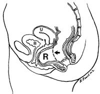
The process of defecation (having a bowel movement) requires the coordinated effort of different muscles. The pelvic floor is made up of several muscles that support the rectum like a hammock. When an individual wants to have a bowel movement the pelvic floor muscles are supposed to relax allowing the rectum to empty. While the pelvic floor muscles are relaxing, muscles of the abdomen contract to help push the stool out of the rectum. Individuals with pelvic floor dysfunction have a tendency to contract instead of relax the pelvic floor muscles. When this happens during an attempted bowel movement, these individuals are effectively pushing against an unyielding muscular wall.

How is pelvic floor dysfunction diagnosed?
The diagnosis of pelvic floor disorder starts with a careful history regarding an individual’s symptoms, medical problems and a history of physical or emotional trauma that may be contributing to their problem. Next the physician examines the patient to identify any physical abnormality. A defecating proctogram is a study commonly used to demonstrate the functional problem in a person with pelvic floor dysfunction. During this study, the patient is given an enema of a thick liquid that can be detected with x-ray. A special x-ray video records the movement of the pelvic floor muscles and the rectum while the individual attempts to empty the liquid from the rectum. Normally the pelvic floor relaxes allowing the rectum to straighten and the liquid to pass out of the rectum. This study will demonstrate if the pelvic floor muscles are not relaxing appropriately and preventing passage of the liquid.

The defecating proctogram is also useful to show if the rectum is folding in on itself (rectal prolapse). Many women have outpouching of the rectum known as a rectocele. Usually a rectocele does not affect the passage of stool. In some instances, however, stool may become trapped in a rectocele causing symptoms of incomplete evacuation. The defecating proctogram helps to identify if liquid is getting trapped in a rectocele when the individual is trying to empty the rectum.

How is pelvic floor dysfunction treated?
Pelvic floor dysfunction due to non-relaxation of the pelvic floor muscles may be treated with specialized physical therapy known as biofeedback. With biofeedback, a therapist helps to improve a person’s rectal sensation and pelvic floor muscle coordination. There are various effective techniques used in biofeedback. Some therapists train patients by teaching them to expel a small balloon placed in the rectum. Another technique uses a small probe placed in the rectum or vagina or electrodes placed on the surface of the skin around the opening to the rectum (anus) and on the abdominal wall. These instruments detect when a muscle is contracting or relaxing and provide visual feedback of the muscle action. This visual feedback helps the individual to understand the muscle movement and aids in improving muscle coordination. Approximately 75% of individuals with pelvic floor dysfunction experience significant improvement with biofeedback.

Abnormalities identified with a defecating proctogram such as rectal prolapse and rectocele may be treated with a surgical procedure.

Fecal (Bowel) Incontinence Information

Download PDF

Bowel Incontinence

What is incontinence?
Incontinence is the impaired ability to control gas or stool. Its severity ranges from mild difficulty with gas control to severe loss of control over liquid and formed stools. Incontinence to stool is a common problem, but often it is not discussed due to embarrassment.

What causes incontinence?
There are many causes of incontinence. Injury during childbirth is one of the most common causes. These injuries may cause a tear in the anal muscles. The nerves supplying the anal muscles may also be injured. While some injuries may be recognized immediately following childbirth, many others may go unnoticed and not become a problem until later in life. In these ¬situations, a prior childbirth may not be recognized as the cause of incontinence.

Anal operations or traumatic injury to the tissue surrounding the anal region similarly can damage the anal muscles and hinder bowel control. Some individuals experience loss of strength in the anal muscles as they age. As a result, a minor control problem in a younger person may become more significant later in life.

Diarrhea may be associated with a feeling of urgency or stool leakage due to the frequent ¬liquid stools passing through the anal opening. If bleeding accompanies lack of bowel control, ¬consult your physician. These symptoms may indicate inflammation within the colon (colitis), a rectal tumor, or rectal prolapse – all conditions that require prompt evaluation by a physician.

How is the cause of incontinence determined?
An initial discussion of the problem with your physician will help establish the degree of control difficulty and its impact on your lifestyle. Many clues to the origin of incontinence may be found in patient histories. For example, a woman’s history of past childbirths is very important. Multiple pregnancies, large weight babies, forceps deliveries, or episiotomies may contribute to muscle or nerve injury at the time of childbirth. In some cases, medical illnesses and medications play a role in problems with control.

A physical exam of the anal region should be performed. It may readily identify an obvious injury to the anal muscles. In addition, an ultrasound probe can be used within the anal area to provide a picture of the muscles and show areas in which the anal muscles have been injured.

Frequently, additional studies are required to define the anal area more completely. In a test called anal manometry, a small catheter is placed into the anus to record pressure as patients relax and tighten the anal muscles. This test can demonstrate how weak or strong the muscle really is. A separate test may also be conducted to determine if the nerves that go to the anal muscles are functioning properly.

What can be done to correct the problem?
Treatment of incontinence may include:

  • Dietary changes
  • Constipating medications
  • Muscle strengthening exercises
  • Biofeedback
  • Surgical muscle repair
  • Artificial anal sphincter

After a careful history, physical examination and testing to determine the cause and severity of the problem, treatment can be addressed. Mild problems may be treated very simply with dietary changes and the use of some constipating medications. Diseases which cause inflammation in the rectum, such as colitis, may contribute to anal control problems. Treating these diseases also may eliminate or improve symptoms of incontinence. Sometimes a change in prescribed medications may help. Your physician also may recommend simple home exercises that may strengthen the anal muscles to help in mild cases. A type of physical therapy called biofeedback can be used to help patients sense when stool is ready to be evacuated and help strengthen the muscles.

Injuries to the anal muscles may be repaired with surgery. Some individuals may benefit from a technique that delivers electrical energy to the skin and muscles surrounding the anus which results in firming and thickening of this area to help with continence.

In certain individuals that have nerve damage or anal muscles that are damaged beyond repair, an artificial sphincter may be implanted. The artificial sphincter is a plastic, fluid filled doughnut that is surgically implanted around the damaged anal sphincter. This artificial sphincter keeps the anal canal closed. When an individual wants to have a bowel movement, the fluid can be pumped out of the doughnut to allow the anal canal to open.

In extreme cases, patients may find that a colostomy is the best option for improving their quality of life.


Fecal (Bowel) Incontinence Treatment Guidelines

Download PDF

Practice Parameters for the Treatment of Fecal Incontinence

Joe J. Tjandra, M.D., Sharon L. Dykes, M.D., Ravin R. Kumar, M.D., C. Neal Ellis, M.D., Sharon G. Gregorcyk, M.D., Neil H. Hyman, M.D., W. Donald Buie, M.D., and the Standards Practice Task Force of The American Society of Colon and Rectal Surgeons

The American Society of Colon and Rectal Surgeons is dedicated to ensuring highquality patient care by advancing the science, prevention, and management of disorders and diseases of the colon, rectum, and anus. The Standards Committee is composed of Society members who are chosen because they have demonstrated expertise in the specialty of colon and rectal surgery. This Committee was created to lead international efforts in defining quality care for conditions related to the colon, rectum, and anus. This is accompanied by developing Clinical Practice Guidelines based on the best available evidence. These guidelines are inclusive, and not prescriptive. Their purpose is to provide information on which decisions can be made, rather than dictate a specific form of treatment. These guidelines are intended for the use of all practitioners, health care workers, and patients who desire information about the management of the conditions addressed by the topics covered in these guidelines.

It should be recognized that these guidelines should not be deemed inclusive of all proper methods of care or exclusive of methods of care reasonably directed to obtaining the same results. The ultimate judgment regarding the propriety of any specific procedure must be made by the physician in light of all of the circumstances presented by the individual patient.

METHODOLOGY
A MEDLINE search was performed, from 1966 to February 2006, using the key words “fecal incontinence,” “anus,” “implants,” “bowel sphincter,” “graciloplasty,” and “artificial sphincter.” Selected embedded references also were reviewed. The Cochrane Database of Systematic Reviews was queried.

STATEMENT OF THE PROBLEM
Fecal incontinence is defined in a consensus conference report in 2001 as Brecurrent uncontrolled passage of fecal material for at least one month, in an individual with a developmental age of at least 4 years. 1 Incontinence to flatus also may cause substantial impairment of quality of life and should be considered in the definition. Because of the lack of standardization, the true prevalence of fecal incontinence is difficult to determine.2 Quoted prevalence rates vary widely from 1.4 to 18 percent, with higher rates in nursing home residents, parous females, patients with cognitive impairment or neurologic disorders, and the elderly.3–5 In the geriatric and institutionalized population, the reported rates of fecal incontinence are approximately 50 percent.5,6 Female patients predominate in most series, although epidemiologic studies do not demonstrate any gender differences.2,7 It is postulated that this is the result of the age and gender of patients who seek treatment.

CONTRIBUTING FACTORS
Continence is dependent on the complex relationships between the anal sphincters, pelvic floor function, stool consistency, rectal compliance, and neurologic function. Disease processes or structural defects that alter any of these aspects can contribute to fecal incontinence. In many cases, the etiology of fecal incontinence is multi-factorial and it is not possible to ascertain the relative contribution of each factor. A full discussion of contributing factors is beyond the scope of a practice parameter.

Obstetric injury is the most commonly cited cause of incontinence in females.2 Sphincter disruption is clinically recognized in approximately 10 percent of all vaginal deliveries, but many other females have occult sphincter damage. Although the incidence of sonographic damage to the sphincters after childbirth varies among studies, it occurs in approximately 30 percent of first vaginal deliveries.8,9 One third of these are associated with new symptoms of incontinence or urgency. Independent risk factors include forceps delivery, occipitoposterior position, and prolonged second stage of labor. The extent of a sphincter defect does not necessarily correlate with the degree of fecal incontinence, highlighting the complexity of obstetricrelated disturbances.

ASSESSMENT
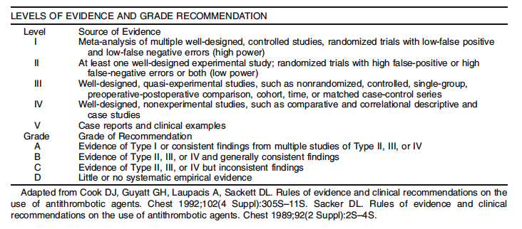
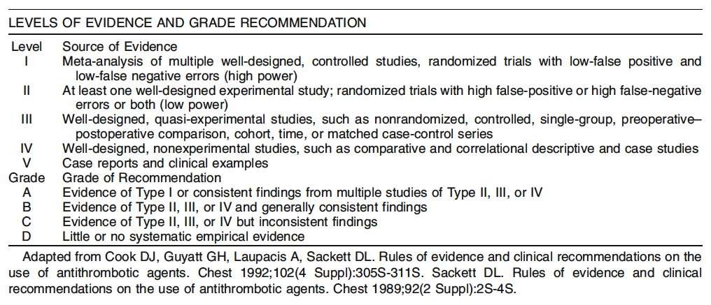
1. Evaluation of fecal incontinence should include consideration of severity and impact. Level of Evidence: Class II; Grade of Recommendation: B.

Severity instruments assess type, frequency, and amount of incontinence. Impact questionnaires address quality of life and attempt to evaluate the effect of incontinence on emotional, occupational, physical, and social function. Both should evaluate these relatively subjective factors with reliability and validity.10

Severity measures can be divided into two types: grading scales and summary scales. Despite the existence of numerous severity scales, there are limited data with regard to their validity and reliability. Grading scales assign numeric scores to degrees of fecal incontinence and/or types of incontinence (flatus to solid stool) but lack assessment of frequency and can be imprecise.11–18 Summary scales have greater validity19–32 and can be used to demonstrate improvement in continence scores after treatment or decline in scores as a consequence of a medical/surgical intervention.20,22,23,29–31 Summary scales also have been shown to correlate with quality of life instruments by evaluating lifestyle scores.19,20,33,34

A validated quality of life instrument for fecal incontinence does exist.35 Furthermore, fecal incontinence is a component of other disease specific quality of life tools.36–38


DIAGNOSIS
1. A problem specific history and physical examination should be performed. Level of Evidence: Class V; Grade of Recommendation: D.

A detailed medical history may help to elicit contributing or exacerbating factors, such as gastrointestinal or neurologic disorders. An obstetric account or history of previous anorectal surgery or perineal trauma can direct/prompt a more focused examination.

Inspection of the perianal skin may reveal excoriation, surgical scars, or fistulas, and the anus may be noted to gape upon spreading the buttocks. Mucosal or full thickness prolapse may be elicited with a Valsalva maneuver. Digital examination may provide a rough estimate of resting and squeeze pressures and is helpful to evaluate for a rectal mass or the presence of impacted stool, which would suggest overflow as a possible mechanism for incontinence. Anoscopy and flexible sigmoidoscopy may help to identify hemorrhoids, inflammatory bowel disease, or neoplasms.

2. Endoanal ultrasound is usually the procedure of choice to diagnose sphincter defects in patients with suspected sphincter injury. Anorectal physiology studies may be helpful in guiding management. Level of Evidence: Class II; Grade of Recommendation: B.

When performed by an experienced clinician, endoanal ultrasound approaches 100 percent sensitivity and specificity in identifying internal and external sphincter defects.39–41 However, as noted previously, the presence of a sphincter defect does not necessarily correlate with incontinence. Of 335 patients with incontinence, 115 patients who were continent, and 18 asymptomatic female volunteers, ultrasonography detected sphincter defects in 65, 43, and 22 percent, respectively.42

Anal manometry is a simple, noninvasive method of measuring internal and external anal sphincter tone, and the length of the high pressure zone of the anal canal. There are few large scale studies that have attempted to validate the role of anorectal physiologic testing in predicting response to treatment options for fecal incontinence. There are significant manometric variations even within
“normal” asymptomatic subjects, dependent on age, gender, and parity. Although the findings of anorectal physiology studies do not consistently correlate with severity of fecal incontinence, they may influence the management decisions that are ultimately selected for treatment.43–45

NONOPERATIVE TREATMENT
1. A trial of increased fiber intake is recommended in milder forms of fecal incontinence to improve symptoms. Level of Evidence: III; Grade of Recommendation: B

Nonoperative therapy is usually the first maneuver to improve the symptoms of fecal incontinence. Most patients with mild fecal incontinence should usually receive an initial trial of nonoperative management.Fecal incontinence is commonly exacerbated by liquid stools or diarrhea.46,47 Stool bulking agents include high fiber diet, psyllium products, or methyl cellulose. An increase in dietary fiber may improve stool consistency by absorbing intraluminal water. Supplementation of diet with psyllium is associated with improved stool consistency and a decrease in symptoms.48 The recommended dose of dietary fiber is 25 to 30 grams per day. Gradual increase of fiber intake during a period of several days can reduce symptoms, such as abdominal bloating and discomfort, that may be associated with increased fiber intake. Fiber supplements in the form of powder, granule, or pill often facilitate this goal. Dairy products are problematic in patients with lactose intolerance.49

2. Anti-diarrheal agents, such as adsorbents or opium derivatives, may reduce fecal incontinence symptoms. Level of Evidence: III; Grade of Recommendation: C.

Adsorbents, such as kaopectate (Pharmacia & Upjohn, Peapack, NJ), act by absorbing excess fluid in the stool. Commonly used opium derivatives are loperamide (Imodium, McNeil Consumer Healthcare, Fort Washington, PA), diphenoxylate hydrochloride plus atropine sulphate (Lomotil, Searle, Chicago, IL), codeine, and tincture of opium. The effects of opioids include decreased intestinal motility, decreased intestinal secretion, and increased absorption. Loperamide can slow bowel motility and increase fluid absorption. In addition, loperamide increases resting anal sphincter pressure.50 The usual dosage of loperamide is 2 to 4 mg followed by titration up to a total of 24 mg per 24 hours in divided doses.

Diphenoxylate can produce central nervous system (CNS) side effects and has greater potential for abuse.51,52 The usual dosage of diphenoxylate is one tablet every three to four hours. Loperamide and codeine may be superior to diphenoxylate in the treatment of symptomatic diarrhea.51 Tincture of opium is less commonly used because of the potential for CNS side effects and addiction.

3. Enemas, laxatives, and suppositories may help to promote more complete bowel emptying in appropriate patients and minimize further post-defecation leakage. Level of Evidence: V; Grade of Recommendation: D.

Bowel management programs often are used to relieve severe constipation in spinal cordinjured patients.53–55 Patients with severe constipation often experience overflow incontinence as a result of constant seepage of stool from the full rectum.56 An enema program in such patients may help to minimize episodes of incontinence. Evaluation and management of abnormal colonic transit also can be helpful.57 Otherwise, there is little evidence to guide clinicians in the use of these therapies for fecal incontinence in patients with an intact spinal cord.

4. Biofeedback is recommended as an initial treatment for motivated patients with incontinence with some voluntary sphincter contraction. Level of Evidence: III; Grade of Recommendation: B.

Biofeedback may be considered a firstline option for many patients with fecal incontinence who have not responded to simple dietary modification or medication.58 The benefit of biofeedback is variable and improvement in as many as 64 to 89 percent of patients has been reported.30,58 Biofeedback is performed to improve sensation, coordination, and strength. Supportive counseling and practical advice regarding diet and skin care can improve the success of biofeedback. Biofeedback may be considered before attempting sphincter repair or for those who have persistent or recurrent symptoms after sphincter repair. It may have a role in the early postpartum period in females with symptomatic sphincter weakness.59 Biofeedback and a pelvic floor exercise program can produce improvement that lasts more than two years.60–62 More than 75 percent of the initial responders to biofeedback had a sustained symptomatic improvement and 83 percent reported an improved quality of life.63

Biofeedback home training is an alternative to ambulatory training programs, especially in the elderly.64 Improved rectal sensation after biofeedback is one of the most consistent predictors of improved continence.65 However, Bstandard care^ (advice and education) alone has been shown in a randomized trial to be as effective as biofeedback therapy.66

5. An anal plug is effective in controlling fecal incontinence in a small minority of patients who can tolerate its use. Level of Evidence: V; Grade of Recommendation: D

The anal plug enables controlled fecal evacuation and helps reduce skin complications.67 However, most patients do not tolerate the anal plug because of discomfort.68 No correlation was found between the results of anorectal physiology studies and the benefit or inconvenience of using the plug.69 A trial of the anal plug in patients quickly reveals whether the patient will find it an effective and acceptable option.70

SURGICAL OPTIONS
1. Sphincter repair is appropriately offered to highly symptomatic patients with a defined defect of the external anal sphincter. Level of Evidence: II; Grade of Recommendation: A.

Anal sphincter repair often confers substantial benefits in patients with localized external sphincter defects. Short term outcomes suggest good to excellent resultsin31to83percentofpatients.71–78 Most of the outcome data on anal sphincter repair pertain to patients with an anterior sphincter defect from obstetric trauma rather than repair of sphincter disruptions from surgical trauma, such as a fistulotomy or sphincterotomy.

However, the benefits of sphincteroplasty tend to deteriorate with long-term followup. In a study from St. Mark_s Hospital, the initial success rate of 76 percent after an overlapping sphincter repair deteriorated dramatically with time.71,79 After five years, no patient was fully continent to flatus and less than 10 percent were fully continent to solid and liquid stool. Similar results have been reported by others. After five to ten years, only 40 to 45 percent of patients were satisfied with the functional outcome.80,81 Similarly, after a median of 69 months, only 14 percent of patients at the Cleveland Clinic were completely continent.82 Adjuvant biofeedback therapy after surgery may improve quality of life and help sustain symptomatic improvement with time.83

No single preoperative manometric variable can predict outcome after a sphincter repair.84 There also is controversy about whether pudendal nerve conduction studies can be used to predict outcome after a sphincter repair.85–89

2. Overlapping or direct sphincter repair yield similar results, as long as adequate mobilization of both ends of the sphincters are performed. Level of Evidence: II; Grade of Recommendation: A.

Overlapping sphincteroplasty, as described by Parks and McPartlin in 197190 and later modified by Slade et al.,91 has been the predominant technique of repair used by colon and rectal surgeons during the last three decades. However, a randomized trial showed no benefit of an overlapping repair compared with a direct repair, as long as both ends of the external sphincter were adequately mobilized and the anorectal ring was plicated.43 In another non-randomized study, the outcome after a sphincter repair was similar among different operative techniques (end to end, overlapping repair, and plication). Severe denervation and pudendal nerve damage often are found in patients who remain incontinent after a sphincter repair.92,93 Dyspareunia might follow sphincteroplasty, although the true incidence has not been well documented.

3. Repeat anal sphincter repair could be considered in patients who have recurrent symptoms and residual anterior sphincter defect after a previous sphincter repair. Level of Evidence: III; Grade of Recommendation: B.

Failure after sphincter repair might be related to persistence or recurrence of the external anal sphincter defect as shown by endoanal ultrasound.94,95 Previous sphincter repair does not seem to affect the clinical outcome of a subsequent repair. In a comparative study,96 the outcome was similar between patients with or without a previous sphincter repair; good results were obtained in 50 and 58 percent of patients, respectively. Nevertheless, the long-term benefit of a repeat sphincter repair was only modest—similar to an initial repair.97

4. Repair of the internal anal sphincter alone has a poor functional outcome and is not generally recommended. Level of Evidence: III; Grade of Recommendation: B.

The role of internal anal sphincter (IAS) repair alone in restoring the resting anal canal pressure is not well defined and this procedure is typically ineffective.98–100 Island anoplasty into an area of IAS defect improved fecal continence in 13 of 14 patients in one report,100 but there was a high incidence of wound breakdown.

5. When passive fecal incontinence caused by internal sphincter dysfunction is the predominant symptom, injectable therapy seems to be effective and safe, although its long-term efficacy has yet to be defined. Level of Evidence: II; Grade of Recommendation: B.

Effective treatment for IAS dysfunction is lacking. Recently, there have been attempts to augment the bulk of the IAS by injection therapy into a defect in the IAS or around an intact, but degenerate, IAS. Of the agents described, injectable silicone biomaterial has been the most studied.45,101,102 The bulking effect of the injected silicone particles with subsequent collagen deposition around the IAS helps to enhance fecal continence. The greatest improvement in fecal continence occurs between one and six months after injection therapy.45 The site of injection could be in the intersphincteric plane or into the submucosa. The latter approach is more likely to be associated with infection, erosions of implants, or anal pain caused by the superficial location of the injected material.103 More precise injection into the intersphincteric space with ultrasound guidance may improve the outcome.45

Other injectable agents currently being evaluated include carboncoated beads. In a preliminary study, injection into the site of the sphincter defect was associated with improvement in the Cleveland Clinic Florida Fecal Incontinence Scale Scores from 11.89 at baseline to 8.07 after a mean followup of 28.5 months.104 Longterm safety and efficacy data are lacking.

6. Sacral nerve stimulation (SNS) is a promising modality for fecal incontinence. Level of Evidence: III; Grade of Recommendation: B.

Sacral nerve stimulation has been associated with encouraging early results with low morbidity. It seems to be better than optimal medical therapy with bulking agents, pelvic floor physiotherapy, and dietary management.44SNS comprises a diagnostic peripheral nerve evaluation (PNE) stage followed by a permanent therapeutic implantation stage. Patients receive a permanent sacral nerve implant if the diagnostic stage produces clinical improvement, marked by a reduction in frequency of episodes or days of fecal incontinence by at least 50 percent based on a twoweek diary.44

Although up to 20 percent of patients do not show adequate response to PNE, this screening test seems to have a 100 percent positive predictive value, because all patients who respond to temporary test stimulation (PNE) seem to benefit from a fullstage SNS.44,105 Eightythree to 100 percent of those achieving permanent implantation maintain an improvement of greater than 50 percent at a mean followup of 10.5 to 24 months.105–110 Although an improvement in continence occurs in most patients, continence to solid and liquid stool sinpatients with a permanent implant ranges from 44 to 73 percent.44,106–110 SNS also has the potential to benefit patients with both fecal and urinary incontinence.111 Patients with limited structural sphincter defects of the internal or external sphincter have been treated with SNS with reported benefit.44,107,109,111 The improvement with SNS in diverse groups of patients reflects the limitations of current knowledge about pelvic floor physiology as well as the precise mechanism of action of SNS.

The incidence of complications with SNS ranges from 5 to 26 percent in various studies. 44,107–110,112 The screening test (PNE) has a very low complication rate.44,109 Complications requiring permanent explanation of the SNS device seem to be uncommon.44,107–110,112 The most common complication in clinical series was pain at the site of the neurostimulator generator, especially in thinner patients.105 Infection of the wound or the sacral nerve implant is rare (<5 percent), if appropriate techniques are used.105,107 Currently, SNS is not FDAapproved for fecal incontinence in the United States, although it is approved for urinary incontinence.

7. Post-anal repair or total pelvic floor repair has a limited role in the treatment of neuropathic fecal incontinence. Level of Evidence: III; Grade of Recommendation: B.

The principle of post-anal repair is to reduce the obtuse anorectal angle113 and has been advocated for patients with weak sphincters but no anatomic sphincter defect114; however, others have failed to show any correlation between the anorectal angle and the outcome of surgery.115 In a study of post-anal repair in a well defined group of patients with neuropathic fecal incontinence, the St. Marks group concluded that the longterm results of post-anal repair successfully relieved fecal incontinence in only 33 percent of patients at five to eight years post operatively.116 Total pelvic floor repair fared slightly better for neuropathic fecal incontinence with 55 percent of patients continent to solid and liquid stools 15 months after the procedure.117

8. Dynamic graciloplasty may have a role in the treatment of severe fecal incontinence when there is irreparable sphincter disruption. Level of Evidence: III; Grade of Recommendation: B.

Dynamic graciloplasty consists of a transposed gracilis muscle used to encircle the anal canal, which is stimulated electrically with an implantable pulse generator.118 Dynamic graciloplasty is most appropriate for patients with extensive sphincter disruption precluding a surgical repair, severe neural damage, or congenital disorders, such as anal atresia.119,120

The results of dynamic graciloplasty have been variable. Although moderately good results are reported from a small number of high volume centers, multi-center trials that included less experienced surgeons have shown a high morbidity and poorer functional outcome. Dynamic graciloplasty restores continence in approximately 35 to 85 percent of patients.119,120 Currently, dynamic graciloplasty is not FDA approved in the United States.

9. The artificial bowel sphincter has a role in the treatment of severe fecal incontinence, especially in patients with significant sphincter disruption. Level of Evidence: III; Grade of Recommendation: B.

The artificial bowel sphincter provides good restoration of continence for solid and liquid stool in patients who retain the device. In a small study, more than 50 percent had occasional loss of flatus, 33 percent experienced involuntary losses of flatus, and 33 percent experienced involuntary loss of flatus and liquid stool.121 More favorable results have been reported by others, with 63 percent achieving complete continence and 79 percent of patients continent to both solid and liquid stool.109 However, the explanation rate is 20 to 37 percent.121–123 In a multi-center cohort study,110 a total of 384 device related or potentially device related adverse events were reported in 112 enrolled patients. Revisional surgery was required in 46 percent of patients.123 A lack of sensation for evacuation has been reported.121,124 Absolute contraindications for this procedure include active perineal sepsis, Crohns disease, radiation proctitis, severe scarring in the perineum, or anoreceptive intercourse. The artificial bowel sphincter seems most applicable to patients with substantial disruption of the anal sphincters.125 As the experience with artificial bowel sphincter has increased, explanation and infection rates seem to have decreased.121,123,124,126

10. The SECCA procedure may be useful for selected patients with moderate fecal incontinence. Level of Evidence: IV; Grade of Recommendation: C.

The SECCA procedure consists of the delivery of temperature controlled radio frequency energy to the anal sphincters. It is believed that the heat generated causes collagen contraction, healing, and remodeling, leading to shorter and tighter muscle fibers.127

In a pilot study of ten patients, the Cleveland Clinic Florida Fecal Incontinence Scale Score improved from 13.8 at baseline to 7.3 at two years, with corresponding improvement in quality of life scales.128 In a multi-center trial of 50 patients, there was a similar improvement in the Incontinence Scale score from 14.5 to 11.1 at six months. Complications included mucosal ulcers and delayed bleeding.129 Otherwise the procedure seems to be safe and may be performed as a day procedure. Long-term results are unknown.

11. A stoma (colostomy or ileostomy) is appropriate for patients with limiting fecal incontinence in which available treatments have failed, are inappropriate because of comorbidities, or when preferred by the patient. Level of Evidence: III; Grade of Recommendation: B.

A stoma is typically successful in controlling the problems of fecal incontinence but may be associated with significant psychosocial issues and stomarelated complications. It is particularly suited for patients with spinal cord injuries or patients who are bedridden.97,130,131 Because a stoma in this situation is usually permanent, appropriate siting of the stoma and counseling is very important.89In patients with severe fecal incontinence in which alternative therapy has failed or is inappropriate, a stoma will usually allow the patient to resume normal activities and improves quality of life.89,132 In a survey, 83 percent of patients with a permanent colostomy reported a significant improvement in lifestyle, and 84 percent of patients would choose to have the stoma again.132

ACKNOWLEDGMENT
Contributing Members of the ASCRS Standards Committee: Gary D. Dunn, M.D., Phillip R. Fleshner, M.D., Clifford Y. Ko, M.D., David H. Levien, M.D., Richard L. Nelson, M.D., Graham L. Newstead, M.D., Charles P. Orsay, M.D., Paul C. Shellito, M.D., Charles A. Ternent, M.D.
REFERENCES
1. Whitehead WE, Wald A, Norton NJ. Treatment options for fecal incontinence. Dis Colon Rectum 2001;44:131–44.
2. Madoff RD, Parker SC, Varma MG, Lowry AC. Faecal incontinence in adults. Lancet 2004;364:621–32.
3. Nelson R, Norton N, Cautley E, Furner S. Communitybased prevalence of anal incontinence. JAMA 1995; 274:559–61.
4. Bharucha AE, Zinsmeister AR, Locke GR. Prevalence and burden of fecal incontinence: a populationbased study in women. Gastroenterology 2005;129:42–9.
5. Kuehn BM. Silence masks prevalence of fecal incontinence. JAMA 2006;295:1362–3.
6. Nelson RL. Epidemiology of fecal incontinence. Gastroenterology 2004;126:S3 – 7.
7. Quander CR, Morris MC, Melson J, Bienias JL, Evans DA. Prevalence of and factors associated with fecal incontinence in a large community study of older individuals. Am J Gastroenterol 2005;100:905 –9.
8. Sultan A, Kamm M, Hudson C, Thomas CM, Bartram CI. Analsphincter disruption during vaginal delivery. N Engl J Med 1993;329:1905–11.
9. Warshaw J. Obstetric anal sphincter injury: incidence, risk factors, and repair. Sem Colon Rectal Surg 2001; 12:90–7.
10. Baxter N, Rothenberger D, Lowry A. Measuring fecal incontinence. Dis Colon Rectum 2003;46:1591 –605.
11. Parks A. Royal Society of Medicine, Section of Proctology meeting, November 27, 1974. President_s address. Anorectal incontinence. Proc R Soc Med 1975;68:681–90.
12. Broden G, Dolk A, Holmstrom B. Recovery of the internal anal sphincter following rectopexy: a possible explanation for continence improvement. Int J Colorectal Dis 1988;3:23–8.
13. Keighley M, Fielding J. Management of faecal incontinence and results of surgical treatment. Br J Surg 1983;70:463–8.
14. Hiltunen K, Matikainen M, Auvinen O, Hietanen P. Clinical and manometric evaluation of anal sphincter function in patients with rectal prolapse. Am J Surg 1986;151:489–92.
15. Corman M. Gracilis muscle transposition for anal incontinence: late results. Br J Surg 1985;72(Suppl):S21–2.
16. Rainey J, Donaldson D, Thomson J. Postanal repair: which patients derive most benefit? J R Coll Surg Edinb 1990;35:101–5.
17. Williams N, Patel J, George B. Development of an electrically stimulated neoanal sphincter. Lancet 1991;338:1166–9.
18. Womack N, Morrison J, Williams N. Prospective study of the effects of postanal repair in neurogenic faecal incontinence. Br J Surg 1988;75:48–52.
19. Rockwood TH, Church JM, Fleshman JW. Patient and surgeon ranking of the severity of symptoms associated with fecal incontinence: the Fecal Incontinence Severity Index. Dis Colon Rectum 1999;42:1525 –32.
20. Hull T, Floruta C, Piedmonte M. Preliminary results of an outcome tool used for evaluation of surgical treatment for fecal incontinence. Dis Colon Rectum 2001;44:799–805.
21. Jorge J, Wexner S. Etiology and management of fecal incontinence. Dis Colon Rectum 1993;36:77 –97.
22. Pescatori M, Anastasio G, Bottini C, Mentasti A. New grading and scoring for anal incontinence: evaluation of 335 patients. Dis Colon Rectum 1992;35:482 –7.
23. VaizeyCJ,CarapetiE,CahillJA, Kamm MA.Prospective comparison of faecal incontinence grading systems. Gut 1999;44:77–80.
24. O_Brien P, Skinner S. Restoring control: the Acticon Neosphincter artificial bowel sphincter in the treatment of anal incontinence. Dis Colon Rectum 2000;43:1213–6.
25. Miller R, Bartolo DC, LockeEdmunds JC, Mortensen NJ. Prospective study of conservative and operative treatment for faecal incontinence. Br J Surg 1988; 75:101–5.
26. Bai Y, Chen H, Hao J. Longterm outcome and quality of life after the Swenson procedure for Hirschsprung_s disease. J Pediatr Surg 2002;37:639 –42.
27. Rothenberger D. Anal incontinence. In: Cameron JL, ed. Current surgical therapy. 3rd ed. Philadelphia: BC Decker, 1989:186–94.
28. Kelly J. Cine radiography in anorectal malformations. J Pediatr Surg 1969;4:538–46.
29. Lunniss PJ, Kamm MA, Phillips RK. Factors affecting continence after surgery for anal fistula. Br J Surg 1994;81:1382 –5.
30. Jensen L, Lowry A. Biofeedback improves functional outcome after sphincteroplasty. Dis Colon Rectum 1997;40:197 –200.
31. Sangalli M, Marti M. Results of sphincter repair in postobstetric fecal incontinence. J Am Coll Surg 1994;179:583 –6.
32. Oliveira L, Pfeifer J, Wexner S. Physiological and clinical outcome of anterior sphincteroplasty. Br J Surg 1996;3:502 –5.
33. Cavanaugh M, Hyman N, Osler T. Fecal incontinence severity index after fistulotomy: a predictor of quality of life. Dis Colon Rectum 2002;45:349 –53.
34. Rothbarth J, Bemelman WA, Meijerink WJ, et al. What is the impact of fecal incontinence on quality of life?Dis Colon Rectum 2001;44;67–71.
35. Rockwood TH, Church JM, Fleshman JW, et al. Fecal incontinence quality of life scale: quality of life instruments for patients with fecal incontinence. Dis Colon Rectum 2000;43:9–16.
36. Temple LK, Bacik J, Savatta SG, et al. The development of a validated instrument to evaluate bowel function after sphincterpreserving surgery for rectal cancer. Dis Colon Rectum 2005;48:1353–65.
37. Aaronson NK, Ahmedzai S, Bergman B, et al. The European Organization for Research and Treatment of Cancer QLPC30: a qualityoflife instrument for use in international clinical trials in oncology. J Natl Cancer Inst 1993;85:365 –76.
38. Sprangers MA, te Velde A, Aaronson NK. The construction and testing of the EORTC colorectal cancerspecific quality of life questionnaire module (QLQCR38). European Organization for Research and Treatment of Cancer Study Group on Quality of Life. Eur J Cancer 1999;35:238–47.
39. Sultan AH, Kamm MA, Talbot IC, Nicholls RJ, Bartram CI. Anal endosonography for identifying external sphincter defects confirmed histologically. Br J Surg 1994;81:463–5.
40. Sultan AH, Nicholls RJ, Kamm MA, Hudson CN, Beynon J, Bartram CI. Anal endosonography and correlation with in vitro and in vivo anatomy. Br J Surg 1993;80:508–11..
41. Berger N, Tjandra JJ, Solomon M. Endoanal and endorectal ultrasound: applications in colorectal surgery. ANZ J Surg 2004;74:71–5.
42. Karoui S, SavoyeCollet C, Koning E, Leroi AM, Denis
P. Prevalence of anal sphincter defects revealed by sonography in 335 incontinent patients and 115 continent patients. AJR Am J Roentgenol 1999;173 389–92.
43. Tjandra JJ, Han WR, Goh J, Carey M, Dwyer P. Direct repair vs. overlapping sphincter repair: A randomized controlled trial. Dis Colon Rectum 2003;46:937–43.
44. Tjandra JJ, Lim JF, Matzel K. Sacral nerve stimulation: an emerging treatment for faecal incontinence. ANZ J Surg 2004;74:1098–106.
45. Tjandra JJ, Lim JF, Hiscock R, Rajendra P. Injectable silicone biomaterial for fecal incontinence due to internal anal sphincter dysfunction is effective. Dis Colon Rectum 2004;47:2138–46.
46. Read NW, Bartolo DC, Read MG. Differences in anal function in patients with incontinence to solids and in patients with liquids. Br J Surg 1984;48:39–42.
47. Bliss DZ, Johnson S, Savik K, Clabots CR, Gerding DN. Fecal incontinence in hospitalized patients who are acutely ill. Nurs Res 2000;49:101–8.
48. Bliss DZ, Jung HJ, Savik K, et al. Supplementation with dietary fiber improves fecal incontinence. Nurs Res 2001;50:203–13.
49. Congilosi Parker S, Thorsen A. Fecal incontinence. Surg Clin North Am 2003;82:1273–90.
50. Read M, Read NW, Barber DC, Duthrie HL. Effects of loperamide on anal sphincter function in patients complaining of chronic diarrhea with fecal incontinence and urgency. Dig Dis Sci 1982;27:807 –14.
51. Palmer KR, Corbett CL, Holdsworth CD. Doubleblind crossover study comparing loperamide, codeine and diphenoxylate in the treatment of chronic diarrhea. Gastroenterology 1980;79:1272–5.
52. Gattuso JM, Kamm MA. Adverse effects of drugs used in the management of constipation and diarrhoea. Drug Safety 1994;10:47–65.
53. Cheetham M, Brazzelli M, Norton C, Glazener CM. Drug treatment for faecal incontinence in adults. Cochrane Database Syst Rev 2003;CD002116.
54. Wiesel PH,NortonC,BrazzelliM.Managementoffaecal incontinence and constipation in adults with central neurological diseases (Cochrane Review). In: The Cochrane Library 4. Oxford: Update Software, 2001.
55. Kirk PM, King RB, Temple R, Bourjaily J, Thomas P. Longterm followup of bowel management after spinal cord injury. SCI Nurs 1997;14:56–63.
56. King JC, Currie DM, Wright E. Bowel training in spina bifida: importance of education, patient compliance, age and anal reflexes. Arch Phys Med Rehabil 1994;75:243–7.
57. Bouchoucha M, Devroede G, Faye A, Arsac M. Importance of colonic transit evaluation in the management of fecal incontinence. Int J Colorectal Dis 2002;17:412 –7.
58. Heymen S, Jones KR, Ringel Y, Scarlett Y, Whitehead WE. Biofeedback treatment of fecal incontinence: a critical review. Dis Colon Rectum 2001;44:728–36.
59. Mahony RT, Malone PA, Nalty J, Behan M, O_Connell PR, O_Herlihy C. Randomized clinical trial of intraanal electromyographic biofeedback physiotherapy with intraanal electromyographic biofeedback augmented with electrical stimulation of the anal sphincter in the early treatment of postpartum fecal incontinence. Am J Obstet Gynecol 2004;191:885–90.
60. Ryn AK, Morren GL, Hallbook O, Sjodahl R. Longterm results of electromyographic biofeedback training for fecal incontinence. Dis Colon Rectum 2000;43:1262–6.
61. Guillemot F, Bouche B, GowerRousseau C, et al. Biofeedback for the treatment of fecal incontinence. Longterm clinical results. Dis Colon Rectum 1995; 38:393–7.
62. Enck P, Daublin G, Heinrich J, Lubke HJ, Strohmeyer G. Longterm efficacy of biofeedback training for fecal incontinence. Dis Colon Rectum 1994;37:997–1001.
63. Pager CK, Solomon MJ, Rex J, Roberts RA. Longterm outcomes of pelvic floor exercise and biofeedback treatment for patients with fecal incontinence. Dis Colon Rectum 2002;45:997 –1003.
64. Musial F, Hinninghofen H, Frieling T, Enck P. Therapy of fecal incontinence in elderly patients: study of a home biofeedback training program. Z Geroltol Geriatr 2000;33:447–53.
65. Prather CM. Physiologic variables that predict the outcome of treatment for fecal incontinence. Gastroenterology 2004;126:135–40.
66. Norton C, Chelvanayagam S, WilsonBarnett J, Radfern S, Kamm MA. Randomized controlled trial of biofeedback for fecal incontinence. Gastroenterology 2003;125:1320–9.
67. Kim J, Shim MC, Choi BY, Ahn SH, Janq SH, Shin HJ. Clinical application of continent anal plug in bedridden patients with intractable diarrhea. Dis Colon Rectum 2001;44:1162–7.
68. Deutekom M, Dobben A. Plugs for containing fecal incontinence. Cochrane Database Syst Rev 2005;20: CD005086.
69. Christiansen J, RoedPetersen K. Clinical assessment of the anal continence plug. Dis Colon Rectum 1993;36:740–2.
70. Norton C, Kamm MA. Anal plug for fecal incontinence. Colorectal Dis 2001;3:323–7.
71. Engel AF, Kamm MA, Sultan AH, Bartram CI, Nicholls RJ. Anterior anal sphincter repair in patients with obstetric trauma. Br J Surg 1994;81:1231–4.
72. Elton C, Stoodley BJ. Anterior anal sphincter repair: results in a district general hospital. Ann R Coll Surg Engl 2002;84:321 –4.
73. Fleshman JW, Peters WR, Shemesh EI, Fry RD, Kodner IJ. Anal sphincter reconstruction anterior overlapping muscle repair. Dis Colon Rectum 1991;34:739–43.
74. Jacobs PP, Scheuer M, Kuijpers JH, Vingerhotts MH. Obstetric fecal incontinence. Role of pelvic floor denervation and results of delayed sphincter repair. Dis Colon Rectum 1990;33:494 –7.
75. Norderval S, Oian P, Revhaug A, Vonen B. Anal incontinence after obstetric sphincter tears: outcome of anatomic primary repairs. Dis Colon Rectum 2005;48:1055–61.
76. Osterberg A, Edebol EegOlofsson K, Graf W. Results of surgical treatment for faecal incontinence. Br J Surg 2000;87:1546–52.
77. Pinta T, KylanpaaBack ML, Salmi T, Jarvinen HJ, Luukkonen P. Delayed sphincter repair for obstetric ruptures: analysis of failure. Colorectal Dis 2003;5:73–8.
78. Sitzler PJ, Thompson JP. Overlap repair of damaged anal sphincter. A single surgeon_s series. Dis Colon Rectum 1996;39:1356 –60.
79. Malouf AJ, Norton CS, Engel AF, Nicholls RJ, Kamm MA. Longterm results of overlapping anterior anal sphincter repair for obstetric trauma. Lancet 2000;355:260–5.
80. Zorcolo L, Covotta L, Bartolo DC. Outcome of anterior sphincter repair for obstetric injury: comparison of early and late results. Dis Colon Rectum 2005;48:524 –31.
81. Bravo Gutierrez A, Madoff RD, Lowry AC, Parker SC, Buie WD, Baxter NN. Longterm results of anterior sphincteroplasty. Dis Colon Rectum 2004;47:727 –31.
82. Halverson AL, Hull TL. Longterm outcome of overlapping anal sphincter repair. Dis Colon Rectum 2002;45:345–8.
83. Davis KJ, Kumar D, Poloniecki J. Adjuvant biofeedback following anal sphincter repair: a randomized study. Aliment Pharmacol Ther 2004;20:539–49.
84. Gearhart S, Hull T, Floruta C, Schroeder T, Hammel J. Anal manometric parameters: predictors of outcome following anal sphincter repair? J Gastrointest Surg 2005;9:115–20.
85. Fleshman JW, Dreznik Z, Fry RD, Kodner IJ. Anal sphincter repair for obstetric injury: manometric evaluation of functional results. Dis Colon Rectum 1991; 34:1061–7.
86. Sangwan YP, Coller JA, Barrett RC, et al. Unilateral pudendal neuropathy. Impact on outcome of anal sphincter repair. Dis Colon Rectum 1996;39:686 –9.
87. Oliveira L, Pfeifer J, Wexner SD. Physiological and clinical outcome of anterior sphincteroplasty. Br J Surg 1996;83:502 –5.
88. Kafka NJ, Coller JA, Barrett RC, et al. Pudendal neuropathy is the only parameter differentiating leakage from solid stool incontinence. Dis Colon Rectum 1997;40:1220 –7.
89. Madoff RD. Surgical treatment options for fecal incontinence. Gastroenterology 2004;126:S48–54.
90. Parks AG, McPartlin JF. Late repair of injuries of the anal sphincter. Proc R Soc Med 1971;74:1187 –9.
91. Slade MS, Goldberg SM, Schottler JL, Balcos EG, Christenson CE. Sphincteroplasty for acquired anal incontinence. Dis Colon Rectum 1977;20:33 –5.
92. Morren GL, Hallbook O, Nystrom PO, et al. Audit of anal sphincter repair. Colorectal Dis 2001;3:17–22.
93. Wexner SD, Marchetti F, Jagelman DG. The role of sphincteroplasty for fecal incontinence reevaluated: a prospective physiologic and functional review. Dis Colon Rectum 1991;34:22–30.
94. Nielsen MB, Hauge C, Rasmussen OO, Pedersen JF, Christiansen J. Anal endosonographic findings in the follow up of primarily sutured sphincteric ruptures. Br J Surg 1992;79:104–6.
95. Pinta T, KylanpaaBack ML, Salmi T, Jarvinen HJ, Luukkonen P. Delayed sphincter repair for obstetric ruptures: analysis of failure. Colorectal Dis 2003;5:73–8.
96. Giordano P, Renzi A, Efron J, et al. Previous sphincter repair does not affect the outcome of repeat repair. Dis Colon Rectum 2002;45:635–40.
97. Vaizey CJ, Norton C, Thornton MJ, Nicholls RJ, Kamm MA. Longterm results of repeat anterior anal sphincter repair. Dis Colon Rectum 2004;47:858–63.
98. Briel JW, de Boer LM, Hop WC, Schouten WR. Clinical outcome of anterior overlapping external anal sphincter repair with internal anal sphincter imbrication. Dis Colon Rectum 1998;41:209–14.
99. Leroi AM, Kamm MA, Weber J, Denis P, Hawley PR. Internal anal sphincter repair. Int J Colorectal Dis 1997;12:243–5.
100. Morgan R, Patel B, Beynon J, Carr ND. Surgical management of anorectal incontinence due to internal anal sphincter deficiency. Br J Surg 1997;84:226–30.
101. Tjandra JJ, Lim JF, Hiscock R, Rajendra P. Injectable silicone biomaterial for fecal incontinence caused by internal anal sphincter dysfunction is effective. Dis Colon Rectum 2004;47:2138 –46.
102. Chan MK, Tjandra JJ. Injectable silicone biomaterial (PTQi) to treat fecal incontinence after hemorrhoidectomy. Dis Colon Rectum 2006;49:433–9.
103. Malouf AJ, Vaizey CJ, Norton CS, Kamm MA. Internal anal sphincter augmentation for fecal incontinence using injectable silicone biomaterial. Dis Colon Rectum 2001;44:595–600.
104. Davis K, Kumar D, Poloniecki J. Preliminary evaluation of an injectable anal sphincter bulking agent (Durasphere) in the management of faecal incontinence. Aliment Pharmacol Ther 2003;18:237–43.
105. Ratto C, Grillo E, Parello A, Petrolino M, Costamagna G, Doglietta GB. Sacral neuromodulation in treatment of fecal incontinence following anterior resection and chemoradiation for rectal cancer. Dis Colon Rectum 2005;48:1027–36.
106. Malouf AJ, Vaizey CJ, Nicholls RJ, Kamm MA. Permanent sacral nerve stimulation for faecal incontinence. Ann Surg 2000;232:143–8.
107. Matzel K, Kamm MA, St¨osser M, et al.MDT301Study Group. Sacral nerve stimulation for faecal incontinence: a multicenter study. Lancet 2004;363: 1270–6.
108. Ganio E, Ratto C, Masin A, et al. Neuromodulation for fecal incontinence: outcome in 16 patients with definitive implant. The initial Italian Sacral Neurostimulation Group (GINS) experience. Dis Colon Rectum 2001;44:965 –70.
109. Kenefick NJ, Vaizey CJ, Cohen RC, et al. Mediumterm results of permanent sacral nerve stimulation for faecal incontinence. Br J Surg 2002;89:896–901.
110. Rasmussen OO, Buntzen S, Sorensen M, Laurberg S, Christiansen J. Sacral nerve stimulation in fecal incontinence. Dis Colon Rectum 2004;47:1158 –62.
111. Leroi AM, Michot F, Grise P, Denis P. Effect of sacral nerve stimulation in patients with faecal and urinary incontinence. Dis Colon Rectum 2001;44:779–89.
112. Rosen H, Urbarz C, Holzer B, Novi G, Schiessel R. Sacral nerve stimulation as a treatment for faecal incontinence. Gastroenterology 2001;121:536 –41.
113. Parks AG. Anorectal incontinence. Proc R Soc Med 1975;68:683–7.
114. Abbas SM, Bissett IP, Neill ME, Parry BR. Longterm outcome of postanal repair in the treatment of faecal incontinence. ANZ J Surg 2005;75:783 –6.
115. Bartolo DC, Roe AM, LockeEdmunds JC. Flapvalve theory of anorectal continence. Br J Surg 1986; 73:1012–4.
116. Setti Carraro P, Kamm MA, Nicholls RJ. Longterm results of postanal repair for neurogenic faecal incontinence. Br J Surg 1994;81:140 –4.
117. Pinho M, Ortiz J, Oya M, Panagamuwa B, Asperer J, Keighley MR. Total pelvic floor repair for treatment of neuropathic faecal incontinence. Am J Surg 1992; 163:340–3.
118. Baeten CG, Geerdes BP, Adang EM, et al.Anal dynamic graciloplasty in the treatment of intractable fecal incontinence. N Engl J Med 1995;332:1600–5.
119. Koch SM, Uludag O, Rongen M, Baeten CG, van Gemert W. Dynamic graciloplasty in patients born with an anorectal malformation. Dis Colon Rectum 2004;47:1711–9.
120. Chapman AE, Geerdes B, Hewett P, et al. Dynamic graciloplasty in the treatment of faecal incontinence. Br J Surg 2002;89:138 –53.
121. Casal E, San Ildefonso A, Carracedo R, Facal C, Sanchez JA. Artificial bowel sphincter in severe anal incontinence. Colorectal Dis 2004;6:180 –4.
122. Michot F, Costaglioli B, Leroi AM, Denis P. Artificial anal sphincter in severe faecal incontinence: outcome of prospective experience with 37 patients in one institution. Ann Surg 2003;237:52 –6.
123. Wong WD, Congliosi SM, Spencer MP, et al. The safety and efficacy of the artificial bowel sphincter for fecal incontinence: results from a multicenter cohort study. Dis Colon Rectum 2002;45:1139 –53.
124. Christiansen J, Rasmussen O, LindorffLarsen K. Longterm results of artificial anal sphincter implantation for severe anal incontinence. Ann Surg 1999;230:45–8.
125. Mundy L, Merlin TL, Maddern GJ, Hiller JE. Systematic review of safety and effectiveness of an artificial bowel sphincter for faecal incontinence. Br J Surg 2004;91:665–72.
126. O_Brien PE, Dixon JB, Skinner S, Laurie C, Khera A, Fonda D. A prospective, randomized, controlled clinical trial of placement of the artifical bowel sphincter (Acticon Neosphincter) for the control of fecal incontinence. Dis Colon Rectum 2004;47:1852–60.
127. Takahashi T, GarciaOsogobio S, Valdovinos MA, et al. Radiofrequency energy delivery to the anal canal for the treatment of fecal incontinence. Dis Colon Rectum 2002;45:915–22.
128. Takahashi T, GarciaOsogobio S, Valdovinos MA, Belmonte C, Barreto C, Velasco L. Extended twoyear results of radiofrequency energy delivery for the treatment of fecal incontinence (the Secca procedure). Dis Colon Rectum 2003;46:711 –5.
129. Efron JE, Corman ML, Fleshman J, et al. Safety and effectiveness of temperaturecontrolled radiofrequency energy delivery to the anal canal (Secca procedure) for the treatment of fecal incontinence. Dis Colon Rectum 2003;46:1606 – 16.
130. Saltzstein RJ, Romano J. The efficacy of colostomy as a bowel management alternative in selected spinal cord injury patients. J Am Paraplegia Soc 1990;13:9–13.
131. Stone JM, Wolfe VA, NinoMurcia M, Perkash I. Colostomy as treatment for complications of spinal cord injury. Arch Phys Med Rehabil 1990;71:514 –8.
132. Norton C, Burch J, Kamm MA. Patients_ views of a colostomy for fecal incontinence. Dis Colon Rectum 2005;48:1062 –9.

Constipation Information

Download PDF

Constipation

What is constipation?
Constipation is a symptom that has different meanings to different individuals. Most commonly, it refers to infrequent bowel movements, but it may also refer to a decrease in the volume or weight of stool, the need to strain to have a movement, a sense of incomplete evacuation, or the need for enemas,suppositories or laxatives in order to maintain regularity.

For most people, it is normal for bowel movements to occur from three times a day to three times a week; other people may go a week or more without experiencing discomfort or harmful effects. Normal bowel habits are affected by diet. The average American diet includes 12 to 15 grams of fiber per day,although 25 to 30 grams of fiber and about 60 to 80 ounces of fluid daily are recommended for proper bowel function. Exercise is also beneficial to proper function of the colon.

About 80 percent of people suffer from constipation at some time during their lives, and brief periods of constipation are normal. Constipation may be diagnosed if bowel movements occur fewer than three times weekly on an ongoing basis. Widespread beliefs, such as the assumption that everyone should have a movement at least once each day, have led to overuse and abuse of laxatives.

Eating foods high in fiber, including bran, shredded wheat, whole grain breads and certain fruits and vegetables will help provide the 25 to 30 grams of fiber per day recommended for proper bowel function.

What causes constipation?
There may be several, possibly simultaneous, causes for constipation, including inadequate fiber and fluid intake, a sedentary lifestyle, and environmental changes. Constipation may be aggravated by travel, pregnancy or change in diet. In some people, it may result from repeatedly ignoring the urge to have a bowel movement.

More serious causes of constipation include growths or areas of narrowing in the colon, so it is wise to seek the advice of a colon and rectal surgeon when constipation persists. Individuals with spinal cord injuries frequently experience problems with constipation. Constipation may be a symptom of diabetes. Constipation may also be associated with scleroderma, or disorders of the nervous or endocrine systems, including thyroid disease, multiple sclerosis, or Parkinson’s disease.

Can medication cause constipation?
Yes, many medications, including pain killers, antidepressants, tranquilizers, and other psychiatric medications, blood pressure medication, diuretics, iron supplements, calcium supplements, and aluminum containing antacids cans low the movement of the colon and worsen constipation.

When should I see a doctor about constipation?
Any persistent change in bowel habit, increase or decrease in frequency or size of stool or an increased difficulty in evacuating warrants evaluation. Whenever constipation symptoms persist for more than three weeks, you should consult your physician. If blood appears in the stool, consult your physician right away.

How can the cause of constipation be determined?
Constipation may have many causes, and it is important to identify them so that treatment can be as simple and specific as possible. Your doctor will want to check for any anatomic causes, such as growths or areas of narrowing in the colon.

Digital examination of the anorectal area is usually the first step, since it is relatively simple and may provide clues to the underlying causes of the problem. Examination of the intestine with either a flexible lighted instrument or barium x-ray study may help pinpoint the problem and exclude serious conditions known to cause constipation, such as polyps, tumors, or diverticular disease. If an anatomic problem is identified, treatment can be directed toward correcting the abnormality.

Other tests may identify specific functional causes to help direct treatment. For example, “marker studies,” in which the patient swallows a capsule containing markers that show up on x-rays taken repeatedly over several days, may provide clues to disorders in muscle function within the intestine. Other physiologic tests evaluate the function of the anus and rectum. These tests may involve evaluating the reflexes of anal muscles that control bowel movements using a small plastic catheter, or x-ray testing to evaluate function of the anus and rectum during defecation.

In many cases, no specific anatomic or functional causes are identified and the cause of constipation is said to be nonspecific.

How is constipation treated?
The vast majority of patients with constipation are successfully treated by adding high fiber foods like bran, shredded wheat, whole grain breads and certain fruits and vegetables to the diet, along with increased fluids. Your physician may also recommend lifestyle changes. Fiber supplements containing indigestible vegetable fiber, such as bran, are often recommended and may provide many benefits in addition to relief of constipation. They may help to lower cholesterol levels, reduce the risk of developing colon polyps and cancer, and help prevent symptomatic hemorrhoids.

Fiber supplements may take several weeks, possibly months, to reach full effectiveness, but they are neither harmful nor habit forming, as some stimulant laxatives may become with overuse or abuse. Other types of laxatives,enemas or suppositories should be used only when recommended and monitored by your colon and rectal surgeon.

Designating a specific time each day to have a bowel movement also may be very helpful to some patients. In some cases, biofeedback may help to retrain poorly functioning anal sphincter muscles. Only in rare circumstances are surgical procedures necessary to treat constipation. Your colon and rectal surgeon can discuss these options with you in greater detail to determine the best treatment for you.

What is a colon and rectal surgeon?
Colon and rectal surgeons are experts in the surgical and nonsurgical treatment of diseases of the colon, rectum and anus. They have completed advanced surgical training in the treatment of these diseases as well as full general surgical training. Board certified colon and rectal surgeons complete residencies in general surgery and colon and rectal surgery, and pass intensive examinations conducted by the American Board of Surgery and the American Board of Colon and Rectal Surgery. They are well versed in the treatment of both benign and malignant diseases of the colon, rectum and anus and are able to perform routine screening examinations and surgically treat conditions if indicated to do so.

Constipation Treatment Guidelines

Download PDF

Practice Parameters for the Evaluation and Management of Constipation

Charles A. Ternent, M.D., Amir L. Bastawrous, M.D., Nancy A. Morin, M.D., C. Neal Ellis, M.D., Neil H. Hyman, M.D., W. Donald Buie, M.D., and The Standards Practice Task Force of The American Society of Colon and Rectal Surgeons

The American Society of Colon and Rectal Surgeons is dedicated to ensuring highquality patient care by advancing the science, prevention, and management of disorders and diseases of the colon, rectum, and anus. The Standards Committee is composed of Society members who are chosen because they have demonstrated expertise in the specialty of colon and rectal surgery. This Committee was created to lead international efforts in defining quality care for conditions related to the colon, rectum, and anus. This is accompanied by developing Clinical Practice Guidelines based on the best available evidence. These guidelines are inclusive, and not prescriptive. Their purpose is to provide information on which decisions can be made, rather than dictate a specific form of treatment. These guidelines are intended for the use of all practitioners, health care workers, and patients who desire information about the management of the conditions addressed by the topics covered in these guidelines.

It should be recognized that these guidelines should not be deemed inclusive of all proper methods of care or exclusive of methods of care reasonably directed to obtaining the same results. The ultimate judgment regarding the propriety of any specific procedure must be made by the physician in light of all of the circumstances presented by the individual patient.

METHODOLOGY
An organized search of MEDLINE, PubMed, and the Cochrane Database of Collected Reviews was performed through October 2006. Keyword combinations included constipation, obstructed defecation, slow transit, surgery, rectocele, rectal intussuception, pelvic dyssynergia, anismus, paradoxical puborectalis, and related articles. Directed searches of the embedded references from the primary articles also were accomplished in selected circumstances.

STATEMENT OF THE PROBLEM
Constipation is a symptom based disorder of unsatisfactory defecation that may be associated with infrequent stools, difficult stool passage, or both.1 The diagnostic criteria for functional constipation according to the Rome III consensus include two or more of the following symptoms: straining, lumpy or hard stools, sensation of incomplete evacuation, sensation of anorectal obstruction, and manual maneuvers to facilitate defecation more than 25 percent of the time, and less than three unassisted defecations per week. These symptoms need to be present for at least three days per month during the previous three months with symptom onset at least six months before diagnosis.2 Loose stools must be rarely present without the use of laxatives, and there must be insufficient criteria for irritable bowel syndrome (IBS).2 The symptoms of chronic constipation frequently overlap with constipation predominant IBS.1 The Rome III diagnostic criteria for IBS include abdominal pain or discomfort at least three days per month in the previous three months (symptom onset more than 3 months before diagnosis) with two or more features: improvement with defecation, onset associated with a change in frequency of stool and/or change in the form of stool.2 Sub-classification into constipation predominant IBS (IBSC) based on the Rome III criteria also requires the presence of Bristol Stool Form Scale Types 1 and 2.2 The numerous possible disorders leading to constipation argue for individualized evaluation and management according to the nature, extent, and chronicity of this common problem.1,3

EVALUATION OF CONSTIPATION
1. A problem specific history and physical examination should be performed in patients with constipation. Level of Evidence: Class IV; Grade of Recommendation: B.

A history and physical examination may identify the presence of alarm symptoms and signs, such as hemochezia, weight loss of more than 10 pounds, family history of colon cancer or inflammatory bowel disease, anemia, change in bowel habits or blood in the stool, which suggest the need for more aggressive endoscopic and/or radiologic evaluation.1,4 An adequate history may help to identify factors associated with constipation, such as immobility, psychiatric illness, contributing medications, endocrine etiologies, such as diabetes and hypothyroidism, previous pelvic surgery, or symptoms consistent with constipation predominant irritable bowel syndrome (IBS).2,5–10 The history may suggest the presence of obstructed defecation if there is straining with bowel movements, incomplete evacuation, sensation of obstructed defecation, and the use of manual maneuvers to aid defecation.11 Nevertheless, symptoms alone may not reliably distinguish slow transit constipation from anorectal dysfunction.1,5

A physical examination, including digital rectal examination, plus the selective use of anoscopy and proctosigmoidoscopy may identify the presence of fecal impaction, stricture, external or internal rectal prolapse, rectocele, paradoxical or nonrelaxing puborectalis activity, or a rectal mass.2,6,12

2. The routine use of blood tests, xray studies, or endoscopy in patients with constipation without alarm symptoms is not indicated. Level of Evidence: Class V; Grade of Recommendation: D.

Evidence to support the routine use of blood tests, radiography, or endoscopy in the routine evaluation of patients with constipation without alarm features is lacking.13 Nevertheless, endoscopic evaluation of the colon is justified for patients who meet criteria for screening colonoscopy or those with alarm features.2,14 Furthermore, blood tests may be helpful to rule out hypercalcemia and/or hypothyroidism.


3. Anorectal physiology and colon transit time investigations may help to identify the underlying etiology and improve the outcome in patients with refractory constipation. Level of Evidence: Class III; Grade of Recommendation: B.

A review of 31 studies of colectomy for constipation found that preoperative physiologic tests, including at least anorectal manometry, defecography, and transit study, resulted in a median satisfaction rate of 89 percent compared with 80 percent for an incomplete physiologic evaluation.15 Studies in which slow colonic transit had been documented before colectomy for refractory constipation also reported an improved rate of good outcomes (90 vs. 67 percent).15,16

The balloon expulsion test is a simple screening procedure to exclude pelvic floor dyssynergia (PFD), because symptoms alone may not be enough to distinguish between slow transit constipation and outlet obstruction.17,18 A prospective study of balloon expulsion in patients with constipation found a specificity and negative predictive value for excluding PFD of 89 and 97 percent, respectively. A nonpathologic balloon expulsion test may avoid the use of other pelvic floor investigations, such as anorectal manometry, surface EMG studies, and defecography.19

Anorectal manometry and surface anal electromyography may help to confirm pelvic floor dyssynergia or anismus.14 The presence of Hirschprungs disease also can be suggested by anorectal manometry when the rectoanal inhibitory reflex is absent.12 Defecography is probably the most useful diagnostic technique for identifying internal rectal intussuception. In the setting of obstructed defecation, defecography may help to detect structural causes, such as intussuception, rectocele with retained stool, pelvic dyssynergia, and extent of rectal emptying. Defecography has been shown to have good interobserver agreement for enterocele and rectocele and fair to moderate inter-observer agreement for intussuception and anismus.20

The measurement of colon transit time using radio paque markers in patients with suspected slow transit constipation is inexpensive, simple, and safe. There are different methodologies that produce similar results,21–25 including the use of radioisotope markers.26–28 The interpretation of colon transit studies may be facilitated by knowledge of the status of the pelvic floor in the patient with constipation.29 Some studies have not found a relationship between smallbowel function and functional results after total abdominal colectomy for colonic inertia.30 However, a longterm, prospective study did suggest that patients with generalized gastrointestinal disorder (GID) have a diminished longterm success rate after colectomy (13 percent GID vs. 90 percent no GID).31 Similarly, a high postoperative morbidity from recurrent smallbowel obstructions (70 percent) exists in patients with GID.32

NONOPERATIVE MANAGEMENT OF CONSTIPATION
1. The initial management of symptomatic constipation is typically dietary modification, including a high fiber diet and fluid supplementation. Level of Evidence: Class II; Grade of Recommendation: B.

Conservative measures should be attempted before surgical intervention for constipation.33 Empiric treatment for constipation with a high fiber diet seems to be an inexpensive and effective therapeutic intervention for addressing constipation related bowel dysfunction.8,34 The daily intake of 25 g of fiber per day has been shown to increase the stool frequency in patients with chronic constipation. Furthermore, increasing fluid intake to 1.5 to 2 liters per day has been shown in a randomized, clinical trial of chronic constipation to increase stool frequency and decrease the need for laxative in individuals already consuming a high fiber diet.34 Increased physical activity also seems to be helpful.35

2. The use of polyethylene glycol, tegaserod, and lubiprostone for the management of chronic constipation is appropriate when dietary management is inadequate. Level of Evidence: Class II; Grade of Recommendation: A.

Polyethylene glycol (PEG) can be used to promote bowel function in patients with chronic constipation. A randomized, clinical trial found that daily therapy with 17 g of PEG laxative for 14 days resulted in significant improvement of bowel movement frequency in patients with constipation compared with placebo at two weeks.36 Prokinetic agents, such as the 5HT4 receptor partial agonist tegaserod maleate, can be used for treatment of constipationpredominant IBS. Seven short term, placebo controlled studies fulfilled the inclusion criteria for the Cochrane review in patients with constipation predominant IBS. Tegaserod improved the number of bowel movements and days without bowel movements compared with placebo.37 Another systematic review found good evidence to support the use of PEG and tegaserod for the treatment of constipation.38 Furthermore, clinical outcome analysis of a single blind, randomized, multi-center trial of the treatment of idiopathic constipation during three months with PEG or lactulose showed that significantly more patients were successfully treated with PEG than lactulose (53 vs. 24 percent) with overall decreased total management costs.38

Lubiprostone (Amitiza) is an oral bicyclic fatty acid that selectively activates Type 2 chloride channels in the apical membrane of the gastrointestinal epithelium, resulting in increased fluid secretion. Two randomized, double blind, multi center, Phase III studies in patients with chronic idiopathic constipation have shown that the frequency of spontaneous bowel movements (SBMs) was significantly greater in patients receiving lubiprostone 24 mg twice per day than in those receiving placebo at each weekly time point throughout both four week studies (P < 0.05). One study found that the mean frequency of SBMs in the lubiprostone group was five per week compared with four per week in the placebo group after seven days (P < 0.0001). Significantly greater improvements occurred with lubiprostone than placebo in the degree of straining, stool consistency, and constipation severity in both studies at all time intervals up to four weeks (P < 0.05).40 3. The use of psyllium supplements and lactulose for the treatment of chronic constipation is appropriate. Level of Evidence: Class II; Grade of Recommendation: B. A systematic review of the literature found that psyllium and lactulose improved symptoms of constipation.38 A prospective, non-randomized trial studied 224 patients with simple constipation who were treated with ispaghula husk and 170 patients who were treated with other laxatives, mostly lactulose, for up to four weeks. The husktreated group produced a higher percentage of normal, wellformed stools and fewer hard stools than other laxatives. The husk was found to be an effective treatment for simple constipation with better stool consistency and lower adverse events compared with lactulose or other laxatives.41 4. The use of common agents, such as milk of magnesia, senna, bisacodyl, and stool softeners, for chronic constipation is reasonable. Level of Evidence: Class III; Grade of Recommendation: C. Various laxatives may be used for chronic constipation but there are inconsistent results in the literature. A metaanalysis38 found 11 large, well controlled, published studies regarding the efficacy of laxatives in constipation. There were 375 patients taking laxatives and 174 patients taking placebo. The treatment group was noted to have a mean increase of 0.9 stools per week and a mean increase in stool weight of 42 g, but these findings were not different than the placebo effect at a four week duration.38,42 Furthermore, long term laxative usage can result in the development of cathartic colon.

INDICATIONS FOR SURGERY

Slow Transit Constipation

1. Patients with refractory slowtransit constipation may benefit from total abdominal colectomy with ileorectal anastomosis (TACIRA). Level of Evidence: Class III; Grade of Recommendation: B.

Clinical improvement with total abdominal colectomy with ileorectal anastomosis (TACIRA) is reported in 50 to 100 percent of patients with slowtransit constipation (STC).43 The results of segmental colon resection for colonic inertia have been disappointing with some small series reporting up to a 100 percent failure rate.15 Similarly, the antegrade colonic enema (ACE) procedure has been described for treatment of intractable constipation. Nevertheless, studies have shown a 33 percent conversion rate to TACIRA with associated stoma complications, wound infection, pain, and psychologic problems in adults.44,45 TACIRA has been reported to have an 8 to 33 percent morbidity from recurrent bowel obstruction and can be associated with diarrhea, incontinence, and recurrence of constipation.43 Patients should be counseled that the abdominal pain and bloating may persist postoperatively even after normalization of bowel frequency.7 A retrospective study of 55 patients after TACIRA for colonic inertia with normal anal manometry identified prolonged postoperative ileus in 24 percent of cases. Good to excellent results were reported in 89 percent of patients and poor results in 11 percent. Postoperative stool frequency was 5, 4, and 3 per day at 1, 2, and 12 months, respectively.46 TACIRA is recommended for carefully selected patients with severe documented colonic inertia and no evidence of severe or correctable pelvic floor dysfunction after nonoperative treatments have failed.15,16,31,46–51 Although constipation is generally relieved after TACIRA, studies have shown that, postoperatively, 41 percent of patients are affected with abdominal pain, 65 percent with bloating, 29 percent require assistance with bowel movements, 47 percent have some incontinence to gas or liquid stool, 52 and 46 percent may be affected with diarrhea.53 Postoperative quality of life assessment after TACIRA showed significantly decreased scores compared with those of the general population.52 Nevertheless, 93 percent of carefully selected patients with TAC would undergo colectomy again for STC given the chance.53 An ileostomy is an alternative consideration in many of these patients. 2. Refractory slowtransit constipation associated with concomitant pelvic outlet obstruction may benefit from correction of the pelvic floor dysfunction and total abdominal colectomy with ileorectal anastomosis. Level of Evidence: Class III; Grade of Recommendation: B. Studies of colectomy for refractory constipation have demonstrated successful outcomes for TACIRA in 89 to 100 percent after preoperative workup, including colon transit study, defecography, and anorectal physiology investigation.15 A thorough preoperative workup may help to exclude patients with constipationpredominant IBS or normaltransit constipation who will be unlikely to benefit from surgical intervention. Furthermore, patients with combined STC and outlet obstruction pathology may be offered individualized management.16,31,47–51 STC and associated pelvic floor dyssynergia can be treated with biofeedback and TACIRA, although this group has been shown to have a higher rate of recurrent defecatory problems and lower satisfaction rates after colectomy.15 STC with rectal intussuception and/or nonemptying rectocele/enterocele can be treated with TACIRA after repair of the anatomic cause of the outlet obstruction.47,50

MANAGEMENT OF PELVIC FLOOR DYSSYNERGIA

1. Biofeedback therapy is appropriately recommended for treatment of symptomatic pelvic floor dyssynergia. Level of Evidence: Class II; Grade of Recommendation: B.

The success rates of biofeedback for the treatment of PFD are reported to be 35 to 90 percent.54–56 A recent, randomized, clinical trial of individuals with chronic severe PFD who had failed management with 20 g per day of fiber plus enemas or suppositories up to twice per week were randomized into five weekly biofeedback sessions (n = 54) or PEG 14.6 to 29.2 g per day plus five weekly sessions in constipation prevention. Stool frequency increased in both groups. However, at six months major improvement was reported in the biofeedback group in 80 percent compared with 22 percent of patients treated with laxatives. These results of biofeedback were sustained at 12 and 24 months along with reductions in straining, sensations of incomplete evacuations, blockage, use of enemas and suppositories, and abdominal pain. Biofeedback patients reporting the major improvement in symptomatology were able to relax the pelvic floor and evacuate a 50ml balloon at 6month and 12month followup. Therefore, biofeedback seems to be the treatment of choice for PFD.57

SURGICAL MANAGEMENT OF OBSTRUCTED DEFECATION

Surgical Procedures Indications for rectocele repair vary but generally include relief of the outlet obstruction symptoms with manual support of the vaginal wall or rectum and lack of rectocele emptying on defecography. Although controversial, some propose that rectoceles should be > 4 cm in size to warrant repair.58

1. Surgical repair of a rectocele may appropriately be performed via a transvaginal approach. Level of Evidence: Class III; Grade of Recommendation: C.

The traditional technique for transvaginal rectocele repair is a nonanatomic, longitudinal plication of the rectovaginal fascia with the repair continuing onto the perineal body in which any injuries to the puborectalis and perineal muscles also are addressed.59 This technique is reported to be successful in preventing vaginal bulging in 80 percent and corrects the need for digital assistance of defecation in 67 percent of patients.58,59 Less favorable clinical results have been reported with a failure to relieve evacuatory difficulty or lower rectal symptoms in 33 percent of patients. Postoperative dyspareunia will occur in 25 percent of patients and at least 10 percent may recur and require reoperation; 36 percent will report a problem with fecal incontinence.60–63 A prospective study of rectocele repair using xenograft has been reported.64 Although significant decreases in rectal emptying difficulties were noted, cure of the rectal emptying difficulties was reported by less than half of the patients at the threeyear followup.64

Recently, the concept of an anatomic defect specific transvaginal rectocele repair has been described. In this technique, the defect in the rectovaginal fascial defect is closed transversely. During the shortterm, results with this technique seem encouraging with the symptom of constipation improved in more than 80 percent of patients and a low incidence of recurrent clinical rectocele or postoperative need for digital assistance of defecation.63,65–68 A pilot study of 30 randomized patients comparing transvaginal to transrectal rectocele repair found that symptoms of outlet obstruction were significantly alleviated by both approaches (93 percent in the vaginal group and 73 percent in the transrectal group), but the transvaginal technique had less recurrent rectoceles than the transrectal approach (7 vs. 40 percent).69 None of the patients developed postoperative de novo dyspareunia in this study; however, the sample size was small.69

2. Surgical repair of a rectocele may appropriately be performed via a transrectal approach. Level of Evidence: Class II; Grade of Recommendation: B.

Although transrectal repairs of rectoceles were described in the mid 1960s, the suboptimal results in terms of bowel and sexual function of the transvaginal repairs led to the rediscovery and popularity of these techniques in the 1980s.70–72 Another benefit of transanal repair is the ability to address the coexistent anorectal pathology that will be present in up to 80 percent of patients.73

The transrectal, anatomic, defectspecific rectocele repair involves the transverse closure of the rectocele by an interrupted plication of the muscularis anteriorly as in a Delorme procedure for rectal prolapse. This method results in a relative foreshortening of the anal canal with diminished internal sphincter function and resting anal pressures leading some to conclude that this procedure is contraindicated in patients with combined fecal incontinence and rectocele.74–76

An alternative is a nonanatomic technique in which the defect is repaired longitudinally by approximating the musculofascial edges of the defect. This repair tends to be under tension but does lengthen the anal canal, which may address the potential for worsening of fecal incontinence with the anatomic repair.77,78

The results with either of these techniques are comparable with evacuatory difficulty improved in 47 to 84 percent, correction of the need for digital assistance of defecation in 54 to 100 percent, and decreased constipation in 48 to 71 percent. Most of the variations in results seem to be related to differences in patient selection and criteria for evaluating the outcomes.

3. The role of transperineal techniques or the use of prosthetic mesh for rectocele repair is uncertain. Level of Evidence: Class III; Grade of Recommendation: D. Transperineal surgery for rectoceles has been recommended in combination with a conventional sphincteroplasty and/or levatorplasty for the management of patients with a symptomatic rectocele and incontinence secondary to a sphincter defect. Short term results of this combined procedure show an improvement in evacuation and continence in 75 percent of patients.79 The transperineal insertion of a prosthetic mesh has been described with a significant reduction in the need for digital assistance of defecation and in the size and amount of barium retained in rectoceles.80 Controlled clinical trials of this technique need to be performed before the role of this procedure in the management of rectoceles can be determined.

4. The role of transrectal stapled repair of rectoceles and rectal intussuception is uncertain. Level of Evidence: Class III; Grade of Recommendation: D.

The repair of rectoceles and internal intussuception using endoanal staplers has been reported and continues to be investigated. Initial results with the stapled rectocele repair are encouraging in terms of evacuatory improvement, but currently there are no studies comparing it to other methods, nor are longterm outcomes known.81–92 There are reports of postoperative bleeding, pain, incontinence, constipation, and rectovaginal fistula using this technique.93,94

5. Surgical repair for rectal intussusception associated with severe, intractable symptoms of obstructed defecation may be considered as a last resort. Level of Evidence: Class III; Grade of Recommendation: C.

A study evaluating the Ivalon rectopexy for treatment of rectal intussuception and outlet obstruction failed to cure defecatory difficulties. Rectopexy was recommended for intussuception associated with ulcer and bleeding but not for those with obstructed defecation symptoms.95 The Delorme repair has been reported in 21 patients with intussuception and outlet obstruction with improvement of symptoms in 71 percent and no recurrent intussuception.96 The Wells rectopexy has been reported to result in defecographic resolution of the intussuception in 92 percent, but complete symptomatic relief was rare.97 A study of rectopexy for treatment of internal intussuception resulted in 70 percent resolution of symptoms and healing of all rectal ulcers.98 The Ripstein procedure was shown to achieve complete resolution of symptoms in 20 percent, partial resolution of outlet obstruction symptoms in 32 percent, and no improvement or worsening symptoms in 48 percent.99

Based on these case series, surgical management of internal intussusception may be considered for those with solitary rectal ulcer and possibly for associated intractable symptoms of outlet obstruction but only after conservative management has failed.

ACKNOWLEDGMENTS
Contributing Members of the ASCRS Standards Committee: Gary Dunn, M.D., Walter Koltun, M.D., Steven Mills, M.D., Terry Phang, M.D., Paul Shellito, M.D., Scott Steele, M.D., Joe Tjandra, M.D.
REFERENCES
1. Brandt LJ, Schoenfeld P, Prather CM, et al.An evidencebased approach to the management of chronic constipation in North America. American College of Gastroenterology Task Force 2005;100:S1–4.
2. Longstreth GF, Thompson WG, Chey WD, et al. Functional bowel disorders. Gastroenteroloy 2006;130: 1480–91.
3. Stewart WF, Liberman JN, Sandler RS, et al. Epidemiology of constipation (EPOC) study in the United States: relation of clinical subtypes to sociodemographic features. Am J Gastrol 1999;94:3530–40.
4. Drossman DA, Corazziari E, Talley NJ, et al.In: Drossman DA. Functional bowel disorders in Rome II: the Functional Gastrointestinal Disorders: diagnosis, pathophysiology, and treatment: a multinational consensus. 2nd ed. McLean, VA; Degnon Associates, 2000:352–97.
5. Beck DE. Initial evaluation of constipation. In: Wexner SD, Bartolo DC, eds. Constipation evaluation and management. Oxford: ButterworthHeinemann, 1995:31–8.
6. Thornton MJ, Lubowski DZ. An overview. In: Wexner SD, Zbar AP, Pescatori M, eds. Complex anorectal disorders: investigation and management. London: SpringerVerlag, 2005:412–28.
7. Pfeifer J. Managing slowtransit constipation. In: Wexner SD, Zbar AP, Pescatori M, eds. Complex anorectal disorders: investigation and management. London: SpringerVerlag, 2005:429–45.
8. Griffenberg L, Morris M, Atkinson N, Levenback C. The effect of dietary fiber on bowel function following radical hysterectomy: a randomized trial. Gynecol Oncol 1997;66:417–24.
9. Preston DM, LennardJones JE. Severe chronic constipation of young women: idiopathic slowtransit constipation. Gut 1986;27:41–8.
10. Walsh PV, PeeblesBrown DA, Watkinson G. Colectomy for slowtransit constipation. Ann R Coll Surg Engl 1987;69:71–5.
11. Mellgren AF, Zetterstrom J, Lopez A. Recocele. In: Wexner SD,ZbarAP, PescatoriM,eds.Complex anorectal disorders: investigation and management. London: SpringerVerlag, 2005:446–60.
12. Rantis PC, Vernava AM, Daniel GL, Longo WE. Chronic constipation: is the workup worth the cost? Dis Colon Rectum 40:280–6
13. Rao SS, Ozturk R, Laine L. Clinical utility of diagnostic tests for constipation in adults: a systematic review. Am J Gastroenterol 2005;100:1605–15.
14. Pignone MP, Rich M, Berg A, et al. Screening for colorectal cancer: a systematic review for the U.S. preventive services task force. Ann Intern Med 2002;137:132–41.
15. Knowles CH, Scott M, Lunniss PJ. Outcome of colectomy for slow transit constipation. Ann Surg 1999;230:627.
16. Wexner SD, Daniel N, Jagelman JG. Colectomy for constipation: physiologic investigation is the key to success. Dis Colon Rectum 1991;34:851–6.
17. Beck DE. Simplified balloon expulsion test. Dis Colon Rectum 1992;35:597–8.
18. Fleshman JW, Dreznik Z, Cohen E, Fry RD, Kodner IJ. Balloon expulsion test facilitates diagnosis of pelvic floor outlet obstruction due to nonrelaxing puborectalis muscle. Dis Colon Rectum 1992;35:1019 –25.
19. Minguez M, Herreros B, Sanchez V, et al. Predictive value of the balloon expulsion test for excluding the diagnosis of pelvic floor dyssynergia in constipation. Gastroenterology 2004;126:57–62.
20. Dobben AC, Wiersma TG, Janssen LW, et al. Prospective assessment of interobserver agreement for defecography in fecal incontinence. AJR Am J Roentgenol 2005;185:1166–72
21. Metcalf AM, Phillips SF, Zinsmeister AR, et al. Simplified assessment of colonic transit. Gastroenterology 1987;92:40 –7.
22. Arhan P, Devroede G, Jehannin B, et al. Segmental colonic transit time. Dis Colon Rectum 1981;24:625 –9.
23. Hinton JM, LennardJones JE, Young AC. A new method for studying gut transit times using radiopaque markers. Gut 1969;10:842 –7
24. Chaussade S, Khyari A, Roche H, et al. Determination of total and segmental colonic transit time in constipated patients. Dig Dis Sci 1989;34:1169 –72.
25. Hutchinson R, Kumar D. Colonic and smallbowel transit studies. In: Wexner SD, Bartolo DC, eds. Constipation: etiology evaluation and management. Oxford: ButterworthHeinemann Ltd., 1995:52–62.
26. Van der Sijp JR, Kamm MA, Nightingale JM, et al. Radioisotope determination of regional colonic transit in severe constipation: comparison with radiopaque markers. Gut 1993;34:402–8.
27. McLean RG, Smart RC, GastonParry D, et al. Colon transit scintigraphy in health and constipation using oral iodine131cellulose. J Nuc Med 1990;31:985–9.
28. Krevsky B, Malmud LS, D_Ercole F, Maurer AH, Fisher RS. Colon transit scintigraphy. A physiologic approach to the measurement of colon transit in humans. Gastroenterology 1986;91:1102 –12.
29. Schmitt SL, Wexner SD, Bartolo DC. Surgical treatment of colonic inertia. In: Wexner SD, Zhar AP, Pescatori M, eds. Complex anorectal disorders: investigation and management. London: SpringerVerlag, 2005:153 –9.
30. Mollen RM, Kuijpers HC, Claassen AT. Colectomy for slowtransit constipation: preoperative functional evaluation is important but not a guarantee for a successful outcome. Dis Colon Rectum 2001;44:577 –80.
31. Redmond JM, Smith GW, Barofsky I, et al. Physiological tests to predict longterm outcome of total abdominal colectomy for intractable constipation. Am J Gastroenterol 1995;90:748–53.
32. Ghosh S, Papachrysostomou M, Batool M, Eastwood MA. Longterm results of subtotal colectomy and evidence of noncolonic involvement in patients with idiopathic slowtransit constipation. Scand J Gastroenterol 1996;31:1083–91.
33. Voderholzer WA, Schatke W, Muhldorfer BE, et al. Clinical response to dietary fiber treatment of chronic constipation. Am J Gastroenterol 1997;92:95 –8.
34. Anti M, Pignataro G, Armuzzi A, et al. Water supplementation enhances the effect of highfiber diet on stool frequency and laxative consumption in adult patients with functional constipation. Hepatogastroenterology 1998;45:727 –32.
35. De Schruver AB, Keulemans YC, Peters HP, et al. Effects of regular physical activity on defecation pattern in middle aged patients complaining of chronic constipation. Scand J Gastroentol 2005;40:422–9.
36. Cleveland MV, Flavin DP, Ruben RA, Epstein RM, Clark GE. New polyethylene glycol laxative for treatment of constipation in adults: a randomized, doubleblind, placebo controlled study. South Med J 2001;94:478–81.
37. Evans BW, Clark WK, et al. Tegaserod for the treatment of irritable bowel syndrome. The Cochrane Database of Systematic Reviews. 2006;Issue 2.
38. Ramkumar D, Rao SS. Efficacy and safety of traditional medical therapies for chronic constipation: systematic review. Am J Gastroenterol 2005;100:936–71.
39. Christie AH, Culbert P, Guest JF. Economic impact of low dose glycol 3350 plus electrolytes compared with lactulose in the management of idiopathic constipation. Pharmacoeconomics 2002;20:49–60.
40. McKeage K, Plosker GL, Siddiqui MA. Lubiprostone. Drug 2006;66:873 –9.
41. Dettmar PW, Sykes J. A multicentre, general practice comparison of ispaghula husk with lactulose and other laxatives in the treatment of simple constipation. Curr Med Res Opin 1998;14:227–33.
42. Jones MP, Talley NJ, Nuyts G, Dubois D. Lack of objective evidence of efficacy of laxatives in chronic constipation. Dig Dis Sci 2002;47:2222 –30.
43. Pikarsky AJ, Singh JJ, Weiss EG, et al. Longterm followup of patients undergoing colectomy for colonic inertia. Dis Colon Rectum 2001;44:1898 –9.
44. Rongen MJ, Van der Hoop AG, Baeten CG. Cecal access for antegrade colon enemas in medically refractory slowtransit constipation: a prospective study. Dis Colon Rectum 2001;44:1644 –9.
45. Gerharz EW, Vik V, Webb G, et al. The value of the MACE (malone antegrade colonic enema) procedure in adult patients. J Am Coll Surg 1997;185:544 –7.
46. Webster C, Dayton M. Results after colectomy for colonic inertia: a sixteenyear experience. Am J Surg 2001;182:639–44.
47. Pemberton JH, Rath DM, Ilstrup DM. Evaluation and surgical treatment of severe chronic constipation. Ann Surg 1991;214:403 –11.
48. Christiansen J, Rasmussen OO. Colectomy for severe slowtransit constipation in strictly selected patients. Scand J Gastroenterol 1996;31:770–3.
49. Lahr SJ, Lahr CJ, Srinivasan A, et al. Operative management of severe constipation. Am Surg 1999;65:1117–21.
50. Zenilman ME, Dunnegan DL, Soper NJ, Becker JM. Successful surgical treatment of idiopathic colonic dysmotility. The role of preoperative evaluation of coloanal motor function. Arch Surg 1989;124:947 –51.
51. Nyam DC, Pemberton JH, Ilstrup DM, Rath DM. Longterm results of surgery for chronic constipation. Dis Colon Rectum 1997;40:273 –9.
52. Thaler K, Dinnewitzer A, Oberwalder M, et al. Quality of life after colectomy for colonic inertia. Tech Coloproctol 2005;9:133 –7.
53. FitzHarris GP, GarciaAguilar J, Parker SC, et al. Quality of life after subtotal colectomy for slow transit constipation: both quality and quantity count. Dis Colon Rectum 2003;46:1720 –1.
54. Wang J, Luo MH, Qi QH, Dong ZL. Prospective study of biofeedback retraining in patients with chronic idiopathic functional constipation. World J Gastroenterol 2003;9:2109–13.
55. Wexner SD, Cheape JD, Jorge JM, et al. A prospective assessment of biofeedback for treatment of paradoxical puborectalis contraction. Dis Colon Rectum 1992;35:145–50.
56. Battaglia E, Serra AM, Buonafede G, et al. Biofeedback for dyssynergia. Dis Colon Rectum 2004;47:90–5.
57. Chiaroni G, Whitehead WE, Pezza V, et al. Biofeedback is superior to laxatives for normal transit constipation
58. Mellgren A, Anzen B, Nilsson BY, et al. Results of rectocele repair. A prospective study. Dis Colon Rectum 1995;38:7–13.
59. Jeffcoate TN. Posterior colpoperineorrhaphy. Am J Obstet Gynecol 1959;77:490–502.
60. Kahn MA, Stanton SI. Posterior colporrhaphy: its effects on bowel and sexual function. Br J Obstet Gynaecol 1997;104:882 –6.
61. Arnold MW, Stewart WR, Aguilar PS. Rectocele repair: a four year_s experience. Dis Colon Rectum 1990; 33:684–7.
62. Yamana T, Takahashi T, Iwadare J. Clinical and physiologic outcomes after transvaginal rectocele repair. Dis Colon Rectum 2006;49:661 –7.
63. Kahn MA, Stanton SL. Techniques of rectocele repair and their effects on bowel function. Int Urogynecol J 1998;9:37–47.
64. Altman D, Zetterstrom J, Mellgren A, et al. A threeyear prospective assessment of rectocele repair using porcine xenograft. Obstet Gynecol 2006;107:59 –65.
65. Glavind K, Madsen H. A prospective study of the discrete fascial defect rectocele repair. Acta Obstet Gynecol Scand 2000;79:145 –7.
66. Kenton K, Shott S, Brubaker L. Outcome after rectovaginal fascia reattachment for rectocele. Am J Obstet Gynecol 1999;181:1360–4.
67. Porter WE, Steele A, Walsh P, Kohli N, Karram MM. The anatomic and functional outcomes of defectspecific rectocele repairs. Am J Obstet Gynecol 1999; 181:1353 – 9.
68. Cundiff GW, Weidner AC, Visco AG, Addison WA, Bump RC. Anatomic and functional assessment of the descrete defect rectocele repair. Am J Obstet Gynecol 1998;179:1451–7.
69. Nieminen K, Hiltunen KM, Laitinen J, et al. Transanal or vaginal approach to rectocele repair: a prospective, randomized pilot study. Dis Colon Rectum 2004;47:1636 –42.
70. Sarles JC, Arnaud A, Selezneff I, Olivier S. Endorectal repair of rectocele. Int J Colorectal Dis 1989;4:167–71.
71. Sehapayak S. Transrectal repair of rectocele: an extended armamentarium of colorectal surgeons. Dis Colon Rectum 1985;28:411–33.
72. Khubchandani IT, Hakki AR, Sheets JA, Stasik JJ. Endorectal repair of rectocele. Dis Colon Rectum 1983; 26:792–6.
73. Pitchford CA. Rectocele: a cause of anorectal pathological changes in women. Dis Colon Rectum 1967;10:464 –6.
74. Sullivan ES, Leaverton GH, Hardwick CE. Transrectal perineal repair: an adjunct to improved function after anorectal surgery. Dis Colon Rectum 1968;11:106–14.
75. Khubchandani IT, Clancy JP, Rosen L, Riether RD, Stasik JJ. Endorectal repair of a rectocele revisited. Br J Surg 1997;8:89–91.
76. Ho YH, Ang M, Nyam D, Tan M, SeowChoen F. Transanal approach to rectocele may compromise anal sphincter pressures. Dis Colon Rectum 1998;41:354 –8.
77. Schouten WR, Gordon PH. Constipation. In: Gordon PH, Nivatvongs S. Principles and practice of surgery for the colon, rectum, and anus. St. Louis: Quality Medical Publishing, 1999:1214–8.
78. Ellis CN. Anterior levatoroplasty for the treatment of chronic anterior anal fissures in women with an associated rectocele. Dis Colon Rectum 2004;47:1170–4.
79. Ayabaca SM, Zbar AP, Pescatori M. Anal continence after rectocele repair. Dis Colon Rectum 2002;45:63–9.
80. Watson SJ, Loder PB, Halligan S, Bartram CI, Kamm MA, Philips RK. Transperineal repair of symptomatic rectocele with Marlex mesh: a clinical, physiological, and radiological assessment of treatment. J Am Coll Surg 1996;183:257–61.
81. Maurel J, Gignoux M. Surgical treatment of supralevator rectoceles. Value of transanal excision with automatic linear stapler. Ann Chir 1993;47:326 –30.
82. Petersen S, Hellmich G, Schuster A, Lehmann D, Albert W, Ludwig K. Stapled transanal rectal resection under laparoscopic surveillance for rectocele and concomitant enterocele. Dis Colon Rectum 2006;49:685–9.
83. Corman ML, Carriero A, Hager T, et al.Consensus conference on the stapled transanal rectal resection (STARR) for disordered defaecation. Colorectal Dis 2006;8:98–101.
84. Ommer A, Albrecht K, Wenger F, Walz MK. Stapled transanal rectal resection (STARR): a new option in the treatment of obstructive defecation syndrome. Langenbecks Arch Surg 2006;391:32–7.
85. Renzi A, Izzo D, Di Sarno G, Izzo G, Di Martino N. Stapled transanal rectal resection to treat obstructed defecation caused by rectal intussusception and rectocele. Int J Colorectal Dis 2006;13:1–7.
86. Binda GA, Pescatori M, Romano G. The dark side of doublestapled transanal rectal resection. Dis Colon Rectum 2005;48:1830–2.
87. Jayne DG, Finan PJ. Stapled transanal rectal resection for obstructed defaecation and evidencebased practice. Br J Surg 2005;92:793–4.
88. Grassi R, Romano S, Micera O, Fioroni C, Boller B. Radiographic findings of postoperative double stapled trans anal rectal resection (STARR) in patient with obstructed defecation syndrome (ODS). Eur J Radiol 2005;53:410 –6.
89. Boccasanta P, Venturi M, Stuto A, et al.Stapled transanal rectal resection for outlet obstruction: a prospective, multicenter trial. Dis Colon Rectum 2004; 47:1285–97.
90. Boccasanta P, Venturi M, Salamina G, Cesana BM, Bernasconi F, Roviaro G. New trends in the surgical treatment of outlet obstruction: clinical and functional results of two novel transanal stapled techniques from a randomised controlled trial. Int J Colorectal Dis 2004; 19:359–369. Epub 2004 Mar 13.
91. D_Avolio M, Ferrara A, Chimenti C. Transanal rectocele repair using EndoGIA: shortterm results of a prospective study. Tech Coloproctol 2005;9:108–114. Epub 2005 Jul 8.
92. Regadas FS, Regadas SM, Rodriguez LV, et al. Transanal repair of rectocele and full rectal mucosectomy with one circular stapler: a novel surgical technique. Tech Coloproctol 2005;9:63 –6.
93. Pescatori M, Dodi G, Salafia C, Zbar AP. Rectovaginal fistula after doublestapled transanal rectotomy (STARR) for obstructed defaecation. Int J Colorectal Dis 2005;20:83–5.
94. Dodi G, Pietroletti R, Milito G, Binda G, Pescatori M. Bleeding, incontinence, pain and constipation after STARR transanal double stapling rectotomy for obstructed defecation. Tech Coloproctol 2003;7:148–53.
95. McCue JL, Thompson JP. Rectopexy for internal rectal intussuception. Br J Surg 1990;77:632–4.
96. Berman IR, Harris MS, Rabeler MR. Delorme_s transrectal excision for internal rectal prolapse. Patient selection, technique, and threeyear followup. Dis Colon Rectum 1990;33:573 –80.
97. Christiansen J, Zhu BW, Rasmussen OO, et al.Internal rectal intussuception: results of surgical repair. Dis Colon Rectum 1992;35:1026–9.
98. Van Tets WF, Kuijpers JH. Internal intussuceptionfact or fancy? Dis Colon Rectum 38:1080 –3.
99. Fleshman JW, Kodner IJ, Fry RD. Internal intussuception of the rectum: a changing perspective. Neth J Surg 1989;41:145–8.

Rectal Prolapse Information

Download PDF

Rectal Prolapse

What is rectal prolapse?
Rectal prolapse is a condition in which the rectum (the lower end of the colon, located just above the anus) becomes stretched out and protrudes out of the anus. Weakness of the anal sphincter muscle is often associated with rectal prolapse at this stage, resulting in leakage of stool or mucus. While the condition occurs in both sexes, it is much more common in women than men.

Why does it occur?
Several factors may contribute to the development of rectal prolapse. It may come from a lifelong habit of straining to have bowel movements or as a late consequence of the childbirth process. Rarely, there may be a genetic predisposition. It seems to be a part of the aging process in many patients who experience stretching of the ligaments that support the rectum inside the pelvis as well as weakening of the anal sphincter muscle. Sometimes rectal prolapse results from generalized pelvic floor dysfunction, in association with urinary incontinence and pelvic organ prolapse as well. Neurological problems, such as spinal cord transection or spinal cord disease, can also lead to prolapse. In most cases, however, no single cause is identified.

Is rectal prolapse the same as hemorrhoids?
Some of the symptoms may be the same: bleeding and/or tissue that protrudes from the rectum. Rectal prolapse, however, involves a segment of the bowel located higher up within the body, while hemorrhoids develop near the anal opening.

How is rectal prolapse diagnosed?
A physician can often diagnose this condition with a careful history and a complete anorectal examination. To demonstrate the prolapse, patients may be asked to sit on a commode and
“strain” as if having a bowel movement.

Occasionally, a rectal prolapse may be “hidden” or internal, making the diagnosis more difficult. In this situation, an x-ray examination called a videodefecogram may be helpful. This examination, which takes x-ray pictures while the patient is having a bowel movement, can also assist the physician in determining whether surgery may be beneficial and which operation may be appropriate. Anorectal manometry may also be used to evaluate the function of the muscles around the rectum as they relate to having a bowel movement.

How is rectal prolapse treated?
Although constipation and straining may contribute to the development of rectal prolapse, simply correcting these problems may not improve the prolapse once it has developed. There are many different ways to surgically correct rectal prolapse.

Abdominal or rectal surgery may be suggested. An abdominal repair may be approached laparoscopically in selected patients. The decision to recommend an abdominal or rectal surgery takes into account many factors, including age, physical condition, extent of prolapse and the results of various tests.

How successful is treatment?
A great majority of patients are completely relieved of symptoms, or are significantly helped, by the appropriate procedure. Success depends on many factors, including the status of a patient’s anal sphincter muscle before surgery, whether the prolapse is internal or external, the overall condition of the patient. If the anal sphincter muscles have been weakened, either because of the rectal prolapse or for some other reason, they have the potential to regain strength after the rectal prolapse has been corrected. It may take up to a year to determine the ultimate impact of the surgery on bowel function. Chronic constipation and straining after surgical correction should be avoided.

What is a colon and rectal surgeon?
Colon and rectal surgeons are experts in the surgical and non-surgical treatment of diseases of the colon, rectum and anus. They have completed advanced surgical training in the treatment of these diseases as well as full general surgical training. Board-certified colon and rectal surgeons complete residencies in general surgery and colon and rectal surgery, and pass intensive examinations conducted by the American Board of Surgery and the American Board of Colon and Rectal Surgery. They are well-versed in the treatment of both benign and malignant diseases of the colon, rectum and anus and are able to perform routine screening examinations and surgically treat conditions if indicated to do so.

© 2012 American Society of Colon & Rectal Surgeons

Rectal Prolapse Treatment Guidelines

Download PDF

Practice Parameters for the Management of Rectal Prolapse

Madhulika Varma, M.D. Janice Rafferty, M.D. W. Donald Buie, M.D.
Prepared by the Standards Practice Task Force of the American Society of Colon and Rectal Surgeons

The American Society of Colon and Rectal Surgeons is dedicated to ensuring high-quality patient care by advancing the science, prevention, and management of disorders and diseases of the colon, rectum, and anus. The Standards Committee is composed of Society members who are chosen because they have demonstrated expertise in the specialty of colon and rectal surgery. This Committee was created to lead international efforts in defining quality care for conditions related to the colon, rec-tum, and anus. This is accompanied by developing Clinical Practice Guidelines based on the best available evidence. These guidelines are inclusive, and not prescriptive. Their purpose is to provide information on which decisions can be made, rather than dictate a specific form of treatment. These guidelines are intended for the use of all practitioners, health care workers, and patients who desire information about the management of the conditions addressed by the topics covered in these guidelines.

It should be recognized that these guidelines should not be deemed inclusive of all proper methods of care or exclusive of methods of care reasonably directed to obtaining the same results. The ultimate judgment regarding the propriety of any specific procedure must be made by the physician in light of all of the circumstances presented by the individual patient.

STATEMENT OF THE PROBLEM
Rectal prolapse, internal intussusception, and solitary rectal ulcer syndrome comprise a spectrum of anatomical ab-normalities involving descent of full- or partial-thickness rectal wall associated with pelvic floor dysfunction. These conditions, although benign, can be extremely debilitating because of the discomfort of prolapsing tissue both internally and externally, associated drainage of mucus or blood, and the common occurrence of fecal incontinence or constipation. In patients with rectal prolapse, diastasis of the levator ani, an abnormally deep cul-de-sac, a redundant sigmoid colon, a patulous anal sphincter, and loss of the rectal sacral attachments are commonly found.1– 6 In times past, restoration of normal anatomy to treat rectal prolapse was considered a definition of success. However, the presence of multiple operations to correct this problem indicates that the achievement of excellent outcomes is somewhat elusive.

Women aged 50 and older are 6 times as likely as men to present with rectal prolapse.7–9 Although it is commonly thought that rectal prolapse is a consequence of multi-parity, approximately one-third of female patients with rectal prolapse are nulliparous. The peak age of incidence is the seventh decade in women, whereas the relatively few men who have this problem may develop pro-lapse at the age of 40 or less. One striking characteristic of younger patients is their increased tendency to have autism, syndromes associated with developmental delay, and psychiatric comorbidities requiring multiple medica-tions.10 Young male patients with rectal prolapse also tend to report significant symptoms related to bowel function, specifically evacuation.

Approximately 50% to 75% of patients with rectal prolapse report fecal incontinence, and 25% to 50% of patients will report constipation.11–15 Incontinence in the setting of rectal prolapse may be explained by the presence of a direct conduit (the prolapse) bypassing the sphincter mechanism, the chronic stretch and trauma to the sphincter caused by the prolapse itself, and continuous stimulation of the rectoanal inhibitory reflex by the prolapsing tissue. Pudendal neuropathy has been demonstrated in up to one-half of patients with prolapse16 and maybe responsible for denervation-related atrophy of the external sphincter musculature.17 Constipation associated with prolapse may result from intussuscepting bowel in the rec-tum creating a blockage that is exacerbated with straining, pelvic floor dyssynergia, and colonic dysmotility.13,14
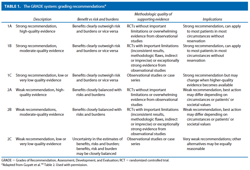
METHODOLOGY
An organized search of MEDLINE/PubMed and the Cochrane Database of Systematic Reviews and Clinical Trials was performed, from 1978 to June 2010, using the key words “rectal prolapse,” “procidentia,” “laparoscopy,” “suture rectopexy,” “mesh rectopexy, resection rectopexy,” “perineal rectosigmoidectomy.” Selected embedded references were also reviewed. All English language manuscripts and studies of adults were reviewed. Recommendations were formulated by the primary authors and reviewed by the entire committee. The final grade of recommendation was performed using the Grades of Recommendation, Assessment, Development, and Evaluation (GRADE) system18 (Table 1) and reviewed by the entire Standards Committee.

RECOMMENDATIONS
Evaluation of Rectal Prolapse
1. The initial evaluation of a patient with rectal prolapse should include a complete history and physical examination. Grade of Recommendation: Strong recommendation based on low quality evidence 1C

Before operative intervention, a careful history and physical examination should be performed. If the diagnosis is suspected from the history, but not detected on physical examination, confirmation can be obtained by asking the patient to reproduce the prolapse by straining while on a toilet with or without use of an enema. Inspection of the perineum with the patient in the sitting or squatting position is helpful for this purpose. A common pitfall in the diagnosis of rectal prolapse is the potential for confusion with prolapsing internal hemorrhoids or rectal mucosal prolapse. Usually, these conditions are easily distinguished by clinical examination. Close inspection of the direction of the prolapsed tissue folds will reveal that in the case of full-thickness rectal prolapse, the folds are always concentric, whereas hemorrhoidal tissue or rectal mucosa develops radial invaginations.

Full inspection of the perineum and complete anorectal examination is equally important. A patulous anus with diminished sphincter tone is usually identified. Proctoscopy reveals a solitary rectal ulcer on the anterior surface of the rectum in 10% to 15% of cases. In the event that the prolapse is still elusive, patients can be asked to photo-graph the prolapse at home. Twenty to thirty-five percent of patients with rectal prolapse report urinary incontinence, and about 15% to 30% have significant vaginal vault prolapse.9,20 These symptoms require evaluation, and potentially, multidisciplinary surgical intervention.

2. Additional tests such as a defecography, colonoscopy, barium enema, and urodynamics can be used selectively to define the diagnosis and identify other important pathology. Grade of Recommendation: Strong recommendation based on moderate quality evidence 1B

If the prolapse cannot be produced during the physical examination, then a defecography may reveal the problem. Defecography may also reveal associated defects such as cystocele, vaginal vault prolapse, and enterocele that may, depending on symptoms, require treatment as well.21,22 Although uncommon, a neoplasm may form the lead point for a rectal intussusception.23 For this reason, and because this problem often occurs in the older population, colonoscopy should be performed based on existing guide-lines of appropriate screening for colorectal cancer. A significant finding on colonoscopic inspection may change the operative approach. For those patients who also have symptoms of vaginal prolapse or urinary incontinence, urodynamics and urogynecologic examination should be considered because surgical intervention may be needed for both the anterior and posterior compartments of the pelvis.24 –26

3. Physiologic testing may be useful to assess functional disorders associated with rectal prolapse, such as constipation or fecal incontinence. Grade of Recommendation: Weak recommendation based on low quality evidence 2C

Do Anorectal physiology studies rarely change the operative strategy for rectal prolapse, but they can often guide treatment for associated functional abnormalities, in particular, in the postoperative period. Patients will often present with rectal prolapse in the setting of lifelong severe constipation. These patients require special consideration in accordance with the ASCRS constipation practice pa-rameter.27 Anorectal physiology testing to assess for pelvic floor dyssynergia and a transit study to rule out colonic inertia should be considered in these situations. Patients with pelvic floor dyssynergia may benefit from postoperative biofeedback , and those who have evidence of surgically amenable slow-transit constipation, and are continent, may be candidates for subtotal colectomy in addition to a rectopexy.28

Chronic dilation of the anal sphincter with diminished internal anal sphincter pressures is a common finding and can lead to fecal incontinence. Again, the evaluation of these patients should be in accordance with the ASCRS practice parameter for fecal incontinence29 and may include endorectal ultrasound to evaluate sphincter defects, and anorectal manometry and pudendal nerve testing, as well. The finding of increased nerve conduction periods (nerve damage) may have postoperative prognostic significance for continence; patients with evidence of nerve damage may have a higher rate of incontinence following surgical correction of the prolapse, although more studies are required to confirm this.30 –32 In general, patients with fecal incontinence secondary to rectal prolapse will have improvement in their symptoms once the prolapse is treated. Unfortunately, in most studies, neither preoperative manometric findings nor nerve conduction velocities have served as reliable predictors of postoperative func-tion.33 Decreased anal squeeze or resting pressures may predate the actual development of the prolapse and con-tribute to the development of the condition.

Nonoperative Management
1. Although many patients who present with rectal pro-lapse are older and have multiple comorbidities, there is little nonoperative treatment available for symptomatic rectal prolapse. Grade: Weak recommendation based on low-quality evidence 2C

Addressing symptoms of constipation using fiber and stool softeners may be of use.34 Table sugar has been used to reduce incarcerated rectal prolapse by absorbing the edema of the rectal , thus making it easier to reduce.35 However, this does not definitively treat the condition. There are no studies that compare surgical and medical management of rectal prolapse.

Operations for Rectal Prolapse
Surgery is the mainstay for treatment of rectal prolapse. However, the number of procedures described in the literature both historically and in recent times continues to increase. Operative repairs include anal encirclement (historical interest only), mucosal resection, perineal proctosigmoidectomy, anterior resection with or without rectopexy, suture rectopexy alone, and a host of procedures involving the use of synthetic meshes affixed to the presacral fascia. Two predominant general approaches, abdominal and perineal, are considered in the operative re-pair of rectal prolapse. The surgical approach is dictated by the comorbidities of the patient, the surgeon’s preference and experience, and the patient’s age and bowel function.36 Although numerous operative approaches to rectal pro-lapse are described using both abdominal and perineal techniques, only a few are actually routinely advocated, and many are of historical interest only. Discussed here are procedures in common practice and routinely reported on in the literature.

Abdominal Procedures for Rectal Prolapse
1. In patients with acceptable risk, procedures incorporating trans-abdominal rectal fixation are typically the procedure of choice for the treatment of rectal prolapse. Grade of Recommendation: Strong recommendation based on moderate quality evidence 1B

In general, it is believed that the perineal approach results in less perioperative morbidity and pain, and a reduced length of hospital stay. However, recurrence rates that are 4 times higher than those for abdominal operations and worse functional outcome as a result of resection of the rectum have prevented this approach from becoming the procedure of choice.11,34,37 Abdominal operations generally have superior overall results and have become the preferred treatment for younger and healthier patients. However, morbidity and mortality is slightly higher with an abdominal approach, making the consideration of patient comorbidities essential in deciding the appropriate repair.33,34

Suture Rectopexy
1. Rectopexy is a key component in the abdominal approach to rectal prolapse. Grade of Recommendation: Strong recommendation based on low-quality evidence 1C

The fixation of the rectum in the pelvis with suture, first described by Cutait38 in 1959, aims to correct the telescoping of the redundant bowel and causes fixation of the rectum from the resultant scarring and fibrosis. The recurrence rates for suture rectopexy are generally re-ported to be from 3% to 9%.39–43 Rectopexy can also pro-duce new-onset or worsened constipation. Fifteen percent of patients experience constipation for the first time fol-lowing rectopexy, and at least 50% of those who are constipated preoperatively are made worse.44 The precise etiology of constipation is unclear. Mechanical as well as functional reasons for constipation should be considered.

2. A sigmoid resection may be added to rectopexy in patients with prolapse and preoperative constipation, but it is not necessary in those without constipation. Grade of Recommendation: Strong recommendation based on moderate-quality evidence 1B

Resection rectopexy is a technique first described by Frykman and Goldberg in 196945 and popularized in the United States in the past 30 years. The appeal of the procedure includes the lack of artificial mesh, ease of operation, and resection of “redundant” sigmoid colon. Recurrence rates are low, ranging from 2% to 5%, and major complication rates range from 0% to 20% and relate either to obstruction or anastomotic leak. The addition of sigmoid-ectomy to the operation was felt to be associated with a lower recurrence rate and improved functional outcome with a minimal increase in morbidity.46,47 It seems to re-duce constipation significantly in those who report this symptom preoperatively in some studies.34,46,48 Others have argued that sigmoidectomy is an inadequate operation for a chronic motility problem that affects the entire bowel, and those patients should be formally evaluated preoperatively and subtotal colectomy recommended if colonic inertia is detected. Although some patients who report incontinence before surgery will have an improvement in symptoms even after a sigmoid resection, resolution of fecal incontinence is less common if sigmoid resection is performed.34 There is increasing evidence that sigmoid resection may not be necessary in those who re-port no history of constipation and whose predominant complaint is fecal incontinence. This particular patient group does not seem to be predisposed to future constipation.49

3. Division of the lateral stalks during rectal dissection may worsen symptoms of constipation postoperatively, but it is associated with decreased recurrence rates. Grade of Recommendation: Weak recommendation based on moderate-quality evidence 2B

Independent of the technique used to perform the rectopexy, the division of the lateral stalks during the rectal dissection has been associated with worsening constipation.6,34,43,47,50 –53 It was theorized that the denervation of the rectum from the neural efferents thought to reside in the lateral ligaments was the cause of this complication. As a result, a revised version of the resection rectopexy advised preservation of the lateral stalks and unilateral fastening of the rectal mesentery to the sacrum at the level of the sacral promontory. However, multiple other studies examining the onset of constipation after preservation of the lateral stalks noted constipation in 18% to 89% of patients in comparison with 14% to 48% of those patients with lateral stalks divided. Furthermore, although there can be some improvement in constipation with preservation of the lateral ligaments, recurrence rates are found to be in-creased.34,51,53

Mesh Rectopexy
1. The Ripstein procedure with fixation of mesh from the anterior rectal wall to the sacral promontory after posterior mobilization may be used for treatment of rectal prolapse, but it is associated with higher morbidity. Grade of Recommendation: Strong recommendation based on low-quality evidence 1C

Prosthetic materials have long been used to affix the rectum to the sacrum to treat rectal prolapse. The Ripstein repair54 (and its many iterations) involves placement of a prosthetic mesh around the mobilized rectum with attach-ment of the mesh to the presacral fascia below the sacral promontory.13 Recurrence rates for this procedure range from 2.3% to 5%. After mobilization of the rectum, Rip-stein originally described using a band of rectangular mesh placed around the anterior aspect of the rectum at the level of the peritoneal reflection. Sutures were used to secure the mesh to the rectum anteriorly and the rectum was pulled upward and posterior. Then, both sides of the mesh were sutured to the presacral fascia. Recurrence rates ranged from 4% to 10%, but complication rates were excessive, up to 50%, primarily because of the placement of a foreign material on the anterior rectal wall.55–57

Because of, including large-bowel obstruction, erosion of the mesh through the bowel, ureteral injury or fibrosis, small-bowel obstruction, rectovaginal fistula, and fecal impaction, Ripstein modified the technique with posterior fixation of the mesh to the sacrum with attachment of the ends of the mesh to the rectum laterally.58 Recurrence rates are similar. Subsequent postoperative morbidity rates are 20%, but most of these complications are minor. Mesh rectopexy results in significant improvement in fecal incontinence in 20% to 60% of patients.9

2. A modified Wells procedure using a variety of foreign materials for posterior fixation of the rectum may be used for treatment of rectal prolapse. Grade of Evidence: Weak recommendation based on moderate-quality evidence 2B

Wells originally described fixation of the rectum using an Ivalon sponge and transection of the lateral ligaments. He reported excellent results with minimal complica-tions.59 A randomized trial of Ivalon (polyvinyl alcohol) sponge vs suture rectopexy found comparable recurrence rates but increased complication rates and postoperative constipation in the Ivalon group and recommended that this technique be abandoned.43 The Ivalon sponge is no longer commercially available. However, the modified Wells technique using other materials such as polyester or polypropylene mesh60,61 continues to be popular, especially for laparoscopic approaches.

3. The ventral mesh rectopexy reduces constipation by avoiding posterolateral mobilization of the rectum and produces results similar to other abdominal approaches. Grade of Recommendation: Weak recommendation based on moderate-quality evidence 2B

D’Hoore and colleagues62 first described the ventral rectopexy repair and its potential advantage in avoiding postoperative constipation. The technique involves mobilization of the anterior wall of the rectum with fixation of mesh to the anterior wall and then fixation of the mesh to the sacrum. This is in contrast to the Orr-Loygue procedure, 63 where the rectum was mobilized both anteriorly and posteriorly, before fixation to the sacrum. A systematic review of 12 non-randomized case series of 728 patients undergoing ventral rectopexy reported a recurrence rate of 3.4, and a weighted decrease in the postoperative constipation rate was estimated to be 23%. However, new onset of constipation was also noted to be 14.4%.64

Anterior Resection
1. The use of anterior resection alone to treat rectal pro-lapse is associated with higher recurrence rates and significant operative and postoperative morbidity; it should not be considered as a first-line treatment. Grade of recommendation: Strong recommendation based on moderate-quality evidence 1B

Anterior resection was described as an alternative strategy to repair prolapse in 1955, and there are some advocates of the technique. Unfortunately, in several retrospective reviews, several shortcomings are evident. In one review of 113 patients, the recurrence rate continued to climb after 2, 5, and 10 years to 3%, 6%, and 12%, with an operative morbidity of 29%, including 3 anastomotic leaks.65 Another review confirmed that, with an average follow-up of 6 years, recurrence occurred in 7% of cases.50 A low pelvic anastomosis in those with borderline continence may cause complete loss of control. Careful selection of patients is necessary for this procedure, and, in general, given the slightly higher recurrence rates and lack of functional advantages, it is not widely practiced.

Adjunctive Operative Techniques for Abdominal Procedures
1. A minimally invasive approach to rectal prolapse by experienced surgeons compares favorably with an open repair. Grade of Evidence: Strong recommendation based on moderate-quality evidence 1B

All abdominal approaches to rectal prolapse have been performed laparoscopically over the past decade with essentially similar results.34,66 – 69 The indications for per-forming a laparoscopic procedure are primarily related to the indications for an abdominal approach; patients with-out previous abdominal surgery are excellent candidates, but prior pelvic surgery is not necessarily an exclusion criterion. The laparoscopic treatment of rectal prolapse was first described in 1992 and involved a rectopexy without sigmoid resection.70 Since that time, numerous series have demonstrated equivalent recurrence rates (4%– 8%) and morbidity (10%–33%) of the laparoscopic repair in comparison with open approaches, but clear benefits in terms of pain control, length of stay, and return of bowel func-tion.15,34,68 In addition, the feasibility of a minimally invasive approach for colorectal resective procedures has been demonstrated in the high-risk patient.71 Wound complication rates have also been found to be decreased in laparoscopic surgery for prolapse. Certainly, those who undergo rectopexy without resection are at very low risk for infection because only trocar incisions are needed. The actual surgical technique to perform laparoscopic rectopexy or resection is the same as that used for open repairs. The goals of surgery remain the same, to eradicate the full-thickness rectal prolapse, improve bowel function and continence, and minimize recurrence rates.15 However, recurrence rates should be judged in light of the length of follow-up, because a significant percentage of recurrences may occur several years after treatment.72–74

Recent applications of robotic surgery for colorectal conditions have focused on pelvic operations because of the ease of maintaining one field for the procedure. Only a few series have been published with small numbers of patients that have demonstrated equivalent outcomes compared with laparoscopic approaches. Disadvantages include longer operating time and cost. However, visualization and ease of suturing and tying account for much of the interest in this technique.75–78

Perineal Operations for Rectal Prolapse
1. Patients with a short, full-thickness rectal prolapse can be treated with a mucosal sleeve resection; but, for a longer prolapse, it is associated with a higher recurrence rate compared with abdominal approaches. Grade of Recommendation: Strong recommendation based on low-quality evidence 1C

For patients with a short, full-thickness rectal prolapse or a mucosal prolapse, a Delorme procedure can be per-formed. It involves a circumferential mucosal sleeve resection and imbrication of the muscularis layer with serial vertical sutures. Recurrence rates are higher than the abdominal approaches in the range of 10% to 15%.79–82 This procedure is advocated for those who are considered “high risk” for an abdominal procedure because of comorbidities or to avoid risk of nerve damage. Complications such as infection, urinary retention, bleeding, and fecal impaction occur in 4% to 12% of cases.79,82 Constipation and fecal incontinence improve following surgery, but urgency and tenesmus do occur. Although restoration of function is not uniform in the series surveyed, in one of the few studies reporting postoperative manometric findings, both mean resting and squeeze pressures were significantly increased by the procedure.81

2. Patients with a full-thickness rectal prolapse who are not candidates for an abdominal operation may be treated with a perineal rectosigmoidectomy but are susceptible to higher recurrence rates in comparison with abdominal approaches. Grade of Recommendation: Strong recommendation based on low-quality evidence 1C

Perineal rectosigmoidectomy involves the full-thickness resection of the rectum and sigmoid colon via the anus with a coloanal anastomosis by the use of sutures or a stapling device. This operation can be performed without general anesthesia, involves a shorter hospital stay, and has lower complication rates (10%), which include bleeding from the staple or suture line, pelvic abscess, and, rarely, an anastomotic leak. As a result, patients undergoing perineal proctosigmoidectomy are generally older with significantly more comorbidities than those who are considered for abdominal repair.11,83 However, recurrence rates have been reported to be as high as 16% to 30%.11,83– 86 Other studies have shown that the use of levatoroplasty to treat levator diastasis can reduce recurrence rates from 21% to 7%.87,88 Only one small randomized controlled trial (n 20) has compared perineal rectosigmoidectomy with an abdominal approach. The recurrence rate was 10% for the perineal group vs 0% for the abdominal group.37

The practice parameters set forth in this document have been developed from sources believed to be reliable. The American Society of Colon and Rectal Surgeons makes no warranty, guarantee, or representation whatsoever as to the absolute validity or sufficiency of any parameter included in this document, and the Society assumes no responsibility for the use of the material contained.

APPENDIX A: CONTRIBUTING MEMBERS OF THE ASCRS STANDARDS COMMITTEE
Donald Buie, M.D., Chair; Janice Rafferty, M.D., Co-Chair; Farshid Araghizadeh, M.D.; Robin Boushey, M.D.; Srihdhar Chalasani, M.D.; George Chang, M.D.; Robert Cima, M.D.; Gary Dunn, M.D.; Daniel Feingold, M.D.; Phillip Fleshner, M.D.; Daniel Geisler, M.D.; Jill Jenua, M.D.; Sharon Gregorcyk, M.D.; Daniel Herzig, M.D.; An-dreas Kaiser, M.D.; Ravin Kumar, M.D.; David Larson, M.D.; Stephen Mills, M.D.; John Monson, M.D.; W. Brian Perry, M.D.; P. Terry Phang, M.D.; David Rivadeneira, M.D.; Howard Ross, M.D.; Peter Senatore, M.D.; Elin Sigurdson, M.D.; Thomas Stahl, M.D.; Scott Steele, M.D.; Scott Strong, M.D.; Charles Ternent, M.D.; Judith Trudel, M.D.; Madhulika Varma, M.D.; and Martin Weiser, M.D.

REFERENCES
1. Broden B, Snellman B. Procidentia of the rectum studied with cineradiography: a contribution to the discussion of causative mechanism. Dis Colon Rectum. 1968;11:330 –347.
2. Kuijpers HC. Treatment of complete rectal prolapse: to narrow, to wrap, to suspend, to fix, to encircle, to plicate or to resect?World J Surg. 1992;16:826 – 830.
3. Nicholls RJ. Rectal prolapse and the solitary ulcer syndrome. Ann Ital Chir. 1994;65:157–162.
4. Parks AG, Swash M, Urich H. Sphincter denervation in anorec-tal incontinence and rectal prolapse. Gut. 1977;18:656 – 665.
5. Roig JV, Buch E, Alos R, et al. Anorectal function in patients with complete rectal prolapse: differences between continent and incontinent individuals. Rev Esp Enferm Dig. 1998;90:794 –805.
6. Yakut M, Kaymakcioglu N, Simsek A, Tan A, Sen D. Surgical treatment of rectal prolapse: a retrospective analysis of 94 cases. Int Surg. 1998;83:53–55.
7. Gourgiotis S, Baratsis S. Rectal prolapse. Int J Colorectal Dis. 2007;22:231–243.
8. Kairaluoma MV, Kellokumpu IH. Epidemiologic aspects of complete rectal prolapse. Scand J Surg. 2005;94:207–210.
9. Madiba TE, Baig MK, Wexner SD. Surgical management of rec-tal prolapse. Arch Surg. 2005;140:63–73.
10. Marceau C, Parc Y, Debroux E, Tiret E, Parc R. Complete rectal prolapse in young patients: psychiatric disease a risk factor of poor outcome. Colorectal Dis. 2005;7:360 –365.
11. Kim DS, Tsang CB, Wong WD, Lowry AC, Goldberg SM, Mad-off RD. Complete rectal prolapse: evolution of management and results. Dis Colon Rectum. 1999;42:460 – 469.
12. Madoff RD, Mellgren A. One hundred years of rectal prolapse surgery. Dis Colon Rectum. 1999;42:441– 450.
13. Schultz I, Mellgren A, Dolk A, Johansson C, Holmstrom B. Long-term results and functional outcome after Ripstein rectopexy. Dis Colon Rectum. 2000;43:35– 43.
14. Schultz I, Mellgren A, Oberg M, Dolk A, Holmstrom B. Whole gut transit is prolonged after Ripstein rectopexy. Eur J Surg. 1999;165:242–247.
15. Senagore AJ. Management of rectal prolapse: the role of laparo-scopic approaches. Semin Laparosc Surg. 2003;10:197–202.
16. Glasgow SC, Birnbaum EH, Kodner IJ, Fleshman JW, Dietz DW. Preoperative anal manometry predicts continence after perineal proctectomy for rectal prolapse. Dis Colon Rectum. 2006;49: 1052–1058.
17. Snooks SJ, Henry MM, Swash M. Anorectal incontinence and rectal prolapse: differential assessment of the innervation to pu-borectalis and external anal sphincter muscles. Gut. 1985;26: 470 – 476.
18. Guyatt G, Gutermen D, Baumann MH, et al. Grading strength of recommendations and quality of evidence in clinical guidelines: report from an American College of Chest Physicians Task Force. Chest. 2006;129:174 –181.
19. Altman D, Zetterstrom J, Schultz I, et al. Pelvic organ prolapse and urinary incontinence in women with surgically managed rectal prolapse: a population-based case-control study. Dis Co-lon Rectum. 2006;49:28 –35.
20. Gonzalez-Argente FX, Jain A, Nogueras JJ, Davila GW, Weiss EG, Wexner SD. Prevalence and severity of urinary incontinence and pelvic genital prolapse in females with anal incontinence or rectal prolapse. Dis Colon Rectum. 2001;44:920 –926.
21. Pescatori M, Spyrou M, Pulvirenti d’Urso A. A prospective eval-uation of occult disorders in obstructed defecation using the ‘iceberg diagram.’ Colorectal Dis. 2006;8:785–789.
22. Renzi A, Izzo D, Di Sarno G, et al. Cinedefecographic findings in patients with obstructed defecation syndrome: a study in 420 cases. Minerva Chir. 2006;61:493– 499.
23. Bounovas A, Polychronidis A, Laftsidis P, Simopoulos C. Sig-moid colon cancer presenting as complete rectal prolapse. Colo-rectal Dis. 2007;9:665– 666.
24. Lim M, Sagar PM, Gonsalves S, Thekkinkattil D, Landon C. Surgical management of pelvic organ prolapse in females: functional outcome of mesh sacrocolpopexy and rectopexy as a com-bined procedure. Dis Colon Rectum. 2007;50:1412–1421.
25. Mellgren A, Johansson C, Dolk A, et al. Enterocele demon-strated by defaecography is associated with other pelvic floor disorders. Int J Colorectal Dis. 1994;9:121–124.
26. Sagar PM, Thekkinkattil DK, Heath RM, Woodfield J, Gon-salves S, Landon CR. Feasibility and functional outcome of laparoscopic sacrocolporectopexy for combined vaginal and rectal prolapse. Dis Colon Rectum. 2008;51:1414 –1420.
27. Ternent CA, Bastawrous AL, Morin NA, Ellis CN, Hyman NH, Buie WD. Practice parameters for the evaluation and manage-ment of constipation. Dis Colon Rectum. 2007;50:2013–2022.
28. Watts JD, Rothenberger DA, Buls JG, Goldberg SM, Nivatvongs
S. The management of procidentia: 30 years’ experience. Dis Colon Rectum. 1985;28:96 –102.
29. Tjandra JJ, Dykes SL, Kumar RR, et al. Practice parameters for the treatment of fecal incontinence. Dis Colon Rectum. 2007;50: 1497–1507.
30. Birnbaum EH, Stamm L, Rafferty JF, Fry RD, Kodner IJ, Flesh-man JW. Pudendal nerve terminal motor latency influences sur-gical outcome in treatment of rectal prolapse. Dis Colon Rectum. 1996;39:1215–1221.
31. Johansen OB, Wexner SD, Daniel N, Nogueras JJ, Jagelman DG. Perineal rectosigmoidectomy in the elderly. Dis Colon Rectum. 1993;36:767–772.
32. Schultz I, Mellgren A, Nilsson BY, Dolk A, Holmstrom B. Pre-operative electrophysiologic assessment cannot predict continence after rectopexy. Dis Colon Rectum. 1998;41:1392–1398.
33. Felt-Bersma RJ, Tiersma ES, Cuesta MA. Rectal prolapse, rectal intussusception, rectocele, solitary rectal ulcer syndrome, and enterocele. Gastroenterol Clin North Am. 2008;37:645– 668.
34. Tou S, Brown SR, Malik AI, Nelson RL. Surgery for complete rectal prolapse in adults. Cochrane Database Syst Rev. 2008: CD001758.
35. Myers JO, Rothenberger DA. Sugar in the reduction of incarcer-ated prolapsed bowel: report of two cases. Dis Colon Rectum. 1991;34:416 – 418.
36. Brown AJ, Anderson JH, McKee RF, Finlay IG. Strategy for se-lection of type of operation for rectal prolapse based on clinical criteria. Dis Colon Rectum. 2004;47:103–107.
37. Deen KI, Grant E, Billingham C, Keighley MR. Abdominal re-section rectopexy with pelvic floor repair versus perineal rec-tosigmoidectomy and pelvic floor repair for full-thickness rectal prolapse. Br J Surg. 1994;81:302–304.
38. Cutait D. Sacro-promontory fixation of the rectum for complete rectal prolapse. Proc R Soc Med. 1959;52:105.
39. Briel JW, Schouten WR, Boerma MO. Long-term results of su-ture rectopexy in patients with fecal incontinence associated with incomplete rectal prolapse. Dis Colon Rectum. 1997;40: 1228 –1232.
40. Carter AE. Rectosacral suture fixation for complete rectal pro-lapse in the elderly, the frail and the demented. Br J Surg. 1983; 70:522–523.
41. Graf W, Karlbom U, Pahlman L, Nilsson S, Ejerblad S. Functional results after abdominal suture rectopexy for rectal pro-lapse or intussusception. Eur J Surg. 1996;162:905–911.
42. Khanna AK, Misra MK, Kumar K. Simplified sutured sacral rectopexy for complete rectal prolapse in adults. Eur J Surg. 1996; 162:143–146.
43. Novell JR, Osborne MJ, Winslet MC, Lewis AA. Prospective ran-domized trial of Ivalon sponge versus sutured rectopexy for full-thickness rectal prolapse. Br J Surg. 1994;81:904 –906.
44. Aitola PT, Hiltunen KM, Matikainen MJ. Functional results of operative treatment of rectal prolapse over an 11-year period: emphasis on transabdominal approach. Dis Colon Rectum. 1999; 42:655– 660.
45. Frykman HM, Goldberg SM. The surgical treatment of rectal procidentia. Surg Gynecol Obstet. 1969;129:1225–1230.
46. Luukkonen P, Mikkonen U, Jarvinen H. Abdominal rectopexy with sigmoidectomy vs. rectopexy alone for rectal prolapse: a prospective, randomized study. Int J Colorectal Dis. 1992;7:219 –222.
47. Sayfan J, Pinho M, Alexander-Williams J, Keighley MR. Sutured posterior abdominal rectopexy with sigmoidectomy compared with Marlex rectopexy for rectal prolapse. Br J Surg. 1990;77: 143–145.
48. McKee RF, Lauder JC, Poon FW, Aitchison MA, Finlay IG. A prospective randomized study of abdominal rectopexy with and without sigmoidectomy in rectal prolapse. Surg Gynecol Obstet. 1992;174:145–148.
49. Hsu A, Brand MI, Saclarides TJ. Laparoscopic rectopexy without resection: a worthwhile treatment for rectal prolapse in patients without prior constipation. Am Surg. 2007;73:858 – 861.
50. Cirocco WC, Brown AC. Anterior resection for the treatment of rectal prolapse: a 20-year experience. Am Surg. 1993;59:265–269.
51. Mollen RM, Kuijpers JH, van Hoek F. Effects of rectal mobiliza-tion and lateral ligaments division on colonic and anorectal function. Dis Colon Rectum. 2000;43:1283–1287.
52. Scaglia M, Fasth S, Hallgren T, Nordgren S, Oresland T, Hulten
L. Abdominal rectopexy for rectal prolapse: influence of surgical technique on functional outcome. Dis Colon Rectum. 1994;37: 805– 813.
53. Speakman CT, Madden MV, Nicholls RJ, Kamm MA. Lateral ligament division during rectopexy causes constipation but pre-vents recurrence: results of a prospective randomized study. Br J Surg. 1991;78:1431–1433.
54. Ripstein CB, Lanter B. Etiology and surgical therapy of massive prolapse of the rectum. Ann Surg. 1963;157:259 –264.
55. Gordon PH, Hoexter B. Complications of the Ripstein procedure. Dis Colon Rectum. 1978;21:277–280.
56. Kupfer CA, Goligher JC. One hundred consecutive cases of complete prolapse of the rectum treated by operation. Br J Surg. 1970;57:482– 487.
57. Roberts PL, Schoetz DJ Jr, Coller JA, Veidenheimer MC. Rip-stein procedure. Lahey Clinic experience: 1963–1985. Arch Surg. 1988;123:554 –557.
58. McMahan JD, Ripstein CB. Rectal prolapse: an update on the rectal sling procedure. Am Surg. 1987;53:37– 40.
59. Wells C. New operation for rectal prolapse. Proc R Soc Med. 1959;52:602– 603.
60. Dulucq JL, Wintringer P, Mahajna A. Clinical and functional outcome of laparoscopic posterior rectopexy (Wells) for full-thickness rectal prolapse: a prospective study. Surg Endosc. 2007; 21:2226 –2230.
61. Madbouly KM, Senagore AJ, Delaney CP, Duepree HJ, Brady KM, Fazio VW. Clinically based management of rectal prolapse. Surg Endosc. 2003;17:99 –103.
62. D’Hoore A, Penninckx F. Laparoscopic ventral recto(col-po)pexy for rectal prolapse: surgical technique and outcome for 109 patients. Surg Endosc. 2006;20:1919 –1923.
63. Loygue J, Nordlinger B, Cunci O, Malafosse M, Huguet C, Parc
R. Rectopexy to the promontory for the treatment of rectal pro-lapse: report of 257 cases. Dis Colon Rectum. 1984;27:356 –359.
64. Samaranayake CB, Luo C, Plank AW, Merrie AE, Plank LD, Bissett IP. Systematic review on ventral rectopexy for rectal pro-lapse and intussusception. Colorectal Dis. 2010;12:504 –512.
65. Schlinkert RT, Beart RW Jr, Wolff BG, Pemberton JH. Anterior resection for complete rectal prolapse. Dis Colon Rectum. 1985; 28:409 – 412.
66. Boccasanta P, Venturi M, Reitano MC, et al. Laparotomic vs. laparoscopic rectopexy in complete rectal prolapse. Dig Surg. 1999;16:415– 419.
67. Kariv Y, Delaney CP, Casillas S, et al. Long-term outcome after laparoscopic and open surgery for rectal prolapse: a case-control study. Surg Endosc. 2006;20:35– 42.
68. Purkayastha S, Tekkis P, Athanasiou T, et al. A comparison of open vs. laparoscopic abdominal rectopexy for full-thickness rectal prolapse: a meta-analysis. Dis Colon Rectum. 2005;48: 1930 –1940.
69. Solomon MJ, Young CJ, Eyers AA, Roberts RA. Randomized clinical trial of laparoscopic versus open abdominal rectopexy for rectal prolapse. Br J Surg. 2002;89:35–39.
70. Berman IR. Sutureless laparoscopic rectopexy for procidentia: technique and implications. Dis Colon Rectum. 1992;35:689 –693.
71. Plocek MD, Geisler DP, Glennon EJ, Kondylis P, Reilly JC. Laparoscopic colorectal surgery in the complicated patient. Am J Surg. 2005;190:882– 885.
72. DiGiuro G, Ignjatovic D, Brogger J, Bergamaschi R. How accu-rate are published recurrence rates after rectal prolapse surgery?A meta-analysis of individual patient data. Am J Surg. 2006;191: 773–778.
73. Raftopoulos Y, Senagore AJ, Di Giuro G, Bergamaschi R. Recurrence rates after abdominal surgery for complete rectal prolapse: a multicenter pooled analysis of 643 individual patient data. Dis Colon Rectum. 2005;48:1200 –1206.
74. Byrne CM, Smith SR, Solomon MJ, Young JM, Eyers AA, Young CJ. Long-term functional outcomes after laparoscopic and open rectopexy for the treatment of rectal prolapse. Dis Colon Rectum. 2008;51:1597–1604.
75. Ayav A, Bresler L, Hubert J, Brunaud L, Boissel P. Robotic-as-sisted pelvic organ prolapse surgery. Surg Endosc. 2005;19:1200 –1203.
76. Delaney CP, Lynch AC, Senagore AJ, Fazio VW. Comparison of robotically performed and traditional laparoscopic colorectal surgery. Dis Colon Rectum. 2003;46:1633–1639.
77. Heemskerk J, de Hoog DE, van Gemert WG, Baeten CG, Greve JW, Bouvy ND. Robot-assisted vs conventional laparoscopic rectopexy for rectal prolapse: a comparative study on costs and time. Dis Colon Rectum. 2007;50:1825–1830.
78. Munz Y, Moorthy K, Kudchadkar R, et al. Robotic assisted rectopexy. Am J Surg. 2004;187:88 –92.
79. Lieberth M, Kondylis LA, Reilly JC, Kondylis PD. The Delorme repair for full-thickness rectal prolapse: a retrospective review. Am J Surg. 2009;197:418 – 423.
80. Senapati A, Nicholls RJ, Thomson JP, Phillips RK. Results of Delorme’s procedure for rectal prolapse. Dis Colon Rectum. 1994;37:456 – 460.
81. Tsunoda A, Yasuda N, Yokoyama N, Kamiyama G, Kusano M. Delorme’s procedure for rectal prolapse: clinical and physiolog-ical analysis. Dis Colon Rectum. 2003;46:1260 –1265.
82. Watkins BP, Landercasper J, Belzer GE, et al. Long-term fol-low-up of the modified Delorme procedure for rectal prolapse. Arch Surg. 2003;138:498 –502.
83. Riansuwan W, Hull TL, Bast J, Hammel JP, Church JM. Comparison of perineal operations with abdominal operations for full-thickness rectal prolapse. World J Surg. 2010;34:1116 –1122.
84. Azimuddin K, Khubchandani IT, Rosen L, Stasik JJ, Riether RD, Reed JF III. Rectal prolapse: a search for the “best” operation. Am Surg. 2001;67:622– 627.
85. Pescatori M, Zbar AP. Tailored surgery for internal and external rectal prolapse: functional results of 268 patients operated upon by a single surgeon over a 21-year period*. Colorectal Dis. 2009; 11:410 – 419.
86. Altomare DF, Binda G, Ganio E, De Nardi P, Giamundo P, Pes-catori M. Long-term outcome of Altemeier’s procedure for rec-tal prolapse. Dis Colon Rectum. 2009;52:698 –703.
87. Chun SW, Pikarsky AJ, You SY, et al. Perineal rectosigmoidec-tomy for rectal prolapse: role of levatorplasty. Tech Coloproctol. 2004;8:3–9.
88. Habr-Gama A, Jacob CE, Jorge JM, et al. Rectal procidentia treatment by perineal rectosigmoidectomy combined with leva-tor ani repair. Hepatogastroenterology. 2006;53:213–217.

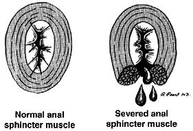
Rectocele Information

Download PDF

Rectocele

What is a rectocele?
A rectocele is a bulging of the front wall of the rectum into the back wall of the vagina. Rectoceles are usually due to thinning of the rectovaginal septum (the tissue between the rectum and vagina) and weakening of the pelvic floor muscles. This is a very common defect; however, most women do not have symptoms. There can also be other pelvic organs that bulge into the vagina, leading to similar symptoms as rectocele, including the bladder (i.e., cystocele) and the small intestines (i.e. enterocele).

What can lead to developing a rectocele?
There are many things that can lead to weakening of the pelvic floor, resulting in a rectocele. These factors include: vaginal deliveries, birthing trauma during vaginal delivery (e.g. forceps delivery, vacuum delivery, tearing with a vaginal delivery, episiotomy during vaginal delivery), history of constipation, history of straining with bowel movements, and history of gynecological (hysterectomy) or rectal surgeries.

What are the symptoms associated with a rectocele?
Most people with a small rectocele do not have symptoms and it is often only discovered during routine physical examination. When the rectocele is large, it most commonly presents with a noticeable bulge into the vagina. Other rectal symptoms may include: difficulty with evacuation during a bowel movement, the need to press against the vagina and/or space between the rectum and the vagina in order to have a bowel movement, straining with bowel movements, constipation, the urge to have multiple bowel movements throughout the day, and rectal pain. Occasionally, the stool becomes stuck in the bulge of the rectum, which is why it is difficult to have a bowel movement. Vaginal symptoms can include: pain with sexual intercourse (dyspareunia), vaginal bleeding, and a sense of fullness in the vagina.

How can a rectocele be diagnosed?
A rectocele is usually found incidentally during a physical examination by your doctor. The evaluation of its severity, and potential relation to constipation symptoms, is hard to assess with physical examination alone. Further testing for a rectocele may include the use of a special x-ray study known as defecography (contrast material instilled into the rectum as an enema, followed by live x-ray imaging during a bowel movement). This study is very specific and can evaluate a rectocele’s size and ability to completely empty.

How can a rectocele be treated?
Rectoceles are not treated merely for their presence, but should only be addressed when they are associated with significant symptoms that interfere with quality of life. Prior to any treatment, there should be a thorough evaluation by your doctor to assess whether all of the complaints can be attributed to the presence of a rectocele alone. There are both medical and surgical treatment options for rectoceles. The majority of symptoms associated with a rectocele can be resolved with medical management; however, treatment depends on the severity of symptoms.

How can a rectocele be treated with medical management only?
It is very important to have a good bowel regimen in order to avoid constipation and straining with bowel movements. A high fiber diet, consisting of 25-30 grams of fiber daily, will help with this goal. This may be achieved with a fiber supplement, high fiber cereal, or high fiber bars. In addition to augmenting fiber intake, increased water intake (typically 6-8 glasses daily) is also highly recommended. This will allow for softer stools that do not require significant straining with bowel movements, thereby reducing your risk for having a bulge associated with a rectocele. Other treatments may include pelvic floor exercises such as Kegel exercises (i.e. biofeedback), stool softeners, hormone replacement therapy, and avoidance of straining with bowel movements. At times, it is also helpful to apply pressure to the back of the vagina during bowel movements.

How can a rectocele be treated with surgical management?
The surgical management of rectoceles should only be considered when symptoms continue despite the use of medical management and are significant enough that they interfere with activities of daily living. There are abdominal, rectal, and vaginal surgeries that can be performed for rectoceles. The choice of procedure depends on the size of the rectocele and its associated symptoms. Most surgeries aim to remove the extra tissue that makes up the rectocele and strengthening the wall between the rectum and vagina with surrounding tissue or use of a mesh (i.e. patch). Colorectal surgeons, as well as gynecologists, are trained in the diagnosis and treatment of this condition. The success rate of the surgery depends upon the specific symptoms and symptom duration. Some of the risks of surgical correction of the rectocele are bleeding, infection, pain during intercourse (dyspareunia), as well as a risk that the rectocele may recur or worsen.