Anorectal Abscess Information


Anal Abscess/Fistula

What is an anal abscess?

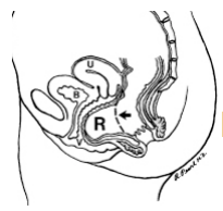
An anal abscess is an infected cavity filled with pus found near the anus or rectum.

What is an anal fistula?

An anal fistula (also called fistula-in-ano) is frequently the result of a previous or current anal abscess, occurring in up to 50% of patients with abscesses. Normal anatomy includes small glands just inside the anus. Occasionally, these glands get clogged and potentially can become infected, leading to an abscess. The fistula is a tunnel that forms under the skin and connects the infected glands to the abscess. A fistula can be present with or without an abscess and may connect just to the skin of the buttocks near the anal opening. Other situations that can result in a fistula include Crohn’s disease, radiation, trauma and malignancy.

How does someone get an anal abscess or a fistula?

The abscess is most often a result of an acute infection in the internal glands of the anus. Occasionally, bacteria, fecal material or foreign matter can clog the anal gland and create a
condition for an abscess cavity to form. Other medical conditions can make these types of
infections more likely.

After an abscess drains on its own or has been drained (opened), a tunnel (fistula) may persist, connecting the infected anal gland to the external skin. This typically will involve some type of drainage from the external opening and occurs in up to 50 % of abscesses. If the opening on the skin heals when a fistula is present, a recurrent abscess may develop.

What are the specific signs or symptoms of an abscess or fistula?

A patient with an abscess may have pain, redness or swelling in the area around the anal area. Fatigue, general malaise, as well as accompanying fever or chills are also common. Similar signs and symptoms may be present when patients have a fistula, with the addition of possible irritation of the perianal skin or drainage from an external opening.

Is any specific testing necessary to diagnose an abscess or fistula?

No. Most anal abscesses or fistula-in-ano are diagnosed and managed on the basis of clinical findings. Occasionally, additional studies such as ultrasound, CT scan, or MRI can assist with the diagnosis of deeper abscesses or the delineation of the fistula tunnel to help guide treatment.

What is the treatment of an anal abscess?

The treatment of an abscess is surgical drainage under most circumstances. An incision is made in the skin near the anus to drain the infection. This can be done in a doctor’s office with local anesthetic or in an operating room under deeper anesthesia. Hospitalization may be required for patients prone to more significant infections such as diabetics or patients with decreased immunity.

Are antibiotics required to treat this type of infection?

Antibiotics alone are a poor alternative to drainage of the infection. For uncomplicated abscesses, the addition of antibiotics to surgical drainage does not improve healing time or reduce the potential for recurrences. There are some conditions in which antibiotics are indicated, such as for patients with compromised or altered immunity, some cardiac valvular conditions or extensive cellulitis. A comprehensive discussion of your past medical history and a physical exam are important to determine if antibiotics are indicated.

What is the treatment of an anal fistula?

Surgery is almost always necessary to cure an anal fistula. Although surgery can be fairly
straightforward, it may also be complicated, occasionally requiring staged or multiple operations. Consider identifying a specialist in colon and rectal surgery who would be familiar with a number of potential operations to treat the fistula.

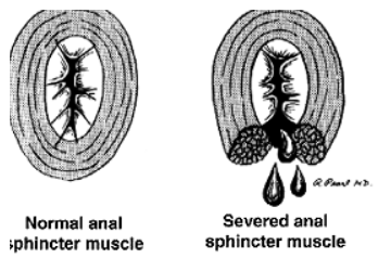
The surgery may be performed at the same time as drainage of an abscess, although sometimes the fistula doesn’t appear until weeks to years after the initial drainage. If the fistula is straightforward, a fistulotomy may be performed. This procedure involves connecting the internal opening within the anal canal to the external opening, creating a groove that will heal from the inside out. This surgery often will require dividing a small portion of the sphincter muscle which has the unlikely potential for affecting the control of bowel movements in a limited number of cases.

Other procedures include placing material within the fistula tract to occlude it or surgically altering the surrounding tissue to accomplish closure of the fistula, with the choice of procedure depending upon the type, length, and location of the fistula. Most of the operations can be performed on an outpatient basis, but may occasionally require hospitalization.

What is the recovery like from surgery?

Pain after surgery is controlled with pain pills, fiber and bulk laxatives. Patients should plan for time at home using sitz baths and attempt to avoid the constipation that can be associated with prescription pain medication. Discuss with your surgeon the specific care and time away from work prior to surgery to prepare yourself for post-operative care.

Can the abscess or fistula recur?

If adequately treated and properly healed, both are unlikely to return. However, despite proper and indicated open or minimally invasive treatment, both abscesses and fistulas can potentially recur. Should similar symptoms arise, suggesting recurrence, it is recommended that you find a colon and rectal surgeon to manage your condition.

© 2012 American Society of Colon & Rectal Surgeons

Anorectal Abscess Treatment Guidelines

Download PDF

Practice Parameters for the Management of Perianal Abscess and Fistula-in-Ano

Scott R. Steele, M.D. • Ravin Kumar, M.D. • Daniel L. Feingold, M.D. • Janice L. Rafferty, M.D. • W. Donald Buie, M.D.
Prepared by the Standards Practice Task Force of the American Society of Colon and Rectal Surgeons

The American Society of Colon and Rectal Surgeons is dedicated to ensuring high quality patient care by advancing the science, prevention, and management of disorders and diseases of the colon, rectum, and anus. The Standards Committee is composed of Society members who are chosen because they have demonstrated expertise in the specialty of colon and rectal surgery. This Committee was created to lead international efforts in defining quality care for conditions related to the colon, rectum, and anus. This is accompanied by developing Clinical Practice Guidelines based on the best available evidence. These guidelines are inclusive, and not prescriptive. Their purpose is to provide information on which decisions can be made, rather than dictate a specific form of treatment. These guidelines are intended for the use of all practitioners, health care workers, and patients who desire information about the management of the conditions addressed by the topics covered in these guidelines.

It should be recognized that these guidelines should not be deemed inclusive of all proper methods of care or exclusive of methods of care reasonably directed to obtaining the same results. The ultimate judgment regarding the propriety of any specific procedure must be made by the physician in light of all the circumstances presented by the individual patient.

STATEMENT OF THE PROBLEM

Anorectal abscess is a potentially debilitating condition most often originating from a cryptoglandular infection in the anal canal,1 and remains one of the more common anorectal conditions encountered in practice. Although the underlying pathogenesis is the same in the majority of patients, these abscesses are classified into perianal, ischiorectal, intersphincteric, and supralevator based on location.2 As such, patients may present with a variety of signs and symptoms ranging from fever, pain, tenderness, erythema, and a fluctuant mass to relatively normal external findings and deep-seated rectal pain.2

In approximately 30% to 50% of patients with an anorectal abscess, a persistent tract, or fistula-in-ano, develops, extending from the anal canal to the perineal skin.3,4 Unfortunately, there is no definitive way to predict who will develop one, or how to prevent one. Patients often report persistent purulent drainage or intermittent perianal swelling and tenderness followed by spontaneous discharge. Fistulas are categorized based on their anatomical course relative to the sphincter complex: intersphincteric, transsphincteric, suprasphincteric, and extrasphincteric.4 Fistulas can also be classified as “simple” or “complex,” with simple fistulas including low transsphincteric and intersphincteric fistulas that cross 30% of the external sphincter.5 Complex fistulas include high transsphincteric fistulas with or without a high blind tract, suprasphincteric and extrasphincteric fistulas, horseshoe fistulas, and those associated with inflammatory bowel disease, radiation, malignancy, preexisting incontinence, or chronic diarrhea, as well.6–8 Given the attenuated nature of the anterior sphincter complex in women, fistulas in this location deserve special consideration and may be considered complex as well. This practice parameter will focus on the evaluation and management of both perianal abscess and fis-tula-in-ano.

METHODOLOGY

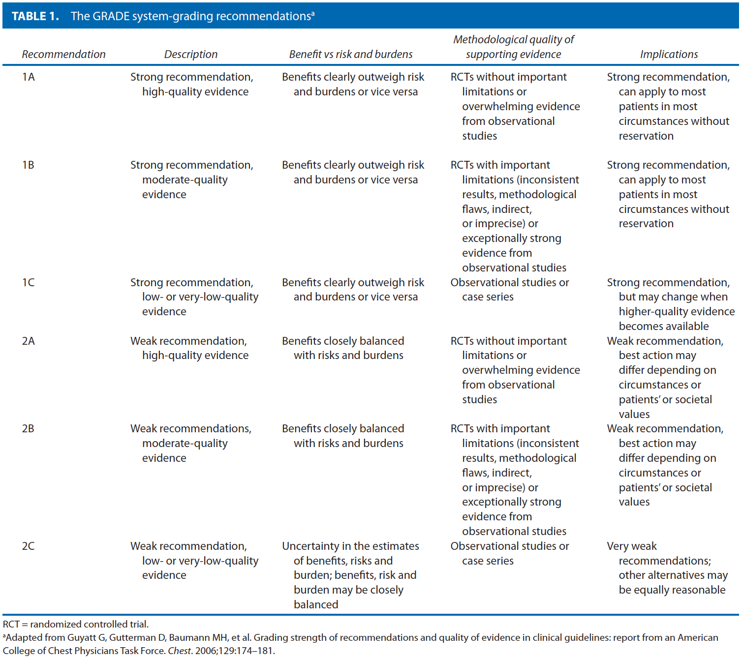
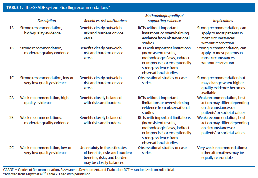
These guidelines are built on the last set of the American Society of Colon and Rectal Surgeons Practice Parameters for treatment of perianal abscess and fistula-in-ano published in 2005.9 An organized search of MEDLINE, PubMed, EMBASE, and the Cochrane Database of Collected Reviews was performed through February 2010. Key word combinations included abscess, fistula, fistula-in-ano, anal, rectal, perianal, perineal, rectovaginal, anovaginal, seton, fistula plug, fibrin glue, advancement flap, and Crohn’s disease. Directed searches of the embedded references from the primary articles were also performed in selected circumstances. Primary authors reviewed all English language manuscripts and studies of adults. Recommendations were formulated by the primary authors and reviewed by the entire Committee. The final grade of recommendation was performed using the Grades of Recommendation, Assessment, Development, and Evaluation (GRADE) system10 and reviewed by the entire Standards Committee (Table 1).11
RECOMMENDATIONS

Initial Evaluation of Perianal Abscess and Fistula-in-Ano

  • 1. A disease-specific history and physical examination should be performed, emphasizing symptoms, risk factors, location, and presence of secondary cellulitis or fistula-in-ano. Grade of Recommendation: Strong recommendation based on low-quality evidence 1C

The diagnosis of anorectal abscess is usually made based on the patient’s history and physical examination. It is important to distinguish anorectal abscess from other perianal suppurative processes such as hidradenitis suppurativa, infected skin furuncles, and infectious processes including herpes simplex virus, HIV, tuberculosis, syphilis, and actinomycosis.12 In addition, features suggestive of Crohn’s disease, including large skin tags or multiple fistu-las, require a more detailed workup and potentially additional medical therapy.13

On examination, a tender, fluctuant mass is almost always present with perianal and ischiorectal abscesses. Patients with intersphincteric or supralevator abscesses may have a paucity of external findings, with only pelvic or rectal tenderness or fluctuance on digital rectal examination. Careful inspection may detect the presence of other anorectal pathology or an external opening suggestive of a fis-tula-in-ano.14,15 Palpation of the perianal area, digital rectal examination, and careful probing of the tract(s) often aids in defining the presence and anatomy of the fistula. Anoscopy and sigmoidoscopy may be performed to try to visualize the internal opening of a fistula and other mucosal abnormalities such as proctitis secondary to Crohn’s disease. In general, laboratory evaluation is not necessary, with the exception of patients with systemic symptoms such as fever, serious underlying medical problems, or an unclear diagnosis.

  • 2. Studies such as fistulography, endoanal ultrasound, CT scan, and MRI may be considered in selected patients to help define the anatomy of an anorectal abscess or fistula-in-ano and to guide management. Grade of Recommendation: Strong recommendation based on low-quality evidence 1C

Although anorectal abscess and fistula-in-ano are most commonly diagnosed and managed on the basis of clinical findings alone, adjunctive radiological studies can occasionally provide valuable information in complex tracts or recurrent disease. The vast majority of fistulas, however, do not require any imaging. Traditionally, fistulography was the method of choice.16 Reported accuracy rates as low as 16% have largely led to this test falling out of favor.17 Endoanal ultrasound is very effective for characterizing anorectal abscess and fistulas with accuracy rates as high as 80% to 89% for delineating fistula tracts, and is especially effective in identifying horseshoe abscess extensions.18 –20 Three-dimensional ultrasound techniques provide even better imaging, especially in patients with complex perianal sepsis or high-riding tracts.21 Combining 3-dimensional ultrasound with hydrogen peroxide injection through the external opening has demonstrated accuracy rates comparable to MRI, with close to 90% concordance.22–24 CT scan can be useful for patients with complex suppurative anorectal conditions, and is especially helpful in identifying supralevator abscesses, or for those patients who would otherwise be difficult to examine without anesthesia.25 In patients with Crohn’s disease who have perianal pathology, CT has proven reliable in helping to delineate fistulas and abscesses from isolated rectal inflammation.26

MRI with or without endoanal coils has reported accuracy rates of more than 90% for mapping fistula tracts and identifying the internal opening.27,28 The majority of studies comparing pelvic MRI with endoanal ultrasound have shown slightly higher20,22,29,30 rather than lower31,32 rates of sensitivity and accuracy— depending, in part, on operator experience (ultrasound) and patient population (ie, recurrent disease, abscess/fistula location, Crohn’s disease).

Perianal Abscess

  • 1. Patients with acute anorectal abscess should be treated in a timely fashion with incision and drainage. Grade of Recommendation: Strong recommendation based on low-quality evidence 1C

The primary treatment of anorectal abscesses remains surgical drainage. In general, the incision should be kept as close as possible to the anal verge to minimize the length of a potential fistula, while still providing adequate drainage. With an adequately sized elliptical incision, postoperative wound packing is usually not necessary. A variation of incision and drainage uses a small latex catheter (eg, 10 –14F Pezzer catheter) placed into the abscess cavity with the use of local anesthesia and a small stab incision. The catheter is removed when the abscess drainage stops and the cavity has closed down around the catheter (usually 3–10 days).33,34
After simple incision and drainage, the overall recurrence rate ranges from 3% to 44%, depending on the abscess location and the length of follow up.35,36 Additional factors associated with recurrence and the need for early repeat drainage include incomplete initial drainage, failure to break up loculations within the abscess, missed abscess, and undiagnosed fistula.37 Horseshoe abscesses have been associated with especially high rates of persistence and recurrence ranging between 18% and 50%,37,38 and often require multiple operations before definitive healing.39

  • 2. Antibiotics have a limited role in the treatment of uncomplicated anorectal abscess. Grade of Recommendation: Strong recommendation based on moderatequality evidence 1B
  • 3. Antibiotics may be considered in patients with significant cellulitis, underlying immunosuppression, or concomitant systemic illness. Grade of Recommendation: Weak recommendation based on low-quality evidence 2C

In general, the addition of antibiotics to routine incision and drainage of uncomplicated anorectal abscess does not improve healing time or reduce recurrences, and it is therefore not indicated.40–42 However, limited data suggest that antibiotics be considered for use in patients with extensive cellulitis, systemic symptoms, or failure to improve with drainage alone.43 In patients with underlying immunosuppression, the data also suggest that antibiotics may play a role. Although patients with a higher absolute neutrophil count (1000/mm3) and fluctuance on examination demonstrate higher resolution rates with incision and drainage, patients with lower neutrophil counts (ANC 500 –1000/mm3) and/or lack of fluctuance on examination have been successfully treated with antibiotics alone in 30% to 88%.44–46

The emergence of community-acquired methicillin-resistant Staphylococcus aureus in otherwise routine anorectal abscesses47 raises the question whether wound culture is indicated after incision and drainage. Although wound culture is rarely helpful, it may be considered in cases of recurrent infection or nonhealing wounds. Patients with underlying HIV infection with either concomitant infections or atypical microbes, including tuberculosis48 may benefit from wound culture and targeted antibiotic treatment.

Finally, recent guidelines from the American Heart Association recommend preoperative antibiotics before incision and drainage of infected tissue in patients with prosthetic valves, previous bacterial endocarditis, congenital heart disease, and heart transplant recipients with valve pathology. Unlike prior guidelines, antibiotic prophylaxis is no longer recommended in patients with routine mitral valve prolapse.49

Fistula-in-Ano
The goal in the treatment of fistula-in-ano is to obliterate the internal fistulous opening and any associated epitheli-alized tracks with minimal sphincter division. Thus, it is imperative to identify the internal opening and the course of all tracts relative to the sphincter muscles. Goodsall’s rule attempts to predict the location of the internal open-ing in relation to its external (secondary) opening. External openings posterior to a transverse line through the anal verge will open into the anal canal in the midline. Con-versely, anterior placed openings will run in a radial direc-tion, analogous to “spokes on a wheel.” Although Good-sall’s rule accurately predicts the location of the internal opening in 49% to 81% of patients, the location of the external opening can be a poor predictor of the location of a fistula, in particular, in patients with long fistula tracts, recurrent fistulas, or Crohn’s disease.14,50 –52 In addition to direct visualization and palpation, the surgeon must be familiar with adjunctive intraoperative measures, includ-ing hydrogen peroxide/methylene blue injection of the ex-ternal opening, to assist in the identification of tract origin, with reported success rates greater than 90% and 80%.50,51 In addition, the etiology should be determined. Approxi-mately 80% of fistulas are secondary to cryptoglandular infection, but other diagnoses such as Crohn’s, trauma, radiation, malignancy, or infection must be considered in fistulas with an unusual appearance or location.

Because no single technique is appropriate for the treatment of all fistulas-in-ano, treatment must be directed by the etiology and anatomy of the fistula, degree of symp-toms, patient comorbidities, and the surgeon’s experience. One should keep in mind the progressive tradeoff between the extent of operative sphincter division, postoperative healing rates, and functional compromise.

Treatment of a Simple Fistula-in-Ano

  • 1. Simple anal fistulas may be treated by fistulotomy. The addition of marsupialization may improve the rate of wound healing. Grade of Recommendation: Strong recom-mendation based on moderate-quality evidence 1B

There is no universal answer to the question of how much muscle can be safely divided. Nevertheless, with proper patient selection, fistulotomy has been associated with success rates of 92% to 97%.53,54 Higher recurrence rates have been associated with complex fistulas, failure to identify the internal opening, and Crohn’s disease.53,55,56

Postoperative alterations in continence are reported in 0% to 73% of patients. This wide range is due to differences in the definition of incontinence, variable follow-up, and the degree of disturbance. Risk factors include preopera-tive incontinence, recurrent disease, female sex, complex fistulas, and prior fistula surgery.53,56 –58 The addition of marsupialization has also been associated with less postop-erative bleeding and accelerated wound healing by approx-imately 4 weeks.59,60 Limited data have shown that fistulectomy, in which the tract is resected, is associated with longer healing times, larger defects, and a higher risk of incontinence, although recurrence rates are similar when compared with fistulotomy.61,62

  • 2. Concomitant fistulotomy with incision and drain-age may be considered in select patients with anorectal ab-scess and fistula. Grade of Recommendation: Weak recom-mendation based on moderate-quality evidence 2B

One of the more controversial topics in dealing with anorectal abscess is the role of primary fistulotomy at the time of initial incision and drainage. Proponents of this practice cite the infected crypt as the origin of the problem and believe that failure to address this leads to higher re-currence rates.35 Opponents counter with the higher rate of continence disturbances in patients undergoing con-comitant fistulotomy and the potential for unnecessary fistulotomy.3,63 Quah and colleagues performed a meta-analysis including 5 trials of 405 patients and demon-strated that sphincter division (via fistulotomy or fistulec-tomy) at the time of incision and drainage was associated with a significant decrease in recurrence (relative risk (RR)  0.17, 95% CI 0.09 – 0.32, P  .001), but higher levels of continence disorders (RR  2.46, 95% CI 0.75–8.06, P  .140).64

Thus, the utility of fistulotomy in conjunction with incision and drainage of an anorectal abscess remains con-troversial. The surgeon should weigh the possible de-creased recurrence rate in light of the potential increased risk of continence disturbances.

  • 3. Simple anal fistulas may be treated with debride-ment and fibrin glue injection. Grade of Recommenda-tion: Weak recommendation based on low-quality evi-dence 2C

Fibrin glue has a number of advantages, including its ease of use, repeatability, and avoidance of sphincter divi-sion, especially in patients with a high risk of incontinence following fistulotomy. However, this must be weighed against the high failure rate. Retrospective and prospective cohort data for fibrin glue use in simple fistulas has dem-onstrated healing rates of 40% to 78%.65– 68 Simple low fistulas have a decreased rate of healing with fibrin glue compared with fistulotomy (50% (3/6) vs 100% (7/7), P  .06) with low rates of incontinence in both groups.

Treatment of Complex Fistula-in-Ano

In select patients, radiographic evaluation may be benefi-cial to identify an occult internal opening and secondary tracts or abscesses, or to help delineate the fistula’s rela-tionship to the sphincter complex.

  • 1. Complex anal fistulas may be treated with de-bridement and fibrin glue injection. Grade of Recom-mendation: Weak recommendation based on low-qual-ity evidence 2C

In the randomized controlled trial by Lindsey et al,69 the authors compared fibrin glue with loose setons followed by flap repair for complex fistulas (n  29). Fibrin glue was associated with higher healing rates (69% (9/13) vs 13% (2/16), P  .003), whereas incontinence rates were similar between the 2 groups (0/13 vs 2/16). Overall heal-ing rates in nonrandomized series using fibrin glue for complex disease has ranged from 10% to 67%.66 –71

Although fibrin glue therapy has a relatively low suc-cess rate in complex disease, for those that eventually heal, it does appear to be a durable repair. Furthermore, given the low morbidity associated with the procedure, it may be considered for initial therapy.

  • 2. Anal fistula plug may be used for treatment of complex anal fistula disease. Grade of Recommendation: Weak recommendation based on moderate-quality evi-dence 2C

The bioprosthetic anal fistula plug is used to close the primary internal anal opening and serves as a matrix for the obliteration of the fistula tract. Although limited data have demonstrated success with the plug in up to 70% to 100% of low-lying fistulas, outcomes in complex disease have been less promising.72–75 In patients with Crohn’s disease, initial reports demonstrated closure rates of 80%,76 with the same group demonstrating persistent clo-sure in 83% of all types of complex fistulas at a median of 12 months.77

Unfortunately, most other studies have not been able to replicate those results, with the majority of studies re-porting 50%.72,73,78 – 80 Lower success rates are associ-ated with longer follow-up periods. The low morbidity, repeatability, and lack of other options warrant consider-ation of this therapy in patients with complex fistulas.

  • 3. Endoanal advancement flaps may be used for treatment of complex anal fistula disease. Grade of Rec-ommendation: Strong recommendation based on mod-erate-quality evidence 1C

Endoanal advancement flap is another sphincter-spar-ing technique that consists of curettage of the tract, and mobilizing a segment of proximal healthy anorectal mu-cosa, submucosa, and muscle to cover the site of the su-tured internal opening. In general, recurrence rates range from 13% to 56%.81– 83 The addition of fibrin glue to obliterate the tract has failed to improve success rates.81,84 Factors associated with failed repair include radiation, underlying Crohn’s disease, active proctitis, rectovaginal fistula, malignancy, and number of prior attempted re-pairs.53,82,85– 88 Although the sphincter is not divided with endoanal advancement flaps, mild or moderate inconti-nence is still reported in up to 7%–38% of patients, with associated decreases reported in both resting and squeeze pressures on postoperative manometry.85,89 –91

  • 4. Complex anal fistulas may be treated by the use of a seton and/or staged fistulotomy. Grade of Recommen-dation: Strong recommendation based on moderate-quality evidence 1B

The seton (ie, suture, rubber band, Silastic vessel loop) is passed through the fistula tract to convert an inflamma-tory process to a foreign body reaction causing perisphinc-teric fibrosis. Setons may be of the cutting type, for which progressive tightening will produce a gradual fistulotomy with scarring of the tract, over the course of weeks. Alter-natively, a loose seton may be placed to promote drainage and avoidance of recurrent perineal sepsis, and may be left in place long-term or removed with ultimate cure. There remains a lack of high-quality data with regard to setons, with only 4 randomized controlled trials to date, all with varying results.69,92–94

In the setting of complex anal fistulas, setons are com-monly used in a staged fashion, with initial seton place-ment to control sepsis followed by a secondary procedure (ie, endoanal advancement flap, fibrin glue, anal plug) weeks later to avoid division of the sphincter muscle.95 Success rates in this setting have ranged from 62% to 100%, depending on the type of secondary procedure.95–99 Changes in continence range from 0% to 54% with pa-tients undergoing 2-staged procedures or cutting setons. Incontinence to flatus is seen more often than liquid or solid stool incontinence.96 –101 Finally, in sepsis secondary to fistula disease that is recalcitrant to other methods, di-version and appropriate drainage may be required.

  • 5. Complex fistulas may be treated with ligation of the intersphincteric fistula tract (LIFT). Grade of Recom-mendation: No recommendation

A relatively new technique called the LIFT procedure involves ligation and division of the fistula tract in the in-tersphincteric space.102–104 The procedure typically in-volves placement of a seton for 8 or more weeks to allow fibrosis of the tract. Using an intersphincteric approach, the tract can be identified, ligated, and divided, with pos-sible closure of the internal opening and widening of the external opening for drainage. Using this approach, there is no sphincter muscle divided and, theoretically, conti-nence is preserved.

Although there are only a few small series to date in the literature, successful closure has been reported in 57% to 94% at a mean follow-up of 3 to 8 months, with a recur-rence rate of 6% to 18%.102–104 In the 3 major series to date, there have been no major changes in continence or morbidity. Unfortunately, data are too preliminary to make a formal recommendation as to their ultimate ex-pected outcomes and place in the treatment algorithm. This parameter will be updated as more evidence becomes available.

Treatment of Perianal Fistula Associated With Crohn’s Disease

Perianal pathology occurs in 40% to 80% of patients with Crohn’s disease with perianal fistula presenting a particular challenge.105 The primary treatment for perianal Crohn’s fistulas is medical; surgery is reserved for the control of sepsis and occasionally as an adjunct for cure. Antibiotics are effective, especially in fistulizing disease, with metroni-dazole and fluoroquinolones demonstrating improved perianal symptoms (at least temporarily) in over 90% of patients.106 Limited data for azathioprine, 6-mercaptopu-rine, cyclosporine, and tacrolimus have also reported some success for fistulizing Crohn’s disease.107–109 Finally, infliximab, a monoclonal antibody against tumor necrosis factor, has been shown to increase the healing rate of perianal fistulas, with complete closure in up to 46% of patients.110 The decision to embark on surgical treatment for perianal Crohn’s disease must be individualized and based on the extent of disease and the severity of symptoms. Unfortunately, despite all efforts, disease may result in proc-tectomy or permanent diversion in patients with severe perianal fistulizing disease.111–114

  • 1. Asymptomatic fistulas in patients with Crohn’s disease do not require surgical treatment. Grade of Recommendation: Strong recommendation based on low-quality evidence 1C

Anal fistulas in patients with perianal Crohn’s disease may be secondary to either Crohn’s disease or cryptoglandular origin. Irrespective of etiology, patients with asymptomatic fistulas and no signs of local sepsis require no surgical intervention.115,116 These fistulas may remain dormant for an extended period of time; therefore, patients need not be subjected to the morbidity of operative intervention.

  • 2. Symptomatic simple low Crohn’s fistulas may be treated by fistulotomy. Grade of Recommendation: Strong recommendation based on low-quality evidence 1C

Fistulotomy is safe and effective in low-lying simple fistulas involving no or minimal external anal sphinc-ter.106,117 Given the chronicity of the disease and high frequency of disease relapse, maximum preservation of sphincter function is essential. Thus, before embarking on any fistulotomy, surgeons should consider all relevant pa-tient factors, in particular, the extent of anorectal disease, sphincter status and continence, rectal compliance, presence of active proctitis, previous anorectal operations, and stool consistency. With proper patient selection, healing rates following fistulotomy are reported in 56% to 100% of patients, with mild incontinence rates of 6% to 12%.54,96,117–119 This may be, in part, secondary to repeated fistulotomy in patients with recurrent low fistulas. Wound healing in this patient population may be delayed by 3 to 6 months.

  • 3. Complex Crohn’s fistulas may be well palliated with long-term draining setons. Grade of Recommendation: Strong recommendation based on low-quality evidence 1C

For complex fistulas associated with Crohn’s disease, long-term (6 wk) placement of loose setons, such as vessel loops or Silastic catheters, can successfully control drainage and allow inflammation to resolve by providing continuous drainage and preventing closure of the external skin opening.105,106,117 Despite this technique, recur-rent sepsis occurs in 20% to 40%, with approximately 8%to 13% of patients experiencing some degree of fecal soil-age.8,99,120 Recent data on the use of concurrent seton drainage and infliximab therapy have reported fistula clo-sure in 24% to 78% of patients following induction therapy, with 25% to 100% of these patients responding to subsequent courses of infliximab therapy.121–123

  • 4. Complex Crohn’s fistulas may be treated with advancement flap closure if the rectal mucosa is grossly normal. Grade of Recommendation: Weak recommendation based on low-quality evidence 2C

Endorectal and anodermal advancement flaps may also be used in complex Crohn’s fistulas in select patients without active proctitis. Short-term success is reported to range between 64% and 75%.82,88,124,125 Recurrence rates increase over time with extended follow-up.113,126 Recto-vaginal fistulas associated with Crohn’s have a short-term success rates of 40% to 50% when treated with a flap.124,127 Treatment with biologics to cause remission of active proctitis may permit the use of an anal flap at a later date.

  • 5. Complex Crohn’s fistulas may require permanent diversion or proctectomy for uncontrollable symptoms. Grade of Recommendation: Strong recommendation based on low-quality evidence 1C

A small percentage of patients with extensive and aggressive disease that is uncontrolled by medical management and long-term seton placement may require diversion or proctectomy to control perianal sepsis.123 For patients with complex perianal Crohn’s disease, diversion rates range from 31% to 49%. Concomitant colonic disease, persistent anal sepsis, prior temporary diversion, fecal incontinence, and anal canal stenosis are reported as pre-dictive factors.112,128 Despite optimal medical and mini-mally invasive therapy, 8% to 40% will require proctec-tomy to control recalcitrant symptoms.106,113,123,129

The practice parameters set forth in this document have been developed from sources believed to be reliable. The American Society of Colon and Rectal Surgeons makes no warranty, guarantee, or representation whatsoever as to the absolute validity or sufficiency of any parameter included in this document, and the Society assumes no responsibility for the use of the material contained.

APPENDIX A: CONTRIBUTING MEMBERS OF THE ASCRS STANDARDS COMMITTEE
W. Donald Buie, M.D., Chair; Janice Rafferty, M.D., Co-chair; Farshid Araghizadeh, M.D.; Robin Boushey, M.D.; Srihdhar Chalasani, M.D.; George Chang, M.D.; Robert Cima, M.D.; Gary Dunn, M.D.; Daniel Feingold, M.D.; Phillip Fleshner, M.D.; Daniel Geisler, M.D.; Jill Jenua, M.D.; Sharon Gregorcyk, M.D.; Daniel Herzig, M.D.; An-dreas Kaiser, M.D.; Ravin Kumar, M.D.; David Larson, M.D.; Stephen Mills, M.D.; John Monson, M.D.; W. Brian Perry, M.D.; P. Terry Phang, M.D.; David Rivadeneira, M.D.; Howard Ross, M.D.; Peter Senatore, M.D.; Elin Sig-urdson, M.D.; Thomas Stahl, M.D.; Scott Steele, M.D.; Scott Strong, M.D.; Charles Ternent, M.D.; Judith Trudel, M.D.; Madhulika Varma, M.D.; Martin Weiser, M.D.
REFERENCES
1. Parks AG. Pathogenesis and treatment of fistula-in-ano. Br Med J. 1961;5224:463– 469.
2. Read DR, Abcarian H. A prospective survey of 474 patients with anorectal abscess. Dis Colon Rectum. 1979;22:566 –568.
3. Ha¨ma¨l¨ainen KP, Sainio AP. Incidence of fistulas after drainage of acute anorectal abscesses. Dis Colon Rectum. 1998;41:1357–1361.
4. Parks AG, Gordon PH, Hardcastle JD. A classification of fistula-in-ano. Br J Surg. 1976;63:1–12.
5. Parks AG, Stitz RW. The treatment of high fistula-in-ano. Dis Colon Rectum. 1976;19:487– 499.
6. Sangwan YP, Rosen L, Riether RD, Stasik JJ, Sheets JA, Khub-chandani IT. Is simple fistula-in-ano simple? Dis Colon Rec-tum. 1994;37:885– 889.
7. Kondylis PD, Shalabi A, Kondylis LA, Reilly JC. Male cryp-toglandular fistula surgery outcomes: a retrospective analysis. Am J Surg. 2009;197:325–330.
8. Galis-Rozen E, Tulchinsky H, Rosen A, et al. Long-term out-come of loose-seton for complex anal fistula: a two-centre study of patients with and without Crohn’s disease [published online ahead of print February 7, 2009]. Colorectal Dis. doi: 10.1111/j.1463-1318.2009.01796.x.
9. Whiteford MH, Kilkenny J III, Hyman N, et al.; The Standards Practice Task Force; The American Society of Colon and Rectal Surgeons. Practice parameters for the treatment of perianal abscess and fistula-in-ano (revised). Dis Colon Rectum. 2005; 48:1337–1342.
10. Schunemann HJ, Jaeschke R, Cook DJ, et al. An official ATS statement: grading the quality of evidence and strength of rec-ommendations in the ATS guidelines and recommendations. Am J Respir Crit Care Med. 2006;174:605– 614.
11. Guyatt G, Gutermen D, Baumann MH, et al. Grading strength of recommendations and quality of evidence in clinical guide-lines: report from an American College of Chest Physicians Task Force. Chest. 2006;129:174 –181.
12. Nelson J, Billingham R. Pilonidal disease and hidradenitis sup-purativa. In: Wolff BG, Fleshman JW, Beck DE, Pemberton JH, Wexner SD, eds. The ASCRS Textbook of Colon and Rectal Sur-gery. New York: Springer; 2007:228 –235.
13. Buchmann P, Keighley MR, Allan RN, Thompson H, Alexan-der-Williams J. Natural history of perianal Crohn’s disease. Ten year follow-up: a plea for conservatism. Am J Surg. 1980; 140:642– 644.
14. Cirocco WC, Reilly JC. Challenging the predictive accuracy of Goodsall’s rule for anal fistulas. Dis Colon Rectum. 1992;35: 537–542.
15. Chinn BT. Outpatient management of pilonidal disease. Semin Colon Rectal Surg. 2003;14:166 –172.
16. Weisman RI, Orsay CP, Pearl RK, Abcarian H. The role of 17. Kuijpers HC, Schulpen T. Fistulography for fistula-in-ano: is it useful? Dis Colon Rectum. 1985;28:103–104.
18. Toyonaga T, Matsushima M, Tanaka Y, et al. Microbiological analysis and endoanal ultrasonography for diagnosis of anal fistula in acute anorectal sepsis. Int J Colorectal Dis. 2007;22: 209 –213.
19. Toyonaga T, Tanaka Y, Song JF, et al. Comparison of accuracy of physical examination and endoanal ultrasonography for preoperative assessment in patients with acute and chronic anal fistula. Tech Coloproctol. 2008;12:217–223.
20. Buchanan GN, Halligan S, Bartram CI, Williams AB, Tarroni D, Cohen CR. Clinical examination, endosonography, and MR imaging in preoperative assessment of fistula in ano: compar-ison with outcome-based reference standard. Radiology. 2004; 233:674 – 681.
21. Santoro GA, Fortling B. The advantages of volume rendering in three-dimensional endosonography of the anorectum. Dis Co-lon Rectum. 2007;50:359 –368.
22. West RL, Zimmerman DD, Dwarkasing S, et al. Prospective comparison of hydrogen peroxide-enhanced three-dimen-sional endoanal ultrasonography and endoanal magnetic reso-nance imaging of perianal fistulas. Dis Colon Rectum. 2003;46: 1407–1415.
23. Buchanan GN, Bartram CI, Williams AB, Halligan S, Cohen CR. Value of hydrogen peroxide enhancement of three-dimen-sional endoanal ultrasound in fistula-in-ano. Dis Colon Rec-tum. 2005;48:141–147.
24. West RL, Dwarkasing S, Felt-Bersma RJ, et al. Hydrogen per-oxide-enhanced three-dimensional endoanal ultrasonography and endoanal magnetic resonance imaging in evaluating peri-anal fistulas: agreement and patient preference. Eur J Gastroen-terol Hepatol. 2004;16:1319 –1324.
25. Guillaumin E, Jeffrey RB Jr, Shea WJ, Asling CW, Goldberg HI. Perirectal inflammatory disease: CT findings. Radiology. 1986; 161:153–157.
26. Yousem DM, Fishman EK, Jones B. Crohn disease: perianal and perirectal findings at CT. Radiology. 1988;167:331–334.
27. Schaefer O, Lohrmann C, Langer M. Assessment of anal fistulas with high-resolution subtraction MR-fistulography: compari-son with surgical findings. J Magn Reson Imaging. 2004;19: 91–98.
28. Maccioni F, Colaiacomo MC, Stasolla A, Manganaro L, Izzo L, Marini M. Value of MRI performed with phased-array coil in the diagnosis and pre-operative classification of perianal and anal fistulas. Radiol Med. 2002;104:58 – 67.
29. Maier AG, Funovics MA, Kreuzer SH, et al. Evaluation of peri-anal sepsis: comparison of anal endosonography and magnetic resonance imaging. J Magn Reson Imaging. 2001;14:254 –260.
30. Sahni VA, Ahmad R, Burling D. Which method is best for imaging of perianal fistula? Abdom Imaging. 2008;33:26 –30.
31. Orsoni P, Barthet M, Portier F, Panuel M, Desjeux A, Grimaud JC. Prospective comparison of endosonography, magnetic res-onance imaging and surgical findings in anorectal fistula and abscess complicating Crohn’s disease. Br J Surg. 1999;86:360 –364.
32. Gustafsson UM, Kahvecioglu B, Astro¨m G, Ahlstro¨m H, Graf
W. Endoanal ultrasound or magnetic resonance imaging for preoperative assessment of anal fistula: a comparative study. Colorectal Dis. 2001;3:189 –197.
33. Beck DE, Fazio VW, Jagelman DG, Lavery IL, Weakley FL. Catheter drainage of ischiorectal abscesses. South Med J. 1988; 81:444 – 446.
34. Vasilveski CA. Anorectal abscesses and fistulas. In: Wolff BG, Fleshman JW, Beck DE, Pemberton JH, Wexner SD, eds. The ASCRS Textbook of Colorectal Surgery. New York: Springer; 2007:192–214.
35. Cox SW, Senagore AJ, Luchtefeld MA, Mazier WP. Outcome after incision and drainage with fistulotomy for ischiorectal abscess. Am Surg. 1997;63:686 – 689.
36. Ramanujam PS, Prasad ML, Abcarian H, Tan AB. Perianal ab-scesses and fistulas: a study of 1023 patients. Dis Colon Rectum. 1984;27:593–597.
37. Onaca N, Hirshberg A, Adar R. Early reoperation for perirectal abscess: a preventable complication. Dis Colon Rectum. 2001; 44:1469 –1473.
38. Held D, Khubchandani I, Sheets J, Stasik J, Rosen L, Riether R. Management of anorectal horseshoe abscess and fistula. Dis Colon Rectum. 1986;29:793–797.
39. Rosen SA, Colquhoun P, Efron J, et al. Horseshoe abscesses and fistulas: how are we doing? Surg Innov. 2006;13:17–21.
40. Llera JL, Levy RC. Treatment of cutaneous abscess: a double-blind clinical study. Ann Emerg Med. 1985;14:15–19.
41. Stewart MP, Laing MR, Krukowski ZH. Treatment of acute abscesses by incision, curettage and primary suture without antibiotics: a controlled clinical trial. Br J Surg. 1985;72:66 – 67.
42. Macfie J, Harvey J. The treatment of acute superficial abscesses: a prospective clinical trial. Br J Surg. 1977;64:264 –266.
43. North JH Jr, Weber TK, Rodriguez-Bigas MA, Meropol NJ, Petrelli NJ. The management of infectious and noninfectious anorectal complications in patients with leukemia. J Am Coll Surg. 1996;183:322–328.
44. Bu¨y¨ukas¸ik Y, Ozcebe OI, Sayinalp N, et al. Perianal infections in patients with leukemia: importance of the course of neutrophil count. Dis Colon Rectum. 1998;41:81– 85.
45. Cohen JS, Paz IB, O’Donnell MR, Ellenhorn JD. Treatment of perianal infection following bone marrow transplantation. Dis Colon Rectum. 1996;39:981–985.
46. Grewal H, Guillem JG, Quan SH, Enker WE, Cohen AM. Ano-rectal disease in neutropenic leukemic patients: operative vs nonoperative management. Dis Colon Rectum. 1994;37:1095–1099.
47. Albright JB, Pidala MJ, Cali JR, Snyder MJ, Voloyiannis T, Bailey HR. MRSA-related perianal abscesses: an underrecognized disease entity. Dis Colon Rectum. 2007;50:996 –1003.
48. Goldberg GS, Orkin BA, Smith LE. Microbiology of human immunodeficiency virus anorectal disease. Dis Colon Rectum. 1994;37:439 – 443.
49. Wilson W, Taubert KA, Gewitz T, et al. Prevention of Infective Endocarditis. Guidelines From the American Heart Associa-tion: A Guideline From the American Heart Association Rheu-matic Fever, Endocarditis, and Kawasaki Disease Committee, Council on Cardiovascular Disease in the Young, and the Council on Clinical Cardiology, Council on Cardiovascular Surgery and Anesthesia, and the Quality of Care and Outcomes Research Interdisciplinary Working Group. Circulation. 2007; 116:1736 –1754.
50. Gonzalez-Ruiz C, Kaiser AM, Vukasin P, Beart RW Jr, Ortega AE. Intraoperative physical diagnosis in the management of anal fistula. Am Surg. 2006;72:11–15.
51. Gunawardhana PA, Deen KI. Comparison of hydrogen peroxide instillation with Goodsall’s rule for fistula-in-ano. ANZ J Surg. 2001;71:472– 474.
52. Coremans G, Dockx S, Wyndaele J, Hendrickx A. Do anal fis-tulas in Crohn’s disease behave differently and defy Goodsall’s rule more frequently than fistulas that are cryptoglandular in origin? Am J Gastroenterol. 2003;98:2732–2735.
53. Garcia-Aguilar J, Belmonte C, Wong WD, Goldberg SM, Mad-off RD. Anal fistula surgery: factors associated with recurrence and incontinence. Dis Colon Rectum. 1996;39:723–729.
54. Davies M, Harris D, Lohana P, et al. The surgical management of fistula-in-ano in a specialist colorectal unit. Int J Colorectal Dis. 2008;23:833– 838.
55. Nwaejike N, Gilliland R. Surgery for fistula-in-ano: an audit of practice of colorectal and general surgeons. Colorectal Dis. 2007;9:749 –753.
56. Jorda´n J, Roig JV, García-Armengol J, García-Granero E, Solana A, Lledo´ S. Risk factors for recurrence and incontinence after anal fistula surgery [published online ahead of print February 7, 2009]. Colorectal Dis. doi: 10.1111/j.1463-1318.2009.01806.x.
57. van Koperen PJ, Wind J, Bemelman WA, Bakx R, Reitsma JB, Slors JF. Long-term functional outcome and risk factors for recurrence after surgical treatment for low and high perianal fistulas of cryptoglandular origin. Dis Colon Rectum. 2008;51: 1475–1481.
58. van Tets WF, Kuijpers HC. Continence disorders after anal fistulotomy. Dis Colon Rectum. 1994;37:1194 –1197.
59. Pescatori M, Ayabaca SM, Cafaro D, Iannello A, Magrini S. Marsupialization of fistulotomy and fistulectomy wounds improves healing and decreases bleeding: a randomized controlled trial. Colorectal Dis. 2006;8:11–14.
60. Ho YH, Tan M, Leong AF, Seow-Choen F. Marsupialization of fistulotomy wounds improves healing: a randomized controlled trial. Br J Surg. 1998;85:105–107.
61. Kronborg O. To lay open or excise a fistula-in-ano: a randomized trial. Br J Surg. 1985;72:970.
62. Belmonte Montes C, Ruiz Galindo GH, Montes Villalobos JL, Decanini Tera´n C. Fistulotomy vs fistulectomy: ultrasono graphic evaluation of lesion of the anal sphincter function [in Spanish]. Rev Gastroenterol Mex. 1999;64:167–170.
63. Schouten WR, van Vroonhoven TJ. Treatment of anorectal abscess with or without primary fistulectomy: results of a prospective randomized trial. Dis Colon Rectum. 1991;34:60 – 63.
64. Quah HM, Tang CL, Eu KW, Chan SY, Samuel M. Meta-anal-ysis of randomized clinical trials comparing drainage alone vs primary sphincter-cutting procedures for anorectal abscess-fistula. Int J Colorectal Dis. 2006;21:602– 609.
65. Adams T, Yang J, Kondylis LA, Kondylis PD. Long-term outlook after successful fibrin glue ablation of cryptoglandular transsphincteric fistula-in-ano. Dis Colon Rectum. 200;51: 1488 –1490.
66. Sentovich SM. Fibrin glue for anal fistulas: long-term results. Dis Colon Rectum. 2003;46:498 – 450.
67. Swinscoe MT, Ventakasubramaniam AK, Jayne DG. Fibrin glue for fistula-in-ano: the evidence reviewed. Tech Coloproc-tol. 2005;9:89 –94.
68. Yeung JM, Simpson JA, Tang SW, Armitage NC, Maxwell-Armstrong C. Fibrin glue for the treatment of fistulae-in-ano: a method worth sticking to? [published online ahead of print February 7, 2009] Colorectal Dis. doi: 10.1111/j.1463-1318. 2009.01801.x.
69. Lindsey I, Smilgin-Humphreys MM, Cunningham C, Mortensen NJ, George BD. A randomized, controlled trial of fibrin glue vs conventional treatment for anal fistula. Dis Colon Rectum. 2002; 45:1608–1615.
70. Loungnarath R, Dietz DW, Mutch MG, Birnbaum EH, Kodner IJ, Fleshman JW. Fibrin glue treatment of complex anal fistulas has low success rate. Dis Colon Rectum. 2004;47:432– 436.
71. de Parades V, Far HS, Etienney I, Zeitoun JD, Atienza P, Bauer
P. Seton drainage and fibrin glue injection for complex anal fistulas[published online ahead of print February 7, 2009]. Colorectal Dis. doi: 10.1111/j.1463-1318.2009.01811.x
72. Song WL, Wang ZJ, Zheng Y, Yang XQ, Peng YP. An anorectal fistula treatment with acellular extracellular matrix: a new technique. World J Gastroenterol. 2008;14:4791– 4794.
73. Ky AJ, Sylla P, Steinhagen R, Steinhagen E, Khaitov S, Ly EK. Collagen fistula plug for the treatment of anal fistulas. Dis Co-lon Rectum. 2008;51:838 – 843.
74. Ellis CN, Rostas JW, Greiner FG. Long-term outcomes with the use of bioprosthetic plugs for the management of complex anal fistulas. Dis Colon Rectum. 2010;53:798 – 802.
75. Zubaidi A, AL-Obeed O. Anal fistula plug in high fistula-in-ano: an early Saudi experience. Dis Colon Rectum. 2009;52: 1584 –1588.
76. O’Connor L, Champagne BJ, Ferguson MA, Orangio GR, Schertzer ME, Armstrong DN. Efficacy of anal fistula plug in closure of Crohn’s anorectal fistulas. Dis Colon Rectum. 2006; 49:1569 –1573.
77. Champagne BJ, O’Connor LM, Ferguson M, Orangio GR, Schertzer ME, Armstrong DN. Efficacy of anal fistula plug in closure of cryptoglandular fistulas: long-term follow-up. Dis Colon Rectum. 2006;49:1817–1821.
78. Schwandner O, Stadler F, Dietl O, Wirsching RP, Fuerst A. Initial experience on efficacy in closure of cryptoglandular and Crohn’s transsphincteric fistulas by the use of the anal fistula plug. Int J Colorectal Dis. 2008;23:319 –324.
79. Safar B, Jobanputra S, Sands D, Weiss EG, Nogueras JJ, Wexner SD. Anal fistula plug: initial experience and outcomes. Dis Co-lon Rectum. 2009;52:248 –252.
80. Christoforidis D, Etzioni DA, Goldberg SM, Madoff RD, Mell-gren A. Treatment of complex anal fistulas with the collagen fistula plug. Dis Colon Rectum. 2008;51:1482–1487.
81. van Koperen PJ, Wind J, Bemelman WA, Slors JF. Fibrin glue and transanal rectal advancement flap for high transsphinc-teric perianal fistulas: is there any advantage? Int J Colorectal Dis. 2008;23:697–701.
82. Mizrahi N, Wexner SD, Zmora O, et al. Endorectal advance-ment flap: are there predictors of failure? Dis Colon Rectum. 2002;45:1616 –1621.
83. Mitalas LE, Gosselink MP, Zimmerman DD, Schouten WR. Repeat transanal advancement flap repair: impact on the over-all healing rate of high transsphincteric fistulas and on fecal continence. Dis Colon Rectum. 2007;50:1508 –1511.
84. Ellis CN, Clark S. Fibrin glue as an adjunct to flap repair of anal fistulas: a randomized, controlled study. Dis Colon Rectum. 2006;49:1736 –1740.
85. Schouten WR, Zimmerman DD, Briel JW. Transanal advance-ment flap repair of transsphincteric fistulas. Dis Colon Rectum. 1999;42:1419 –1422.
86. Zimmerman DD, Briel JW, Gosselink MP, Schouten WR. Ano-cutaneous advancement flap repair of transsphincteric fistulas. Dis Colon Rectum. 2001;44:1474 –1480.
87. Jones IT, Fazio VW, Jagelman DG. The use of transanal rectal advancement flaps in the management of fistulas involving the anorectum. Dis Colon Rectum. 1987;30:919 –923.
88. Sonoda T, Hull T, Piedmonte MR, Fazio VW. Outcomes of primary repair of anorectal and rectovaginal fistulas using the endorectal advancement flap. Dis Colon Rectum. 2002;45: 1622–1628.
89. Athanasiadis S, Helmes C, Yazigi R, Ko¨hler A. The direct clo-sure of the internal fistula opening without advancement flap for transsphincteric fistulas-in-ano. Dis Colon Rectum. 2004; 47:1174 –1180.
90. Perez F, Arroyo A, Serrano P, Sa´nchez A, et al. Randomized clinical and manometric study of advancement flap versus fis-tulotomy with sphincter reconstruction in the management of complex fistula-in-ano. Am J Surg. 2006;192:34 – 40.
91. Uribe N, Milla´n M, Minguez M, et al. Clinical and manometric results of endorectal advancement flaps for complex anal fis-tula. Int J Colorectal Dis. 2007;22:259 –264.
92. Zbar AP, Ramesh J, Beer-Gabel M, Salazar R, Pescatori M. Conventional cutting vs. internal anal sphincter-preserving se-ton for high trans-sphincteric fistula: a prospective random-ized manometric and clinical trial. Tech Coloproctol. 2003;7: 89 –94.
93. Shukla N. Multicentric randomized controlled clinical trial of Kshaarasootra (Ayurvedic medicated thread) in the manage-ment of fistula-in-ano. Indian Council of Medical Research. Indian J Med Res. 1991;94:177–185.
94. Ho KS, Tsang C, Seow-Choen F, et al. Prospective randomised trial comparing ayurvedic cutting seton and fistulotomy for low fistula-in-ano. Tech Coloproctol. 2001;5:137–141.
95. Tyler KM, Aarons CB, Sentovich SM. Successful sphincter-sparing surgery for all anal fistulas. Dis Colon Rectum. 2007;50: 1535–1539.
96. Williams JG, MacLeod CA, Rothenberger DA, Goldberg SM. Seton treatment of high anal fistulae. Br J Surg. 1991;78:1159 –1161.
97. Isbister WH, Al Sanea N. The cutting seton: an experience at King Faisal Specialist Hospital. Dis Colon Rectum. 2001;44: 722–727.
98. Mentes BB, Oktemer S, Tezcaner T, Azili C, Leventog˘lu S, Og˘uz
M. Elastic one-stage cutting seton for the treatment of high anal fistulas: preliminary results. Tech Coloproctol. 2004;8:159 –162.
99. Eitan A, Koliada M, Bickel A. The use of the loose seton tech-nique as a definitive treatment for recurrent and persistent high trans-sphincteric anal fistulas: a long-term outcome [pub-lished online ahead of print February 24, 2009]. J Gastrointest Surg. doi: 10.1007/s11605-009-0826-6.
100. Theerapol A, So BY, Ngoi SS. Routine use of setons for the treatment of anal fistulae. Singapore Med J. 2002;43:305–307.
101. Chuang-Wei C, Chang-Chieh W, Cheng-Wen H, Tsai-Yu L, Chun-Che F, Shu-Wen J. Cutting seton for complex anal fistu-las. Surgeon. 2008;6:185–188.
102. Bleier JL, Moloo H, Goldberg SM. Ligation of the intersphinc-teric fistula tract: an effective new technique for complex fistu-las. Dis Colon Rectum. 2010;53:43– 46.
103. Shanwani A, Nor AM, Amri N. Ligation of the intersphincteric fistula tract (LIFT): a sphincter-saving technique for fistula-in-ano. Dis Colon Rectum. 2010;53:39 – 42.
104. Rojanasakul A, Pattanaarun J, Sahakitrungruang C, Tan-tiphlachiva K. Total anal sphincter saving technique for fistula-in-ano: the ligation of the intersphincteric fistula tract. J Med Assoc Thai. 2007;90:581–586.
105. Makowiec F, Jehle EC, Becker HD, Starlinger M. Perianal ab-scess in Crohn’s disease. Dis Colon Rectum. 1997;40:443– 450.
106. McKee RF, Keenan RA. Perianal Crohn’s disease: is it all bad news? Dis Colon Rectum. 1996;39:136 –142.
107. Korelitz BI, Present DH. Favorable effect of 6-mercaptopurine on fistulae of Crohn’s disease. Dig Dis Sci. 1985;30:58 – 64.
108. Sandborn WJ, Present DH, Isaacs KL, et al. Tacrolimus for the treatment of fistulas in patients with Crohn’s disease: a ran-domized, placebo-controlled trial. Gastroenterology. 2003;125: 380 –388.
109. Present DH, Lichtiger S. Efficacy of cyclosporine in treatment of fistula of Crohn’s disease. Dig Dis Sci. 1994;39:374 –380.
110. Present DH, Rutgeerts P, Targan S, et al. Infliximab for the treatment of fistulas in patients with Crohn’s disease. N Engl J Med. 1999;340:1398 –1405.
111. Yamamoto T, Allan RN, Keighley MR. Effect of fecal diversion alone on perianal Crohn’s disease. World J Surg. 2000;24:1258 –1262.
112. Galandiuk S, Kimberling J, Al-Mishlab TG, Stromberg AJ. Perianal Crohn disease: predictors of need for permanent diversion. Ann Surg. 2005;241:796 – 801.
113. Lo¨ffler T, Welsch T, Mu¨hl S, Hinz U, Schmidt J, Kienle P. Long-term success rate after surgical treatment of anorectal and rectovaginal fistulas in Crohn’s disease [published online ahead of print January 27, 2009]. Int J Colorectal Dis. doi: 10.1007/s00384-009-0638-x.
114. Gaertner WB, Decanini A, Mellgren A, et al. Does infliximab infusion impact results of operative treatment for Crohn’s perianal fistulas? Dis Colon Rectum. 2007;50:1754 –1760.
115. Solomon MJ. Fistulae and abscesses in symptomatic perineal Crohn’s disease. Int J Colorectal Dis. 1996;11:222–226.
116. Mardini HE, Schwartz DA. Treatment of perianal fistula and abscess: Crohn’s and non-Crohn’s. Curr Treat Options Gastro-enterol. 2007;10:211–220.
117. Sangwan YP, Schoetz DJ Jr, Murray JJ, et al. Perianal Crohn’s disease: results of local surgical treatment. Dis Colon Rectum. 1996;39:529 –535.
118. Williamson PR, Hellinger MD, Larach SW, Ferrara A. Twenty year review of the surgical management of perianal Crohn’s disease. Dis Colon Rectum. 1995;38:389 –392.
119. Pescatori M, Interisano A, Basso L, et al. Management of perianal Crohn’s disease: results of a multicenter study in Italy. Dis Colon Rectum. 1995;38:121–124.
120. Takesue Y, Ohge H, Yokoyama T, Murakami Y, Imamura Y, Sueda T. Long-term results of seton drainage on complex anal fistulae in patients with Crohn’s disease. J Gastroenterol. 2002; 37:912–915.
121. Topstad DR, Panaccione R, Heine JA, Johnson DR, MacLean AR, Buie WD. Combined seton placement, infliximab infusion, and maintenance immunosuppressives improve healing rate in fistulizing anorectal Crohn’s disease: a single center experience. Dis Colon Rectum. 2003;46:577–583.
122. Guidi L, Ratto C, Semeraro S, et al. Combined therapy with infliximab and seton drainage for perianal fistulizing Crohn’s disease with anal endosonographic monitoring: a single centre experience. Tech Coloproctol. 2008;12:1111–1117.
123. Hyder SA, Travis SP, Jewell DP, McC Mortensen NJ, George BD. Fistulating anal Crohn’s disease: results of combined surgical and infliximab treatment. Dis Colon Rectum. 2006;49: 1837–1841.
124. Hull TL, Fazio VW. Surgical approaches to low anovaginal fistula in Crohn’s disease. Am J Surg. 1997;173:95–98.
125. Joo JS, Weiss EG, Nogueras JJ, Wexner SD. Endorectal advancement flap in perianal Crohn’s disease. Am Surg. 1998;64: 147–150.
126. Ozuner G, Hull TL, Cartmill J, Fazio VW. Long-term analysis of the use of transanal rectal advancement flaps for complicated anorectal/vaginal fistulas. Dis Colon Rectum. 1996;39: 10 –14.
127. Hyman N. Endoanal advancement flap repair for complex anorectal fistulas. Am J Surg. 1999;178:337–340.
128. Mueller MH, Geis M, Glatzle J, et al. Risk of fecal diversion in complicated perianal Crohn’s disease. J Gastrointest Surg. 2007; 11:529 –537.
129. Bell SJ, Williams AB, Wiesel P, Wilkinson K, Cohen RC, Kamm MA. The clinical course of fistulating Crohn’s disease. Aliment Pharmacol Ther. 2003;17:1145–1151.

Anal Cancer Treatment Guidelines

Download PDF

Practice Parameters for Anal Squamous Neoplasms

Scott R. Steele, M.D., Madhulika G. Varma, M.D., Genevieve B. Melton, M.D.,
Howard M. Ross, M.D., Janice F. Rafferty, M.D., W. Donald Buie, M.D., on behalf of the Standards Practice Task Force of the American Society of Colon and Rectal Surgeons


The American Society of Colon and Rectal Surgeons is dedicated to ensuring high-quality patient care by advancing the science, prevention, and management of disorders and diseases of the colon, rectum, and anus. The Standards Committee is composed of Society members who are chosen because they have demonstrated expertise in the specialty of colon and rectal surgery. This Committee was created to lead international efforts in defining quality care for conditions related to the colon, rectum, and anus. This is accompanied by developing Clinical Practice Guidelines based on the best available evidence. These guidelines are inclusive and not prescriptive. Their purpose is to provide information on which decisions can be made, rather than dictate a specific form of treatment. These guidelines are intended for the use of all practitioners, health care workers, and patients who desire information about the management of the conditions addressed by the topics covered in these guidelines.
It should be recognized that these guidelines should not be deemed inclusive of all proper methods of care or exclusive of methods of care reasonably directed to obtaining the same results. The ultimate judgment regarding the propriety of any specific procedure must be made by the physician in light of all the circumstances presented by the individual patient.

Statement Of The Problem
Squamous-cell carcinoma (SCC) of the anus remains a relatively rare malignancy, with the American Cancer Society estimating approximately 5260 new cases (3260 in women; 2000 in men) and 720 deaths in the United States for 2010 alone.1 Although accounting for only ~2% of all colorectal malignancies, the incidence continues to steadily rise, up from ~4600 cases in 2006.2 Anal cancer has several histological variants, including cloacogenic, basaloid, epidermoid, and mucoepidermoid carcinomas, with squamous cell comprising the vast majority.3 Because of their similar presentation, response to treatment, and eventual outcome, anal cancers are often grouped together.4 Large population-based data suggest that approximately half of patients present with localized disease, one-third with regional nodal disease, and 10% to 15% with distant metastases.5,6 Epidemiologic studies have also demonstrated a strong, putatively causal, relationship with infection with the human papillomavirus (HPV), particularly serotypes 16 and 18.7 Although the overall relative infrequency of this disease has historically led to a limited number of adequately powered studies on which to base high-grade recommendations for diagnosis and treatment, this emerging epidemic allows sufficient data upon which to develop evidence-based clinical practice guidelines. In addition, there has been an evolution of not only the terminology of these lesions, but also particular attention to strict location definitions to provide a more uniform assessment of outcomes.
Anal intraepithelial neoplasia (AIN) is a precursor lesion to anal SCC and is further stratified into 3 grades: AIN I, AIN II , and AIN III , which indicate low-, moderate-, and high-grade dysplasia, and correspond to the histological findings, including cytologic changes, mitotic activity, nuclear membrane changes, and abnormalities in squamous-cell maturation/differentiation.8,9 Bowen disease is synonymous with carcinoma in situ, but, as a term, it should be avoided to decrease confusion. Similarly, the terms high-grade squamous intraepithelial lesion (HSIL) and low-grade squamous intraepithelial lesion (LSIL) have been suggested. In this classification, the term LSIL corresponds to AIN I, and HSIL encompasses AIN II and AIN III (carcinoma in situ and Bowen disease).10 Because these terms are more appropriately reserved for the cytologic abnormalities rather than the true histology, more recently the terms high-grade (HGAIN) and low-grade (LGAIN) anal intraepithelial neoplasia have been proposed that correspond to AIN IIIIIIIII and AIN I/IIII, respectively. Although terminology in the perianal region continues to evolve over time, this practice parameter will use the terms HGAIN and LGAIN, which, like anal cancer, has also demonstrated a similar increase in incidence, especially in immunosuppressed
patients.6,11–16 Although various tumors can arise in the anal canal and perianal region, this practice parameter will focus strictly on LGAIN/HGAIN and squamous-cell anal neoplasms. For the purposes of this parameter, anal canal cancers are defined as tumors that originate from mucosa of the anus, in contrast to those that arise within skin or distal to the mucocutaneous junction that are termed anal margin tumors. Anatomically, per American Joint Commission on Cancer (AJCC) definitions, the anal canal “begins where the rectum enters the puborectalis sling at the apex of the anal sphincter complex (palpable as the anorectal ring on digital examination and approximately 1–2 cm proximal to the dentate line) and ends at the squamous mucosa blending with the perianal skin.”17 Anal margin tumors’ boundaries are indistinct, but generally extend radially 5 to 6 cm from the squamous mucocutaneous junction, although this may vary slightly based on individual body habitus.1


Methodology
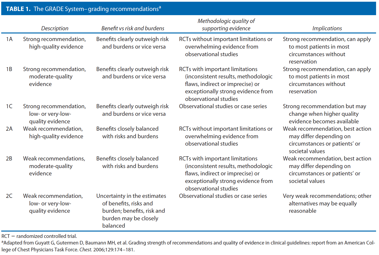
These guidelines are built on the last set of the American Society of Colon and Rectal Surgeons Practice Parameters for treatment of anal squamous neoplasms published in 2008.18 An updated organized search of MEDLINE, PubMed, Embase, and the Cochrane Database of Collected Reviews was performed through June 2011. Key-word combinations included “AIN,” “anal cancer,” “anal malignancy,” “anal carcinoma,” “anal intraepithelial neoplasia,” “Bowen’s disease,” “anal margin,” “anal canal,” “LSIL,’; “HSIL,” “Nigro protocol,” “HPV,” “human papilloma virus,” “vaccination,” “anal surgery,” “chemotherapy,” “radiation therapy,” and “squamous-cell cancer.” Directed searches of the embedded references from the primary articles were also performed in selected circumstances. Although not exclusionary, primary authors focused on all English language articles and studies of adults. Recommendations were formulated by the primary authors and reviewed by the entire Standards Committee. The final grade of recommendation was performed with the use of the Grades of Recommendation, Assessment, Development, and Evaluation (GRADE) system (Table 1).19

Anal Canal Squamous-cell Carcinoma
Pretreatment Evaluation

    A. A disease-specific history should be performed, emphasizing symptoms, risk factors, and signs of advanced disease. Grade of Recommendation: Strong recommendation based on low-quality evidence, 1C.

Most patients present with a slow-growing mass located in the intraanal or perianal position.20 Pain and bleeding are common, occurring in approximately half of patients, although up to 20% of patients may be asymptomatic.21,22 Diagnosis of SCC may often be delayed, because the nonspecific symptoms are initially attributed to other benign anorectal pathology such as hemorrhoids in 70% to 80% of patients.20,23 With lymphatic spread primarily to the inguinal region, groin pain may indicate regional involvement. Risk factors associated with anal SCC include infection with the HPV, HIV seropositivity, history of other HPV-related genital malignancies (cervical cancer, cervical intraepithelial neoplasia (CIN), vulvar cancer, or vulvar intraepithelial neoplasia)13,14; previous sexually acquired diseases, cigarette smoking, anoreceptive intercourse, multiple sexual partners, history of solid organ transplant, and other forms of immunosuppression.14,16,24–28 Because the incidence of anal cancer is higher in men practicing anoreceptive intercourse with men and those with HIV positivity, a high index of suspicion should be maintained in these patients presenting with anorectal complaints.29 Although potentially sensitive and difficult, inquiry to establish the presence or absence of these risk factors is important. Certain factors such as previous radiation, general medical issues, or inadequately controlled HIV may prove to be limiting or contraindications to chemoradiation therapy (CRT) or radical surgery, and are important to determine at the time of diagnosis.

    B. A disease-specific physical examination should be performed to determine size, possible lymph node involvement, or metastatic disease. Grade of Recommendation: Strong recommendation based on moderate-quality evidence, 1B.

Physical examination should focus primarily on the anorectal examination and evaluation of the inguinal and femoral nodes.30 Digital rectal examination should be performed to identify the lesion location and to evaluate for fixation and the presence of sphincter invasion. Perirectal lymph nodes may be sometimes palpable. Anoscopy or proctoscopy with biopsy is essential to establish the size of the lesion, to determine the location within the anal canal, and to confirm diagnosis. Presence of palpable inguinal lymphadenopathy can suggest the need for fine-needle aspiration or core biopsy to confirm malignant involvement and help guide radiation fields. In general, metastatic disease is difficult to detect on physical examination, although a complete physical examination should be performed to help identify any potential sites of distant spread that may warrant further evaluation.
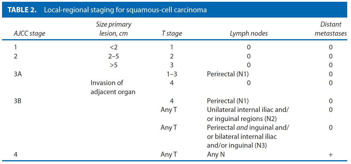
The AJCC17 local-regional staging for SCC of the anal canal focuses on the primary lesion size, the existence of local invasion, and the presence or absence of regional node disease. As such, clinical evaluation including size is critically important to determine proper staging (Table 2 ). Invasion of the anal sphincter or perianal skin does not constitute a T4 lesion; however, this should be determined to aid in potential alterations in treatment.31

    C. Endoscopic and radiologic evaluation should be performed to help determine staging, and concomitant or metastatic disease. Grade of Recommendation: Strong recommendation based on moderate-quality evidence, 1B.


Biopsy should be performed under direct vision or through an anoscope or sigmoidoscope. Although anal cancer is not a risk factor for the development of colon cancer, colorectal neoplasms have been demonstrated in up to 15% of patients with anal cancer; therefore, colonoscopy should be performed to rule out synchronous colorectal neoplasms based on standard age and risk profile assessment.32,33 Chest, abdomen, and pelvic CT should be performed to evaluate for lymphadenopathy, in particular, inguinal lymph node radiographic abnormalities that may warrant biopsy, and to exclude lung and liver metastases.4 Because SCC can metastasize not infrequently to the brain, head CT may be performed if clinical symptoms suggest involvement. In addition, evaluation of the primary tumor may be considered. Endoanal ultrasound (EAUS) and MRI are presently the 2 most accepted modalities and may be useful in determining primary tumor depth, evaluating sphincter involvement, and evaluating perirectal lymph node involvement, with data reporting increased accuracy and sensitivity over physical examination alone.34,35 MRI is comparable to EAUS for primary tumor size and nodal status and may be considered.36 Although not typically a part of the routine evaluation, FDG-PET/CT has been shown to identify distant metastases that are not detected by physical examination or other imaging modalities in 17% to 25%,37,38 resulting in a reported change in treatment (ie, radiotherapy) in up to 5% to 19% of cases.39–41

    D. Sentinel lymph node evaluation for detection of regional nodal metastases is still investigational. Grade of Recommendation: Weak recommendation based on low-quality evidence, 2C.

Small case series have identified sentinel lymph node (SLN) evaluation of inguinal nodes to be superior to physical examination, CT, and PET/CT for the detection and staging of regional nodes.42,43 Both indocyanine green and technetium-(99)m sulfur colloid injection along with blue dye methods have been described with successful results in identifying nodal metastases.44 Overall detection rates of SLN have ranged from 73% to 100%.44,45 There are a few reports of patients with false-negative SLN not receiving radiation therapy and subsequent development of nodal disease, although the false-negative rate is generally low.45 Although clearly technically feasible at present, the exact role of SLN evaluation remains to be determined.

Treatment
Primary Treatment

    1. The primary treatment for most SCCs of the anal canal should be combined modality CRT. Grade of Recommendation: Strong recommendation based on high-quality evidence, 1A.

Historically, anal canal cancer was treated by abdominoperineal resection (APR). However, since Nigro’s demonstration of complete tumor response with equivalent rates of disease-free and overall survival along with added benefit of sphincter preservation, combined modality CRT has become the primary treatment for most patients.46,47 The radiation fields typically include the primary tumor bed and the inguinal nodal basins. Unfortunately, despite the improved outcomes, national cancer registry data have shown ~25% of patients did not receive primary CRT as recently as 2003 to 2005.48 Whereas Nigro’s protocol originally consisted of 5-fluorouracil (5-FU), mitomycin C (MMC), and 3000 cGy of external beam radiation to the pelvis, several different treatment regimens have followed.49,50 Multiple prospective, multi-institutional, randomized controlled trials have demonstrated benefits including lower rates of local failure, tumor recurrence, and need for colostomy when using combined CRT over radiation therapy alone for the first-line therapy of anal cancer.51–55 Although there seems to be no significant change in overall survival, a higher disease-free survival has been reported with combined CRT. Combined therapy, however, has been associated with an increased incidence of hematological toxicity.56 Radiation therapy alone may be considered in patients who cannot tolerate the additional toxicity of chemotherapy.56,57 Local excision is an appropriate consideration only for small superficial lesions outside the anal canal at the anal margin in most instances.58 For those early lesions with node-negative disease and close margins (<1 mm) or residual microscopic disease on biopsy, consideration may be given to additional low-dose, reduced-volume CRT.59

    1b. Intensity-modulated radiation therapy-based chemoradiotherapy (IMRT) may be considered to decrease treatment-related toxicity. Grade of Recommendation: Weak recommendation based on moderate-quality evidence, 2B.

One of the drawbacks to CRT, especially with the more recent use of dose-escalating radiation is the treatment-related side effects and resulting breaks in therapy—occurring in up to 40% to 60% of patients.60–62 IMRT involves delivery of radiation in a manner that still provides total radiation doses of up to 60 Gy for the primary lesion, while tailoring the high-dose radiation to individual tumors to minimize side effects. With the use of this regimen, all grades of radiation-induced, especially acute, toxicity (ie, hematological, skin, diarrhea) occur at significantly lower rates, while decreasing breaks in treatment to18% to 42% lasting on average 4 to 5 days.62,63 Complete primary tumor response with IMRT occurs in more than 90% of patients.62 Overall survival, local-regional survival, and colostomy-free survival rates are similar to rates of the use of traditional radiation therapy methods.63 This technique typically involves a longer treatment time, increased monitoring requirement, and an overall increase in the volume of healthy tissue exposed to radiation. In addition, IMRT has an as-yet undetermined potential risk of future secondary malignancies, and the long-term toxicities are still undetermined.64,65

    2. Multidrug chemotherapy including MCC and 5-FU along with radiation is usually preferable to other chemotherapy regimens with radiation. Grade of Recommendation: Strong recommendation based on high-quality evidence, 1A.

Although modifications to Nigro’s original protocol have been made trying to identify the ideal regimen, the combination of MMC with 5-FU along with radiation remains the most common combination.51,54,66 A recent multicenter randomized controlled trial (RTOG 98-11) compared 5-FU/MMC with radiation versus induction chemotherapy with 5-FU/cisplatin followed by 5-FU/cisplatin with radiation and demonstrated similar rates of 5-year disease-free survival (60% vs 70%, p = 0.17), overall survival (75% vs 70%, p = 0.10). However, the MMC-based arm without induction chemotherapy was associated with a lower 5-year local-regional recurrence (25% vs 33%) and distant metastasis rates (15% vs 19%) in comparison with cisplatin-based therapy.67 This study has been criticized because induction chemotherapy was only given in the 5-FU/cisplatin arm, and thus the treatment strategies were not equivalent. However, based on these results, MMC/
5-FU-based chemoradiation remains a common first line for anal squamous carcinoma. MMC was also associated with lower rates of colostomy (10% vs 19%, p = 0.02) at the cost of higher rates of hematological toxicity. Secondary analysis from this US Intergroup trial identified large tumor size (>5 cm) as the only pretreatment variable predictive for colostomy need.68 More recently, the ACT II phase III study randomly assigned patients to 5-FU/MMC vs 5-FU/cisplatin.69 This study demonstrated similar rates of disease control and survival, colostomy, and overall toxicity between the 2 treatment arms. Furthermore, the 5-FU/MMC arm was still associated with a 60% rate of severe hematological toxicity.
Recent single-institutional data with median follow-up of 83 months reported similar overall and disease-free survival and colostomy-free rates with 5-FU/MMC with 5-FU/cisplatin therapy.70 Combined 5-FU/cisplatin may also be beneficial as an induction regimen for patients with poor-prognosis SCC (T3/T4 and/or N2/N3 disease).71
Additional phase II data from European trials have demonstrated a small beneficial effect of MMC combined with cisplatin and radiotherapy over 5-FU/MMC for locally advanced disease (>4 cm N0 or node positive), although overall toxicity profiles were generally worse in the MMC/cisplatin group.72,73

    3. Higher doses of radiation therapy without prolonged breaks in treatment are preferable when tolerated. Grade of Recommendation: Weak recommendation based on moderate-quality evidence, 2B.

Ideally, an uninterrupted treatment regimen is preferred for both patient convenience and improved outcomes. Longer total treatment duration has been associated with higher rates of local regional failure (HR = 1.96) and colostomy rates (HR = 1.57) in pooled multi-institutional and retrospective data,74–76 although other reports have not found a consistent correlation between overall treatment time and local failure.77 In some instances, grade 3 and higher toxicities will result in a necessary treatment break for recovery. In contrast to treatment breaks, split-course therapy has a planned hiatus in therapy. Limiting treatment breaks has demonstrated similar local control and colostomy-free survival rates in comparison with interrupted treatment cycles.78 Others have reported worse disease-free survival with prolonged (>7day) breaks in treatment.79 The increased utilization of IMRT-based CRT regimens may ultimately decrease these treatment-related toxicities while maintaining similar outcomes.80
Higher radiation boost doses to the primary tumor to yield of 56 to 60 GY and up to 65 GY for T3/T4 lesions along with concurrent chemotherapy have demonstrated improved locoregional control in single-institution retrospective series.81 Additional doses after the completion of initial therapy do not appear to affect locoregional control and may increase the overall incidence of late morbidity,77 although limited data suggest that boost dosing with brachytherapy may be superior to external beam radiotherapy in select patients.75

Treatment of Recurrent or Persistent Disease

    1. Abdominoperineal resection is effective salvage therapy for persistent or recurrent disease: Grade of Recommendation: Strong recommendation based on moderate-quality evidence, 1B.

Unfortunately, ~20% to 30% of patients will have persistent or recurrent disease following primary CRT.82 Predictors of recurrent/persistent locoregional disease after definitive CRT include higher T and N stage at original presentation, HIV-positive status, and the inability to complete CRT.82,83 Patients with persistent disease (present within 6 months of completing CRT) have a poorer prognosis than those with recurrent disease (occurrence >6 months after CRT).84–86 APR is recommended for persistent or recurrent disease for salvage therapy, with achievable 5-year local-regional control in up to 30% to 77%87–90 and overall survival rates reported in 24% to 69%.85–91 Positive margins, either microscopic or macroscopic, following resection, male sex, and higher Charlson comorbidity index portend a worse prognosis, whereas younger age (<55 years), T1-2, and N0-1 disease have been associated with higher overall survival following APR.87,92 Major wound complications are common, reported in 36% to 80%,87 although muscle flap use at the time of reconstruction has been associated with significantly lower rates.90

    2. Systemic chemotherapy should be considered in patients with extrapelvic metastasis or recurrence following surgical salvage. Grade of Recommendation: Strong recommendation based on low-quality evidence, 1C.

Whereas disease localized to the perianal region, anal canal, and regional nodes has a more structured treatment strategy, metastatic disease has no uniform treatment regimen. Platinum-based salvage chemotherapy is commonly offered, although multiple agents have been used.93–95 Limited data with the use of a combination of 5-FU and cisplatin has been reported with response rates of 66%, with median survival of 35 months (actuarial survival at 1 and 5 years of 62% and 32%).96 Biological agents and surgical resection of metastases are largely anecdotal.97 Small case series have demonstrated some success with the use of cetuximab, a monoclonal antibody against epidermal growth factor, alone or in combination with irinotecan for metastatic disease.95,98 Despite these reports, distant metastases are notoriously difficult to treat, and overall median survival is approximately 9 months.99

Management of Inguinal Lymph Node Disease

    1. Chemoradiation is the treatment of choice for inguinal lymph node disease. Grade of Recommendation: Strong recommendation based on low-quality evidence, 1C.

Similar to management of the primary anal lesion, the mainstay of treatment for concomitant disease of the perirectal or inguinal nodes is chemoradiation. A complete response has been reported in 79% to 92%.51,54,62,72,100 With the identification of any positive inguinal lymph node, bilateral inguinal basins should be incorporated into the radiation fields with the addition of a boost technique. Metachronous lymph nodes are seen in 10% to 20% of patients, normally within 6 months of completing treatment of the primary lesion.101 These metachronous nodes should also be treated with CRT, and typically respond well.102 Elective prophylactic lymphadenectomy is generally not warranted and is associated with high wound complication rates as well as lower-extremity complications.103 Selective inguinal node dissection may be considered for persistent disease following CRT. In small case series, long-term survival has been reported after successful removal of disease.104

Anal Cancer in HIV-Positive Patients

    1. CD4 counts may be used to predict the outcome and tolerance of CRT in HIV-positive patients: Grade of Recommendation: Weak recommendation based on low-quality evidence, 2C.

The incidence of anal carcinoma is higher in individuals who are HIV-positive105 and continues to rise despite the widespread use of highly active antiretroviral therapy.106,107 Patients with underlying immunosuppression provide additional concerns regarding the ability to tolerate CRT as well as wound healing deficiencies with surgical therapy, and may only tolerate lower doses of chemoradiation.108 Studies comparing immunocompetent and immunosuppressed SCC patients, however, have reported similar overall survival, disease-free survival and colostomy-free survival rates by the use of standard multimodality CRT with MMC and 5-FU when the course of treatment is completed.109,110 There is some controversy regarding CD4 count and correlation with outcome and toxicity, but, in general, this may be used for HIV-positive patients as a general guide along with clinical assessment.109 Patients with CD4 counts >200 cells/mL should typically be treated similarly to non-HIV-infected patients with anal cancer with the use of chemoradiation. CD4 counts <200 cells/mL have been shown to have higher toxicity, and treatment should be individualized.111 Treatment with highly active antiretroviral therapy allows patients to better tolerate CRT and may improve local control,112,113 although higher overall rates of both acute and long-term toxicities have been reported.114,115

Posttreatment Surveillance

    1. Follow-up examination should typically include anorectal examination including digital rectal examination, anoscopy, and inguinal palpation. Grade of Recommendation: Strong recommendation based on low-quality evidence, 1C.

Anal cancers regress both during and after CRT; therefore, follow-up should generally commence 6 to 12 weeks after the completion of treatment.116,117 Patients should normally be followed up at 3 to 6 months for the first 2 years, 6 to 12 months until 5 years, and annually thereafter, with varying intervals dictated by clinical findings.118 Recurrences are often amenable to further treatment that may result in cure.119 In general, at a minimum anorectal examination should consist of visual inspection, digital rectal examination, and anoscopy, along with inguinal palpation.8 Lesions occurring 3 or more months after the completion of primary CRT are concerning for persistent disease and should be biopsied because digital examination alone is unreliable for confirming residual malignancy.116

    2. Imaging studies such as EAUS, CT, MRI, and FDG-PET/CT should be considered for posttreatment surveillance to assess for persistent or recurrent disease. Grade of Recommendation: Strong recommendation based on low-quality evidence, 1C.

In addition to physical examination, EAUS has been used to determine response to therapy.120,121 There is some evidence to suggest that EAUS is better than physical examination alone at detecting recurrent disease, with 3-dimensional evaluation more sensitive than traditional 2-dimensional EAUS.122 MRI has not been demonstrated to be a good predictor of clinical response in early (6–8 week) follow-up of CRT,123 whereas longer-term (6–12 months) evaluation demonstrating decreased tumor size and a reduction/stabilization of signal intensity has been associated with improved outcomes in small series.124 There is some evidence that high signal intensity comparable to skeletal muscle may indicate recurrent disease.125 FDG-PET/CT has demonstrated excellent outcomes in the post-CRT setting in differentiating partial response with a complete response126,127 to help guide the need for biopsy for histological confirmation of persistent disease, as well as for the identification of PET-avid recurrences both locally and distant.128 Several laboratory parameters including hemoglobin and biomarkers such as the tumor suppressor genes p53 and p21 have shown some promise in outcome correlation, although, at present, they have a limited role in follow-up.129,130

Anal Margin Squamous-cell Carcinoma

    A disease-specific history and physical examination should be performed, emphasizing risk factors, tumor size, location, and signs of advanced disease. Grade of Recommendation: Strong recommendation based on low-quality evidence, 1C.

Anal margin SCC is essentially a skin malignancy arising between the distal end of the anal canal and a 5-cm margin surrounding the anal verge.17 Patients present with symptoms similar to anal canal tumors, including bleeding, pruritus, or a mass that may resemble skin lesions elsewhere, but often has characteristic rolled everted edges with a central ulceration.4 Staging of anal margin cancers by AJCC criteria follows that of skin cancer elsewhere, based on tumor size and lymph node involvement. Evaluation should consist of a perianal examination, including digital rectal examination, anoscopy, and palpation of the femoral and inguinal lymph node basins.8 T1-3 are staged the same manner as SCC of the anal canal, but T4 signifies invasion of deep extradermal structures such as bone, nerve, striated muscle, or cartilage. N0 and N1 refer to no regional or regional lymph node spread.17 In general, CT of the chest, abdomen, and pelvis should be done to assess for distant metastasis.8
Treatment of anal margin SCC varies depending on size and depth of invasion. In general, T1 and early T2 lesions can be adequately treated with wide local excision (WLE) with a 1-cm margin, although close proximity to the anal canal may make this difficult.4,131,132 Definitive treatment by WLE alone for these early lesions has been associated with 5-year survival rates up to an 88%.133 Primary radiation, combined with chemotherapy, is also an option, albeit less effective than appropriate excision. Small series with radiation alone have demonstrated failure rates of up to 36%.134 Larger cancers usually should be treated with upfront radiation to the inguinal nodal basins along with radiation or excision of the primary tumor. For T3 and T4 lesions, radiation to both inguinal regions and the pelvis along with chemotherapy with the use of 5-FU and MMC or cisplatin should normally be added. APR may be required for larger and deeper, less favorable lesions (T2-4 or N1), those involving the sphincter muscles, patients with incontinence, or patients with multiple recurrences after local excision.132,133 Salvage therapy for treatment failure after local excision can include repeat local excision, APR, and/or radiation therapy with or without chemotherapy. Accounting for size and location, tumors of the anal margin, especially larger ones, are associated with lower rates of overall and colostomy-free survival in comparison with anal canal lesions.31

Low-grade And High-grade Anal Intraepithelial Neoplasia
Despite the predominance of terms for this entity, LGAIN/HGAIN is used because of its similarity to CIN—even sharing pathologic grading criteria for classification.135 It is believed to be a precursor to anal cancer.136 Although a number of risk factors are associated with the development of LGAIN/HGAIN, infection with HPV is the most common, with others including receptive anal intercourse, HIV seropositivity, cigarette smoking, low CD4 counts, and immunosuppression.137–144

Pretreatment Evaluation

    1. A disease-specific history and physical examination should be performed for LGAIN/HGAIN, emphasizing symptoms, risk factors, and location of disease. Grade of Recommendation: Strong recommendation based on low-quality evidence, 1C.

LGAIN/HGAIN is often found incidentally during surgery for other unrelated problems such as hemorrhoids. However, high-risk populations include men who have sex with men (MSM), HIV-negative women with a history of anal receptive intercourse and/or other HPV-related anogenital malignancies, and immunosuppression such as in patients who have undergone organ transplantation.145 HIV positivity represents not only another high-risk cohort for the existence of LGAIN/HGAIN, approaching 60% in some studies,146 but it also is a risk factor for the progression of the disease to higher grades.147 The pathway of HPV infection leading to CIN, and ultimately, cervical cancer in females parallels that for HPV, LGAIN/HGAIN, and anal squamous-cell cancer.148 This association of HPV with LGAIN/HGAIN has been demonstrated in both males and females.149,150 There are limited data regarding the natural history of untreated LGAIN/HGAIN in the HIV-negative population. Once established in the anal epithelium, it seems that LGAIN/HGAIN rarely regresses, even in HIV-negative individuals.137 The natural history in HIV-positive patients is more ominous. Progression to higher grades (LGAIN to HGAIN) occurs in more than 50% of HIV-positive homosexual males within 2 years.147,151– 153 The risk for progression to invasive cancer ranges from 10% to 50% among HIV-positive patients.152–154

    2. Anal Papanicolaou smear cytological examination may be useful in the detection and follow-up of LGAIN/HGAIN. Grade of Recommendation: Strong recommendation based on low-quality evidence, 1C.

Screening procedures for LGAIN/HGAIN include anal cytology, colposcopy, biopsy, and high-resolution anoscopy (HRA). Based on numerous similarities between LGAIN/HGAIN and CIN, anal Papanicolaou (Pap) smear cytology consists of using anal swabs to sample cells from the canal and has been instituted for both screening high-risk individuals and as surveillance after treatment for LGAIN/HGAIN. The sensitivity of anal Pap smear evaluation compared with HRA-directed biopsies ranges from 69% to 93% and specificity ranges from 32% to 59%.155–157 Unfortunately, anal cytology in high-risk cohorts such as MSM has false-negative cytology in up to 23% of HIV-negative and 45% for HIV-positive patients.146 Although some economic modeling studies have suggested that frequent anal cytology may be a cost-effective method to prevent anal cancer,158,159 there have not been any randomized or cohort studies to demonstrate improved survival or outcomes. HRA typically involves the application of 3% acetic acid and Lugol iodine solution to the anal canal with evaluation under high-resolution microscopy to help differentiate normal from abnormal tissue. Directed biopsies are performed for any suspicious areas145,160 and for identification of areas that need further treatment.

Treatment

    1. Observation alone with close clinical follow-up may be considered in select cases for the management of LGAIN/HGAIN. Grade of Recommendation: Weak recommendation based on low-quality evidence, 2C.

There has been considerable debate in the past regarding the optimal treatment strategy for LGAIN/HGAIN. Because of the increased risk of recurrence following treatment, especially in high-risk cohorts such as HIV-positive patients, some have advocated the strategy of expectant management with surveillance every 4 to 6 months.154 Supporters of this “watch and wait” strategy cite overall low rates of disease progression and malignant potential (especially for LGAIN), and the increased morbidity associated with excision and repeated focal destruction. On the other hand, a comprehensive approach with cytology, HRA, targeted biopsies, and directed therapy has reported clearance of HGAIN in up to 80%, with progression to higher-grade disease and invasive cancer in less than 5%.161,162 Therefore, expectant management with close follow-up may be considered in select cases depending on risk factors, comorbidities, and available resources. However, because of the high prevalence of concomitant CIN, a referral to gynecology is recommended to complete the evaluation.163

    2. Topical 5% imiquimod cream with close long-term follow-up is an appropriate therapy for LGAIN/HGAIN of the anal margin. Grade of Recommendation: Strong recommendation based on low-quality evidence, 1C.

Imiquimod is an immune response modifier with both anti-HPV and antitumor effects. Use of topical 5% imiquimod cream has support from cohort and case series. Pooled analysis has demonstrated a complete response in 48% of patients, with additional partial response in 34% at a mean follow-up of 11 to 39 months.164 Individual series vary, with some series reporting a complete clinical response in 77% to 86%,165,166 despite most of the data being in the high-risk cohort of HIV-positive MSM. Side effects include irritation, burning, and erosions, which may adversely affect patient compliance.167,168 Yet, overall withdrawal rates remain <5%,169 and treatment can often be successfully reinitiated after a break in therapy. Overall recurrence remains a problem, as well as new LGAIN/HGAIN lesions from different HPV serotypes in untreated areas. Therefore, long-term follow-up is essential.170

    3. Topical 5% 5-FU cream with close long-term follow-up is an appropriate therapy for LGAIN/HGAIN. Grade of Recommendation: Strong recommendation based on low-quality evidence, 1C.

Topical 5-FU was originally used for extramammary Paget disease and has been described for use in LGAIN/HGAIN for 25 years.171–173 Treatment periods typically range from 9 to 16 weeks, although recurrence may be minimized with protracted application.172,174 Initial clinical response rates have been reported in up to 90%, although recurrence may occur in up to 50%.174 Risk factors for recurrence include poorly defined areas of involvement, follicular involvement, poor immune response, dense scar tissue, and recurrent or persistent HPV infection and poor compliance with therapy.175 Local side effects are very common, occurring in up to 85%, and include skin irritation and hypopigmentation, yet rarely result in discontinuation of therapy.174

    4. Photodynamic therapy with close long-term follow-up may be appropriate therapy for select patients with LGAIN/HGAIN. Grade of Recommendation: Weak recommendation based on low-quality evidence, 2C.

Similar to use in other types of skin cancers, photodynamic therapy has been described in patients with LGAIN/HGAIN since 1992.176 In this process, photosensitizing agents such as 5-aminolevulinic acid creams are applied to the affected area followed by treatment with a specific nanometer wavelength laser. Studies have thus far been limited to case reports and small series, and its ultimate role for patients with LGAIN/HGAIN remains to be determined.177–181

    5. Targeted destruction and close clinical long-term follow-up is appropriate therapy for LGAIN/HGAIN. Grade of Recommendation: Strong recommendation based on low-quality evidence, 1C.

A variety of approaches to destruction of LGAIN/HGAIN have been described with the goal of preventing disease progression, including WLE and targeted therapy with the use of HRA. Wide local excision is guided by frozen sections of the affected areas, although total excision of all disease is often difficult. One-centimeter margins are used, with large skin and mucosal defects closed with the aid of local flaps. Although anal mapping was once routine and is still used, it is generally not required.182,183 WLE is associated with recurrence rates of 13% to 63%,183–185 and high rates of local wound complications such as stenosis and incontinence.184 Targeted destruction guided by HRA is effective to identify, biopsy, and destroy LGAIN/HGAIN without the morbidity associated with WLE. However, there remains an overall high risk of persistent or recurrent disease, especially among HIV-positive patients, reported in up to 20% to 80%.151,160,186 Surgical complications such as incontinence and stenosis are generally not reported.160 Infrared coagulation has also been used as an effective ablative device and may be associated with less pain in comparison with other tissue destruction techniques. Although individual lesions are successfully destroyed in up to 72% to 81% following initial treatment,187 persistent local disease has been reported in 65% of patients, with similar high recurrence rates, especially in HIV-positive patients.188 Effectiveness in preventing progression to invasive cancer has been demonstrated with either WLE or targeted destruction.188,189

    6. Patients with LGAIN/HGAIN should be offered close long-term clinical follow-up. Grade of Recommendation: Strong recommendation based on low-quality evidence, 1C.

Patients with LGAIN/HGAIN should typically be monitored for the development of recurrence, persistence, or progression to anal cancer. Surveillance examinations may be performed at 3- to 6-month intervals as long as dysplasia is present.150,154,189 This approach allows for the treatment of recurrent or persistent dysplasia or the detection of invasive anal SCC. Follow-up generally includes digital rectal examination, anoscopic examination, with or without the aid of magnification or the application of acetic acid and Lugol solution, and can be performed in an office setting.190 Anorectal cytology and/or biopsy may also be included, as indicated.161 The importance of close follow-up should be particularly emphasized among high-risk cohorts such as HIV-positive patients, patients wit a history of other HPV-related genital malignancies, recipients of solid organ transplants, or MSM who have been shown to have higher risk of persistence or recurrence of high-grade dysplasia (up to 80%) regardless of treatment modality.160,191

    7. Vaccination against HPV 16/18 may be considered in high-risk patients such as HIV-positive patients and MSM. Grade of Recommendation: Weak recommendation based on low-quality evidence, 2C.

Approximately 80% of anal SCC is believed to be secondary to HPV serotypes 16 and 18.192 Targeted vaccination against these serotypes, especially in high-risk cohorts, may be able to prevent the development of malignancy.6 Several early reports have documented the safety and efficacy of available vaccines, even in HIV-positive patients.193,194 Similar to studies of HPV in adolescent girls,195 models utilizing vaccination in MSM beginning at age 12 without previous HPV exposure has demonstrated the cost-effectiveness of this approach to prevent infection with HPV, genital warts, and ultimately anal SCC.195 At present, the vaccine make-up, timing of administration, and overall indications continue to be a source of investigation and discussion. Although highly controversial, its ultimate role remains to be determined.

Appendix A: Contributing Members Of The Ascrs Standards Committee
Committee Members: W. Donald Buie, M.D., Chair, Janice Rafferty, M.D., Co-chair, Jose Guillem, M.D., Council Representative, Robin Boushey, M.D., George Chang, M.D., Daniel Feingold, M.D., Philip Fleshner, M.D., Jill Genua, M.D., Kerry Hammond, M.D., William Harb, M.D., Samantha Hendren, M.D., Daniel Herzig, M.D., Andreas Kaiser, M.D., David Larson, M.D., Sang Lee, M.D., James McCormick, D.O., Genevieve Melton-Meaux, M.D., Steven Mills, M.D., John Monson, M.D., Harvey Moore III , M.D., W. Brian Perry, M.D., P. Terry Phang, M.D., David Rivadeneira, M.D., Howard Ross, M.D., Scott Steele, M.D., Scott Strong, M.D., Charles Ternent, M.D., Madhulika Varma, M.D., Martin Weiser, M.D., Kirsten Wilkins, M.D.


References

1. American Cancer Society. Cancer Facts & Figures 2010. Atlanta, GA: American Cancer Society; 2010. Available at http://www.cancer.org/acs/groups/content/@epidemiologysurveilance/documents/document/acspc-026238.pdf Accessed November 11, 2011.
2. Jemal A, Siegel R, Ward E, Murray T, Xu J, Thun MJ. Cancer statistics, 2007. CA Cancer J Clin. 2007;57:43–66.
3. U Uronis HE, Bendell JC. Anal cancer: an overview. Oncologist. 2007;12:524–534.
4. Gordon PH. Current status: perianal and anal canal neoplasms. Dis Colon Rectum. 1990;33:799–808.
5. Johnson LG, Madeleine MM, Newcomer LM, Schwartz SM, Daling JR. Anal cancer incidence and survival: the surveillance, epidemiology, and end results experience, 1973–2000. Cancer. 2004;101:281–288.
6. Abbas A, Yang G, Fakih M. Management of anal cancer in 2010. Part 1: Overview, screening, and diagnosis. Oncology (Williston Park). 2010;24:364–369.
7. Frisch M, Glimelius B, van den Brule AJC, et al. Sexually transmitted infection as a cause of anal cancer. N Engl J Med. 1997;337:1350–1358.
8. Welton ML, Varma MG. Anal cancer. In: Wolff BG, Fleshman JW, Beck DE, et al, eds. The ASCRS Textbook of Colon and Rectal Surgery. New York, NY: Springer Science+Business Media, LLC; 2007:482–500.
9. Bean SM, Chhieng DC. Anal-rectal cytology: a review. Diagn Cytopathol. 2010;38:538–546.
10. Bullard Dunn KM, Rothenberger DA. Colon, rectum, and anus. Chapter 29. In: Brunicardi FC, Andersen DK, Billiar TR, Dunn DL, Hunter JG, Matthews JB, Pollock RE, eds. Schwartz’s Principles of Surgery. 9th ed. http://www.accessmedicine.com. Accessed May 10, 2012.
11. Martin F, Bower M. Anal intraepithelial neoplasia in HIV positive people. Sex Transm Infect. 2001;77:327–331.
12. Nahas CS, Lin O, Weiser MR, Temple LK, Wong WD, Stier EA. Prevalence of perianal intraepithelial neoplasia in HIV-infected patients referred for high-resolution anoscopy. Dis Colon Rectum. 2006;49:1581–1586.
13. Santoso JT, Long M, Crigger M, Wan JY, Haefner HK. Anal intraepithelial neoplasia in women with genital intraepithelial neoplasia. Obstet Gynecol. 2010;116:578–582.
14. Park IU , Ogilvie JW Jr, Anderson KE, Li ZZ , Darrah L, Madoff R, Downs L Jr. Anal human papillomavirus infection and abnormal anal cytology in women with genital neoplasia. Gynecol Oncol. 2009;114:399–403.
15. Patel HS, Silver AR, Levine T, Williams G, Northover JM. Human papillomavirus infection and anal dysplasia in renal transplant recipients. Br J Surg. 2010;97:1716–1721.
16. Ogilvie JW Jr, Park IU , Downs LS, Anderson KE, Hansberger J, Madoff RD. Anal dysplasia in kidney transplant recipients. J Am Coll Surg. 2008;207:914–921.
17. Anus. In: Edge SB, Byrd DR, Compton CC, Fritz AG, Greene FL, Trotti A, eds. AJCC Cancer Staging Manual. 7th ed., New York, NY: Springer; 2010:165–173.
18. Fleshner PR, Chalasani S, Chang GJ, Levien DH, Hyman NH, Buie WD, and the Standards Practice Task Force of The American Society of Colon and Rectal Surgeons. Practice Parameters for anal squamous neoplasms. Dis Colon Rectum. 2008;51:2–9.
19. Guyatt G, Gutterman D, Baumann MH, et al. Grading strength of recommendations and quality of evidence in clinical guidelines: report from an American College of Chest Physicians Task Force. Chest. 2006;129:174–181.
20. Klas JV, Rothenberger DA, Wong WD, et al. Malignant tumors of the anal canal: the spectrum of disease, treatment, and outcomes. Cancer. 1999;85:1686–1693.
21. Ryan DP, Compton CC, Mayer RJ. Carcinoma of the anal canal. N Engl J Med. 2000;342:792–800.
22. Robb BW, Mutch MG. Epidermoid carcinoma of the anal canal. Clin Colon Rectal Surg. 2006;19:54–60.
23. Tanum G, Tveit K, Karlsen KO. Diagnosis of anal carcinoma: doctor’s finger still the best? Oncology. 1991;48:383–386.
24. Welton ML, Sharkey FE, Kahlenberg MS. The etiology and epidemiology of anal cancer. Surg Oncol Clin N Am. 2004;13:263–275.
25. Frisch M, Glimelius B, van den Brule AJ, et al. Sexually transmitted infection as a cause of anal cancer. N Engl J Med. 1997;337:1350 –1358.
26. Daling JR, Weiss NS, Hislop TG et al. Sexual practices, sexually transmitted diseases, and the incidence of anal cancer. N Engl J Med. 1987;317:973–977.
27. Daling JR, Sherman KJ, Hislop TG, et al. Cigarette smoking and the risk of anogenital cancer. Am J Epidemiol. 1992;135:180–189.
28. Chiao EY, Krown SE, Stier EA, et al. A population-based analysis of temporal trends in the incidence of squamous anal canal cancer in relation to the HIV epidemic. J Acquir Immune Defic Syndr. 2005;40:451–455.
29. Palefsky JM, Holly EA, Ralston ML, et al. High incidence of anal high grade squamous intra-epithelial lesions among HIV-positive and HIV negative homosexual and bisexual men. AIDS. 1998;12:495–503.
30. Anal canal. In: Greene FL, Page DL, Fleming ID, et al, eds. AJCC Cancer Staging Manual. 6th ed. New York, NY: Springer; 2002:125–130.
31. Grabenbauer GG, Kessler H, Matzel KE, Sauer R, Hohenberger W, Schneider IH. Tumor site predicts outcome after radiochemotherapy in squamous-cell carcinoma of the anal region: long-term results of 101 patients. Dis Colon Rectum. 2005;48:1742–1751.
32. Wasvary HJ, Barkel DC, Klein SN. Is total colonic evaluation for anal cancer necessary? Am Surg. 2000;66:592–594.
33. Klompje J, Petrelli NJ, Herrera L, Mittelman A. Synchronous and metachronous colon lesions in squamous cell carcinoma of the anal canal. J Surg Oncol. 1987;35:86–88.
34. Giovannini M, Bardou VJ, Barclay R, et al. Anal carcinoma: prognostic value of endorectal ultrasound (ERUS). Results of a prospective multicenter study. Endoscopy. 2001;33:231–236.
35. Jacopo M. Endoanal ultrasound for anal cancer staging. Int J Colorectal Dis. 2011;26:385–386.
36. Otto SD, Lee L, Buhr HJ, Frericks B, Höcht S, Kroesen AJ. Staging anal cancer: prospective comparison of transanal endoscopic ultrasound and magnetic resonance imaging. Gastrointest Surg. 2009;13:1292–1298.
37. Trautmann TG, Zuger JH. Positron emission tomography for pretreatment staging and posttreatment evaluation in cancer of the anal canal. Mol Imaging Biol. 2005;7:309–313.
38. Cotter SE, Grigsby PW, Siegel BA, et al.. FDG-PET/CT in the evaluation of anal carcinoma. Int J Radiat Oncol Biol Phys. 2006;65:720–725.
39. Krengli M, Milia ME, Turri L, et al. FDG-PET/CT imaging for staging and target volume delineation in conformal radiotherapy of anal carcinoma. Radiat Oncol. 2010;5:10.
40. Nguyen BT, Joon DL, Khoo V, et al. Assessing the impact of FDG-PET in the management of anal cancer. Radiother Oncol. 2008;87:376–382.
41. Kidd EA, Dehdashti F, Siegel BA, Grigsby PW. Anal cancer maximum F-18 fluorodeoxyglucose uptake on positron emission tomography is correlated with prognosis. Radiother Oncol. 2010;95:288–291.
42. Mistrangelo M, Pelosi E, Bellò M, et al. Comparison of positron emission tomography scanning and sentinel node biopsy in the detection of inguinal node metastases in patients with anal cancer. Int J Radiat Oncol Biol Phys. 2010;77:73–78.
43. De Nardi P, Carvello M, Canevari C, Passoni P, Staudacher C. Sentinel node biopsy in squamous-cell carcinoma of the anal canal. Ann Surg Oncol. 2011;18:365–370.
44. Hirche C, Dresel S, Krempien R, Hünerbein M. Sentinel node biopsy by indocyanine green retention fluorescence detection for inguinal lymph node staging of anal cancer: preliminary experience. Ann Surg Oncol. 2010;17:2357–2362.
45. de Jong JS, Beukema JC, van Dam GM, Slart R, Lemstra C, Wiggers T. Limited value of staging squamous cell carcinoma of the anal margin and canal using the sentinel lymph node procedure: a prospective study with long-term follow-up. Ann Surg Oncol. 2010;17:2656–2662.
46. Buroker TR, Nigro N, Bradley G, et al. Combined therapy for cancer of the anal canal: a follow-up report. Dis Colon Rectum. 1977;20:677–678.
47. Nigro ND, Vaitkevicius VK, Buroker T, et al. Combined therapy for cancer of the anal canal. Dis Colon Rectum. 1981;24:73–75.
48. Bilimoria KY, Bentrem DJ, Ko CY, Stewart AK, Winchester DP, Talamonti MS, Halverson AL. Squamous cell carcinoma of the anal canal: utilization and outcomes of recommended treatment in the United States. Ann Surg Oncol. 2008;15:1948–1958.
49. Y Young SC, Solomon MJ, Hruby G, Frizelle FA. Review of 120 anal cancer patients. Colorectal Dis. 2009;11:909–914.
50. Glynne-Jones R, Meadows H, Wan S, et al. National Cancer Research Institute Anal Sub Group and Colorectal Clinical Oncology Group. EXTRA–a multicenter phase II study of chemoradiation using a 5 day per week oral regimen of capecitabine and intravenous mitomycin C in anal cancer. Int J Radiat Oncol Biol Phys. 2008;72:119–126.
51. Cummings BJ, Keane TJ, O’Sullivan B, Wong CS, Catton CN. Epidermoid anal cancer: treatment by radiation alone or by radiation and 5-fluorouracil with and without mitomycin C. Int J Rad Oncol Biol Phys. 1991;21:1115–1125.
52. Raffetto N, Monaco A, Banelli E. Radiotherapy and chemotherapy in the conservative treatment of anal canal carcinoma. Anticancer Res. 2008;28(2B):1335–1339.
53. Arnott SJ, Cunningham JD, Gallagher J, et al. UKCCCR Anal Cancer Trial Working Party. Epidermoid anal cancer: results from the UKCCCR randomized trial of radiotherapy alone versus radiotherapy, 5-fluorouracil, and mitomycin. Lancet. 1996;348:1049–1054.
54. Flam M, John M, Pajak TF, et al. Role of mitomycin in combination with fluorouracil and radiotherapy, and of salvage chemoradiation in the definitive nonsurgical treatment of epidermoid carcinoma of the anal canal: results of a phase 3 randomized intergroup study. J Clin Oncol. 1996;14:2527–2539.
55. Bartelink H, Roelofsen F, Eschwege F, et al. Concomitant radiotherapy and chemotherapy is superior to radiotherapy alone in the treatment of locally advanced anal cancer: results of Phase 3 randomized trial of the European Organization for Research and Treatment of Cancer Radiotherapy and Gastrointestinal Cooperative Groups. J Clin Oncol. 1997;15:2040–2049.
56. Rabbani AN, Zlotecki RA, Kirwan J, George TJ Jr, Morris CG, Rout WR, Mendenhall WM. Definitive radiotherapy for squamous cell carcinoma of the anal canal. Am J Clin Oncol. 2010;33:47–51.
57. Balamucki CJ, Zlotecki RA, Rout WR, et al. Squamous cell carcinoma of the anal margin: the University of Florida experience. Am J Clin Oncol. 2011;34:406–410.
58. Fuchshuber PR, Rodriguez-Bigas M, Weber T, Petrelli NJ. Anal canal and perianal epidermoid cancers. J Am Coll Surg. 1997;185:494–505.
59. Hatfield P, Cooper R, Sebag-Montefiore D. Involved-field, low-dose chemoradiotherapy for early-stage anal carcinoma. Int J Radiat Oncol Biol Phys. 2008;70:419–424.
60. Chen YW, Yen SH, Chen SY, et al. Anus-preservation treatment for anal cancer: retrospective analysis at a single institution. J Surg Oncol. 2007;96:374–380.
61. Hodges JC, Das P, Eng C, et al. Intensity-modulated radiation therapy for the treatment of squamous cell anal cancer with para-aortic nodal involvement. Int J Radiat Oncol Biol Phys. 2009;75:791–794.
62. Salama JK, Mell LK, Schomas DA, et al. Concurrent chemotherapy and intensity-modulated radiation therapy for anal canal cancer patients: a multicenter experience. J Clin Oncol. 2007;25:4581–4586.
63. Pepek JM, Willett CG, Wu QJ, et al. Intensity-modulated radiation therapy for anal malignancies: a preliminary toxicity and disease outcomes analysis. Int J Radiat Oncol Biol Phys. 2010;78:1413–1419.
64. Lisbona A, Averbeck D, Supiot S, et al. IM1T combined to IGRT: increase of the irradiated volume. Consequences? [in French]. Cancer Radiother. 2010;14:563–570.
65. Hall EJ. Intensity-modulated radiation therapy, protons, and the risk of second cancers. Int J Radiat Oncol Biol Phys. 2006;65:1–7.
66. Jiang Y, Mackley H, Cheng H, Ajani JA. Anal carcinoma therapy: can we improve on 5 fluorouracil/mitomycin/radiotherapy? J Natl Compr Canc Netw. 2010;8:135–144.
67. Ajani JA, Winter KA, Gunderson LL, et al. Fluorouracil, mitomycin, and radiotherapy vs fluorouracil, cisplatin, and radiotherapy for carcinoma of the anal canal: a randomized controlled trial. JAMA. 2008;299:1914–1921.
68. Ajani JA, Winter KA, Gunderson LL, et al. US intergroup anal carcinoma trial: tumor diameter predicts for colostomy. J Clin Oncol. 2009;27:1116–1121.
69. James R, Wan S, Glynne-Jones R, et al. A randomized trial of chemoradiation using mitomycin or cisplatin with of without maintenance cisplatin/5FU in squamous cell carcinoma of the anus (ACT II ). J Clin Oncol. 2009;27;1149.
70. Olivatto LO, Cabral V, Rosa A, et al. Mitomycin-C- or cisplatin-based chemoradiotherapy for anal canal carcinoma: long-term results. Int J Radiat Oncol Biol Phys. 2011;79:490–495.
71. Meropol NJ, Niedzwiecki D, Shank B, et al. Induction therapy for poor-prognosis anal canal carcinoma: a phase II study of the cancer and Leukemia Group B (CALGB 9281). J Clin Oncol. 2008; 26: 3229–3234.
72. Matzinger O, Roelofsen F, Mineur L, et al; EORTC Radiation Oncology and Gastrointestinal Tract Cancer Groups. Mitomycin C with continuous fluorouracil or with cisplatin in combination with radiotherapy for locally advanced anal cancer (European Organisation for Research and Treatment of Cancer phase II study 22011-40014). Eur J Cancer. 2009;45:2782–2791.
73. Crehange G, Bosset M, Lorchel F, et al. Combining cisplatin and mitomycin with radiotherapy in anal carcinoma. Dis Colon Rectum. 2007;50:43–49.
74. Ben-Josef E, Moughan J, Ajani JA, et al. Impact of overall treatment time on survival and local control in patients with anal cancer: a pooled data analysis of Radiation Therapy Oncology Group Trials 87-04 and 98-11. J Clin Oncol. 2010 28:5061–5066.
75. Hannoun-Levi JM, Ortholan C, Resbeut M, et al. High-dose split-course radiation therapy for anal cancer: outcome analysis regarding the boost strategy (CORS-03 Study). Int J Radiat Oncol Biol Phys. 2011;80:712–720.
76. Widder J, Kastenberger R, Fercher E, et al. Radiation dose associated with local control in advanced anal cancer: retrospective analysis of 129 patients. Radiother Oncol. 2008;87:367–375.
77. Glynne-Jones R, Sebag-Montefiore D, Adams R, et al. For the UKCCCR Anal Cancer Trial Working Party. “Mind the Gap”—the impact of variations in the duration of the treatment gap and overall treatment time in the first UK Anal Cancer Trial (ACT I). Int J Radiat Oncol Biol Phys. 2011;81:1488–1494.
78. Janssen S, Meier zu Eissen J, Kolbert G, Bremer M, Karstens JH, Meyer A. Anal cancer treated with radio-chemotherapy: correlation between length of treatment interruption and outcome. Int J Colorectal Dis. 2009;24:1421–1428.
79. Oehler C, Provencher S, Donath D, Bahary JP, Lütolf UM, Ciernik IF. Chemo-radiation with or without mandatory split in anal carcinoma: experiences of two institutions and review of the literature. Radiat Oncol. 2010;5:36.
80. Z Zagar TM, Willett CG, Czito BG. Intensity-modulated radiation therapy for anal cancer: toxicity versus outcomes. Oncology (Williston Park). 2010;24:815–823, 828.
81. Engineer R, Mallik S, Mahantshetty U, Shrivastava S. Impact of radiation dose on locoregional control and survival on squamous cell carcinoma of anal canal. Radiother Oncol. 2010;95:283–287.
82. Roohipour R, Patil S, Goodman KA, et al. Squamous-cell carcinoma of the anal canal: predictors of treatment outcome. Dis Colon Rectum. 2008;51:147–153.
83. Das P, Bhatia S, Eng C, Ajani JA, et al. Predictors and patterns of recurrence after definitive chemoradiation for anal cancer. Int J Radiat Oncol Biol Phys. 2007;68:794–800.
84. Ferenschild FT, Vermaas M, Hofer SO, Verhoef C, Eggermon AM, de Wilt JH. Salvage abdominoperineal resection and perineal wound healing in local recurrent or persistent anal cancer. World J Surg. 2005;29:1452–1457.
85. Papaconstantinou HT, Bullard KM, Rothenberger DA, Madoff RD. Salvage abdominoperineal resection after failed Nigro protocol: modest success, major morbidity. Colorectal Dis. 2006;8:124–129
86. Stewart D, Yan Y, Kodner IJ, et al. Salvage surgery after failed chemoradiation for anal canal cancer: should the paradigm be changed for high-risk tumors? J Gastrointest Surg. 2007;11:1744–1751.
87. Schiller DE, Cummings BJ, Rai S, et al. Outcomes of salvage surgery for squamous cell carcinoma of the anal canal. Ann Surg Oncol. 2007;14:2780–2789.
88. Mullen JT, Rodriguez-Bigas MA, Chang GJ, et al. Results of surgical salvage after failed chemoradiation therapy for epidermoid carcinoma of the anal canal. Ann Surg Oncol. 2007;14:478–483.
89. Goéré D, Bonnet S, Pocard M, Deutsch E, Lasser P, Elias D. Oncologic and functional results after abdominoperineal resection plus pseudocontinent perineal colostomy for epidermoid carcinoma of the anus. Dis Colon Rectum. 2009;52:958–963.
90. Lefevre JH, Parc Y, Kernéis S, et al. Abdomino-perineal resection for anal cancer: impact of a vertical rectus abdominis myocutaneous flap on survival, recurrence, morbidity, and wound healing. Ann Surg. 2009;250:707–711.
91. Vorob’ev GI, Shelygin IuA, Nechushkin MI, Rybakov EG. Results of surgical treatment of residual and recurrent anal tumors [in Russian] Khirurgiia (Mosk). 2008;(8):4–9.
92. Mariani P, Ghanneme A, De la Rochefordière A, Girodet J, Falcou MC, Salmon RJ. Abdominoperineal resection for anal cancer. Dis Colon Rectum. 2008;51:1495–1501.
93. Alcindor T. Activity of paclitaxel in metastatic squamous anal carcinoma. Int J Colorectal Dis. 2008;23:717.
94. Grifaichi F, Padovani A, Romeo F, Trinca C, Moscetti L, Cortesi E. Response of metastatic epidermoid anal cancer to single agent irinotecan: a case report. Tumori. 2001; 87:58–59.
95. Phan LK, Hoff PM. Evidence of clinical activity for cetuximab combined with irinotecan in a patient with refractory anal canal squamous-cell carcinoma: report of a case. Dis Colon Rectum. 2007;50:395–398.
96. Faivre C, Rougier P, Ducreux M, et al. 5-fluorouracile and cisplatinum combination chemotherapy for metastatic squamous-cell anal cancer [in French]. Bull Cancer. 1999;86:861–865.
97. Eng C, Pathak P. Treatment options in metastatic squamous cell carcinoma of the anal canal. Curr Treat Options Oncol. 2008;9:400–407.
98. Lukan N, Ströbel P, Willer A, et al. Cetuximab-based treatment of metastatic anal cancer: correlation of response with KRAS mutational status. Oncology. 2009;77:293–299.
99. Nivatvongs S. Perianal and anal canal neoplasms. In: Gordon P, Nivatvongs S, eds. Principles and Practice of Surgery of the Colon, Rectum, and Anus. 2nd ed. St. Louis, MO: Quality Medical Publishing, Inc.; 1999:447–471.
100. Sischy B. The use of radiation therapy combined with chemotherapy in the management of squamous cell carcinoma of the anus and marginally respectable adenocarcinoma of the rectum. Int J Rad Oncol Biol Phys. 1985;11:1587–1593.
101. Golden GT, Horseley JS. Surgical management of epidermoid carcinoma of the anus. Am J Surg. 1976; 131:275–280.
102. Wright JL, Patil SM, Temple LK, Minsky BD, Saltz LB, Goodman KA. Squamous cell carcinoma of the anal canal: patterns and predictors of failure and implications for intensity-modulated radiation treatment planning. Int J Radiat Oncol Biol Phys. 2010;78:1064–1072.
103. Quan SH. Anal and perianal tumors. Surg Clin North Am. 1978;58:549–603.
104. Akbari RP, Paty PB, Guillem JG, et al. Oncologic outcomes of salvage surgery for epidermoid carcinoma of the anus initially managed with combined modality therapy. Dis Colon Rectum. 2004;47:1136–1144.
105. Melbye M, Cote T, Kessler L, et al. High incidence of anal cancer among AIDS patients. The AIDS/Cancer Working Group. Lancet. 1994;343:636–639.
106. Crum-Cianflone NF, Hullsiek KH, Marconi VC, et al. Infectious Disease Clinical Research Program HIV Working Group. Anal cancers among HIV-infected persons: HAART is not slowing rising incidence. AIDS. 2010;24:535–543.
107. Piketty C, Selinger-Leneman H, Grabar S, et al. Marked increase in the incidence of invasive anal cancer among HIV-infected patients despite treatment with combination antiretroviral therapy. AIDS. 2008;22:1203–1211.
108. Hammad N, Heilbrun LK, Gupta S, et al. Squamous cell cancer of the anal canal in HIV-infected patients receiving highly active antiretroviral therapy: a single institution experience. Am J Clin Oncol. 2011;34:135–139.
109. Seo Y, Kinsella MT, Reynolds HL, Chipman G, Remick SC, Kinsella TJ. Outcomes of chemoradiotherapy with 5-fluorouracil and mitomycin C for anal cancer in immunocompetent versus immunodeficient patients. Int J Radiat Oncol Biol Phys 2009;75:143–149.
110. Myerson RJ, Outlaw ED, Chang A, et al. Radiotherapy for epidermoid carcinoma of the anus: thirty years’ experience. Int J Radiat Oncol Biol Phys. 2009;75:428–435.
111. Hoffman R, Welton M, Klencke B, Weinberg V, Krieg R. The significance of pretreatment CD4 count on the outcome and treatment tolerance of HIV-positive patients with anal cancer. Int J Rad Oncol Biol Phys. 1999;44:127–131.
112. Cleator S, Fife K, Nelson M, Gazzard B, Phillips R, Bower M. Treatment of HIV-associated invasive anal cancer with combined chemoradiation. Eur J Cancer. 2000;36:754–758.
113. Place RJ, Gregorcyk SG, Huber PJ, Simmang CL. Outcome analysis of HIV-positive patients with anal squamous cell carcinoma. Dis Colon Rectum. 2001;44:506–512.
114. Wexler A, Berson AM, Goldstone SE, et al. Invasive anal squamous-cell carcinoma in the HIV-positive patient: outcome in the era of highly active antiretroviral therapy. Dis Colon Rectum. 2008;51:73–81.
115. Oehler-Jänne C, Huguet F, Provencher S, et al. HIV-specific differences in outcome of squamous cell carcinoma of the anal canal: a multicentric cohort study of HIV-positive patients receiving highly active antiretroviral therapy. J Clin Oncol. 2008;26:2550–2557.
116. Papillon J, Mayer M, Montbarbon JF, Gerard JP, Chassard JL, Bailly C. A new approach to the management of epidermoid carcinoma of the anal canal. Cancer. 1983;51:1830–1837.
117. Longo WE, Vernava AM, Wade TP, et al. Recurrent squamous cell carcinoma of the anal canal: predictors of initial treatment failure and results of salvage therapy. Ann Surg. 1994;220:40–49.
118. Glynne-Jones R, Northover JM, Cervantes A. ESMO Guidelines Working Group. Anal cancer: ESMO Clinical Practice Guidelines for diagnosis, treatment and follow-up. Ann Oncol. 2010;21(suppl 5):v87–v92.
119. Haboubi NY, Edilbe MW, Hill J. Justification for staging of epidermoid anal carcinoma after salvage surgery: a pathological guideline. Colorectal Dis. 2007;9:238–244.
120. Martellucci J, Naldini G, Colosimo C, Cionini L, Rossi M. Accuracy of endoanal ultrasound in the follow-up assessment for squamous cell carcinoma of the anal canal treated with radiochemotherapy. Surg Endosc. 2009;23:1054–1057.
121. Tarantino D, Bernstein MA. Endoanal ultrasound in the staging and management of squamous-cell carcinoma of the anal canal: potential implications of a new ultrasound staging system. Dis Colon Rectum. 2002; 45: 16–22.
122. Christensen AF, Nielsen MB, Svendsen LB, Engelholm SA. Three-dimensional anal endosonography may improve detection of recurrent anal cancer. Dis Colon Rectum. 2006;49: 1527–1532.
123. Goh V, Gollub FK, Liaw J, et al. Magnetic resonance imaging assessment of squamous cell carcinoma of the anal canal before and after chemoradiation: can MRI predict for eventual clinical outcome? Int J Radiat Oncol Biol Phys. 2010;78:715–721.
124. Koh DM, Dzik-Jurasz A, O’Neill B, Tait D, Husband JE, Brown G. Pelvic phased-array MR imaging of anal carcinoma before and after chemoradiation. Br J Radiol. 2008;81:91–98.
125. Roach SC, Hulse PA, Moulding FJ, Wilson R, Carrington BM. Magnetic resonance imaging of anal cancer. Clin Radiol. 2005; 60:1111–1119.
126. Schwarz JK, Siegel BA, Dehdashti F, Myerson RJ, Fleshman JW, Grigsby PW. Tumor response and survival predicted by post-therapy FDG-PET/CT in anal cancer. Int J Radiat Oncol Biol Phys. 2008;71:180–186.
127. Vercellino L, Montravers F, de Parades V, et al. Impact of FDG PET/CT in the staging and the follow-up of anal carcinoma. Int J Colorectal Dis. 2011;26:201–210.
128. Nguyen BT, Joon DL, Khoo V, et al. Assessing the impact of FDG-PET in the management of anal cancer. Radiother Oncol. 2008;87:376–382.
129. Roldán GB, Chan AK, Buckner M, Magliocco AM, Doll CM. The prognostic value of hemoglobin in patients with anal cancer treated with chemoradiotherapy. Dis Colon Rectum. 2010;53:1127–1134.
130. Lampejo T, Kavanagh D, Clark J, et al. Prognostic biomarkers in squamous cell carcinoma of the anus: a systematic review. Br J Cancer. 2010;103:1858–1869.
131. Beahrs OH, Wilson SM. Carcinoma of the anus. Ann Surg. 1976;184:422–428.
132. Moore HG, Guillem JG. Anal neoplasms. Surg Clin North Am. 2002;82:1233–1251.
133. Greenall MJ, Quan SH, Stearns MW, et al. Epidermoid cancer of the anal margin: pathologic features, treatment, and clinical results. Am J Surg. 1985;149:95–101.
134. Papillon J, Chassard JL. Respective roles of radiotherapy and surgery in the management of epidermoid carcinoma of the anal margin: series of 57 patients. Dis Colon Rectum. 1992;35:422–429.
135. Cleary RK, Schaldenbrand JD, Fowler JJ, Schuler JM, Lampman RM. Perianal Bowen’s disease and anal intraepithelial neoplasia: review of the literature. Dis Colon Rectum. 1999;42:945–951.
136. Darragh TM, Winkler B. The ABCs of anal-rectal cytology. College of American Pathologists. Available at: http://www.cap.org/apps/cap.portal. Accessed November 28, 2010.
137. Palefsky, JM, Holly, EA, Ralston, ML, et al. Anal squamous intraepithelial lesions in HIV-positive and HIV-negative homosexual and bisexual men. J Acquir Immune Defic Syndr Hum Retrovirol. 1998;17:320–326.
138. Palefsky JM, Shiboski S, Moss A. Risk factors for anal human papilloma virus infection and anal cytologic abnormalities in HIV-positive and HIV-negative homosexual men. J Acquir Immune Defic Syndr. 1994;7:599–606.
139. Moscicki, AB, Hills, NK, Shiboski, S, et al. Risk factors for abnormal anal cytology in young heterosexual women. Cancer Epidemiol Biomarkers Prev. 1999;8:173.
140. Holmes F, Borek D, Owen-Kummer M, et al. Anal cancer in women. Gastroenterology. 1988;95:107–111.
141. Caussy D, Goedert JJ, Palefsky J, et al. Interaction of human immunodeficiency and papilloma viruses: association with anal epithelial abnormality in homosexual men. Int J Cancer. 1990; 46:214–219.
142. Friedman HB, Saah AJ, Sherman ME, et al. Human papillomavirus, anal squamous intraepithelial lesions, and human immunodeficiency virus in a cohort of gay men. J Infect Dis. 1998; 178:45–52.
143. Ogunbiyi OA, Scholefield JH, Raftery AT, et al. Prevalence of anal human papillomavirus infection and intraepithelial neoplasia in renal allograft recipients. Br J Surg. 1994;81:365–367.
144. Daling JR, Sherman KJ, Hislop TG, et al. Cigarette smoking and the risk of anogenital cancer. Am J Epidemiol. 1992;135:180–189.
145. Palefsky JM, Rubin M. The epidemiology of anal human papillomavirus and related neoplasia. Obstet Gynecol Clin North Am. 2009;36:187–200.
146. Chin-Hong PV, Berry JM, Cheng SC, et al. Comparison of patient- and clinician-collected anal cytology samples to screen for human papillomavirus-associated anal intraepithelial neoplasia in men who have sex with men. Ann Intern Med. 2008;149:300–306.
147. Palefsky J, Holly EA, Hogeboom CJ, et al. Virologic, immunologic, and clinical parameters in the incidence and progression of anal squamous intraepithelial lesions in HIV-positive and HIV-negative homosexual men. J Acquir Immune Defic Syndr Hum Retrovirol. 1998; 17:314–319.
148. Schiffman MH, Castle P. Epidemiologic studies of a necessary causal risk factor: human papillomavirus infection and cervical neoplasia. J Natl Cancer Inst. 2003;95:E2.
149. Holly EA, Ralston ML, Darragh TM, Greenblatt RM, Jay N, Palefsky JM. Prevalence and risk factors for anal squamous intraepithelial lesions in women. J Natl Cancer Inst. 2001;93:843–849.
150. Chin-Hong PV, Vittinghoff E, Cranston RD, et al. Age related prevalence of anal cancer precursors in homosexual men: the EXPLORE study. J Natl Cancer Inst. 2005; 97:896–905.
151. Pineda CE, Welton ML. Controversies in the management of anal high-grade squamous intraepithelial lesions. Minerva Chir. 2008;63:389–399.
152. Watson AJ, Smith BB, Whitehead MR, et al. Malignant progression of anal intra-epithelial neoplasia. ANZ J Surg. 2006;76:715–717.
153. Scholefield JH, Castle MT, Watson NF. Malignant transformation of high-grade anal intraepithelial neoplasia. Br J Surg. 2005; 92:1133–1136.
154. Devaraj B, Cosman BC. Expectant management of anal squamous dysplasia in patients with HIV. Dis Colon Rectum. 2006;49:36–40.
155. Arain S, Walts AE, Thomas P, Bose S. The anal Pap smear: cytomorphology of squamous intraepithelial lesions. Cytojournal. 2005; 2:4.
156. Palefsky JM, Holly EA, Hogeboom CJ, Berry JM, Jay N, Darragh TM. Anal cytology as a screening tool for anal squamous intraepithelial lesions. J Acquir Immune Defic Syndr Hum Retrovirol. 1997;14:415–422.
157. Fox PA, Seet JE, Stebbing J, et al. The value of anal cytology and human papillomavirus typing in the detection of anal intraepithelial neoplasia: a review of cases from an anoscopy clinic. Sex Transm Infect. 2005;81:142–146.
158. Goldie SJ, Kuntz KM, Weinstein MC, Freedberg KA, Welton ML, Palefsky JM. The clinical effectiveness and cost-effectiveness of screening for anal squamous intraepithelial lesions in homosexual and bisexual HIV-positive men. JAMA. 1999;281:1822–1829.
159. Goldie SJ, Kuntz KM, Weinstein MC, Freedberg KA, Palefsky JM. Cost-effectiveness of screening for anal squamous intraepithelial lesions and anal cancer in human immunodeficiency virus-negative homosexual and bisexual men. Am J Med. 2000;108:634–641.
160. Chang GJ, Berry JM, Jay N, et al. Surgical treatment of high-grade anal squamous intraepithelial lesions: a prospective study. Dis Colon Rectum. 2002;45:453–458.
161. Pineda CE, Berry JM, Jay N, Palefsky JM, Welton ML. High resolution anoscopy in the planned staged treatment of anal squamous intraepithelial lesions in HIV-negative patients. J Gastrointest Surg. 2007;11:1410–1416.
162. Pineda CE, Berry JM, Jay N, Palefsky JM, Welton ML. High-resolution anoscopy targeted surgical destruction of anal high-grade squamous intraepithelial lesions: a ten-year experience. Dis Colon Rectum. 2008;51:829–837.
163. Valari O, Koliopoulos G, Karakitsos P, et al. Human papillomavirus DNA and mRNA positivity of the anal canal in women with lower genital tract HPV lesions: predictors and clinical implications. Gynecol Oncol. 2011;122:505–508.
164. Mahto M, Nathan M, O’Mahony C. More than a decade on: review of the use of imiquimod in lower anogenital intraepithelial neoplasia. Int J STD AIDS. 2010;21:8–16.
165. Wieland U, Brockmeyer NH, Weissenborn SJ, et al. Imiquimod treatment of anal intraepithelial neoplasia in HIV-positive men. Arch Dermatol. 2006;142:1438–1444.
166. Rosen T, Harting M, Gibson M. Treatment of Bowen’s disease with topical 5% imiquimod cream: retrospective study. Dermatol Surg. 2007;33:427–432.
167. Wendling J, Saiag P, Berville-Levy S, Bourgault-Villada I, Clerici T, Moyal-Barracco M. Treatment of undifferentiated vulvar intraepithelial neoplasia with 5% imiquimod cream: a prospective study of 12 cases. Arch Dermatol 2004; 140:1220–1224.
168. Beutner KR, Spruance SL, Hougham AJ, Fox TL, Owens ML, Douglas JM Jr. Treatment of genital warts with an immune-response modifier (imiquimod). J Am Acad Dermatol. 1998;38:230–239.
169. Moore RA, Edwards JE, Hopwood J, Hicks D. Imiquimod for the treatment of genital warts: a quantitative systematic review. BMC Infect Dis. 2001;1:3.
170. Kreuter A, Potthoff A, Brockmeyer NH, et al. German Competence Network HIV/AIDS. Imiquimod leads to a decrease of human papillomavirus DNA and to a sustained clearance of anal intraepithelial neoplasia in HIV-infected men. J Invest Dermatol. 2008;128:2078–2083.
171. Schmidt KU, Wiskemann A, Mensing H. Etretinate and 5-fluorouracil in intra-anal Bowen’s disease [in German] Hautarzt. 1986;37:278–280.
172. Bargman H, Hochman J. Topical treatment of Bowens disease with 5-fluorouracil. J Cutan Med Surg. 2003;7:101–105.
173. Cox NH, Eedy DJ, Morton CA. Guidelines for management of Bowens disease: 2006 update. Br J Dermatol. 2007;156:11–21.
174. Richel O, Wieland U, De Vries HJ, et al. Topical 5-fluorouracil treatment of anal intraepithelial neoplasia in human immunodeficiency virus-positive men. Br J Dermatol. 2010;163:1301–1307.
175. Graham BD, Jetmore AB, Foote JE, et al. Topical 5-fluorouracil in the management of extensive anal Bowen’s disease: a preferred approach. Dis Colon Rectum. 2005;48:444–450.
176. Petrelli NJ, Cebollero JA, Rodriguez-Bigas M, Mang T. Photodynamic therapy in the management of neoplasms of the perianal skin. Arch Surg. 1992;127:1436–1438.
177. Allison RR, Sheng C, Cuenca R, Bagnato VS, Austerlitz C, Sibata CH. Photodynamic therapy for anal cancer. Photodiagnosis Photodyn Ther. 2010;7:115–119.
178. Kruijt B, van der Snoek EM, Sterenborg HJ, Amelink A, Robinson DJ. A dedicated applicator for light delivery and monitoring of PDT of intra-anal intraepithelial neoplasia. Photodiagnosis Photodyn Ther. 2010;7:3–9.
179. van der Snoek EM, Amelink A, van der Ende ME, et al. Photodynamic therapy with topical metatetrahydroxychlorin (Fosgel) is ineffective for the treatment of anal intraepithelial neoplasia, grade III . J Acquir Immune Defic Syndr. 2009;52:141–143.
180. Hamdan KA, Tait IS, Nadeau V, Padgett M, Carey F, Steele RJ. Treatment of grade III anal intraepithelial neoplasia with photodynamic therapy: report of a case. Dis Colon Rectum. 2003;46:1555–1559.
181. Scholefield JH. Treatment of grade III anal intraepithelial neoplasia with photodynamic therapy: report of a case. Dis Colon Rectum., 2003;46:1555–1559.
182. Strauss RJ, Fazio VW. Bowen’s disease of the anal and perianal area: a report and analysis of twelve cases. Am J Surg. 1979;137:231–234.
183. Margenthaler JA, Dietz DW, Mutch MG, et al. Outcomes, risk of other malignancies, and need for formal mapping procedures in patients with perianal Bowen’s disease. Dis Colon Rectum. 2004; 47:1655–1661.
184. Brown SR, Skinner P, Tidy J, Smith JH, Sharp F, Hosie KB. Outcome after surgical resection for high-grade anal intraepithelial neoplasia (Bowen’s disease). Br J Surg. 1999; 8:1063–1066.
185. Marchesa P, Fazio VW, Oliart S, Goldblum JR, Lavery IC. Perianal Bowen’s disease: a clinicopathologic study of 47 patients. Dis Colon Rectum. 1997;40:1286–1293.
186. Chung AP, Rosenfeld DB. Intraoperative high-resolution anoscopy: a minimally invasive approach in the treatment of patients with Bowen’s disease and results in a private practice setting. Am Surg. 2007;73:1279–1283.
187. Goldstone SE, Hundert JS, Huyett JW. Infrared coagulator ablation of high-grade anal squamous intraepithelial lesions in HIV-negative males who have sex with males. Dis Colon Rectum. 2007; 50:565–575.
188. Goldstone SE, Kawalek AZ, Huyett JW. Infrared coagulator: a useful tool for treating anal squamous intraepithelial lesions. Dis Colon Rectum. 2005;48:1042–1054.
189. Chang GJ, Welton ML. Anal neoplasia. Semin Colon Rectal Surg. 2003;14:111–118.
190. Jay N, Berry JM, Hogeboom CJ, Holly EA, Darragh TM, Palefsky JM. Colposcopic appearance of anal squamous intraepithelial lesions: relationship to histopathology. Dis Colon Rectum. 1997;40:919–928.
191. Chin-Hong PV, Palefsky JM. Natural history and clinical management of anal human papillomavirus disease in men and women infected with human immunodeficiency virus. Clin Infect Dis. 2002;35:1127–1134.
192. Kim JJ. Targeted human papillomavirus vaccination of men who have sex with men in the USA: a cost-effectiveness modelling analysis. Lancet Infect Dis. 2010;10:845–852.
193. Wilkin T, Lee JY, Lensing SY, et al. Safety and immunogenicity of the quadrivalent human papillomavirus vaccine in HIV-1-infected men. J Infect Dis. 2010;202:1246–1253.
194. Anderson JS, Hoy J, Hillman R, et al. A randomized, placebo-controlled, dose-escalation study to determine the safety, tolerability, and immunogenicity of an HPV-16 therapeutic vaccine in HIV-positive participants with oncogenic HPV infection of the anus. J Acquir Immune Defic Syndr. 2009;52:371–381.
195. Kim JJ, Goldie SJ. Health and economic implications of HPV vaccination in the United States. N Engl J Med. 2008;359:821–832.

Anal Fissure Information

Download PDF

Anal Fissure

What Is An Anal Fissure?

An anal fissure (fissure-in-ano) is a small, oval shaped tear in skin that lines the opening of the anus. Fissures typically cause severe pain and bleeding with bowel movements. Fissures are quite common in the general population, but are often confused with other causes of pain and bleeding, such as hemorrhoids.

What Are The Symptoms Of An Anal Fissure?

The typical symptoms of an anal fissure include severe pain during, and especially after, a bowel movement, lasting from several minutes to a few hours. Patients may also notice bright red blood from the anus that can be seen on the toilet paper or on the stool. Between bowel movements, patients with anal fissures are often relatively symptom-free. Many patients are fearful of having a bowel movement and may try to avoid defecation secondary to the pain.

What Causes An Anal Fissure?

Fissures are usually caused by trauma to the inner lining of the anus. Patients with tight anal sphincter muscles (i.e., increased muscle tone) are more prone to developing anal fissures. A hard, dry bowel movement is typically responsible, but loose stools and diarrhea can also be the cause. Following a bowel movement, severe anal pain can produce spasm of the anal sphincter muscle, resulting in a decrease in blood flow to the site of the injury, thus impairing healing of the wound. The next bowel movement results in more pain, anal spasm, decreased blood flow to the area, and the cycle continues. Treatments are aimed at interrupting this cycle by relaxing the anal sphincter muscle to promote healing of the fissure. Other, less common, causes include inflammatory conditions and certain anal infections or tumors. Anal fissures may be acute (recent onset) or chronic (present for a long period of time). Chronic fissures may be more difficult to treat, and may also have an external lump associated with the tear, called a sentinel pile or skin tag, as well as extra tissue just inside the anal canal (hypertrophied papilla) .

What Is The Treatment Of Anal Fissures?

The majority of anal fissures do not require surgery. The most common treatment for an acute anal fissure consists of making the stool more formed and bulky with a diet high in fiber and utilization of over-the-counter fiber supplementation (totaling 25-35 grams of fiber/day). Stool softeners and increasing water intake may be necessary to promote soft bowel movements and aid in the healing process. Topical anesthetics for pain and warm tub baths (sitz baths) for 10-20 minutes several times a day (especially after bowel movements) are soothing and promote relaxation of the anal muscles, which may help the healing process. Other medications (such as nitroglycerin, nifedipine, or diltiazem) may be prescribed that allow relaxation of the anal sphincter muscles. Your surgeon will go over benefits and side-effects of each of these with you. Narcotic pain medications are not recommended for anal fissures, as they promote constipation. Chronic fissures are generally more difficultto treat, and your surgeon may advise surgical treatment.

Will The Problem Return?

Fissures can recur easily, and it is quite common for a fully healed fissure to recur after a hard bowel movement or other trauma. Even when the pain and bleeding have subsided, it is very important to continue good bowel habits and a diet high in fiber as a lifestyle change. If the problem returns without an obvious cause, further assessment is warranted.

What Can Be Done If The Fissure Does Not Heal?

A fissure that fails to respond to conservative measures should be re-examined. Persistent hard or loose bowel movements, scarring, or spasm of the internal anal muscle all contribute to delayed healing. Other medical problems such as inflammatory bowel disease (Crohn’s disease), infections, or anal tumors can cause symptoms similar to anal fissures. Patients suffering from persistent anal pain should be examined to exclude these symptoms. This may include a colonoscopy or an exam in the operating room under anesthesia.

What Does Surgery Involve?

Surgical options for treating anal fissure include Botulinum toxin (Botox®) injection into the anal sphincter and surgical division of a portion of the internal anal sphincter (lateral internal sphincterotomy). Both of these are performed typically as outpatient, same-day procedures, or occasionally in the office setting. The goal of these surgical options is to promote relaxation of the anal sphincter, thereby decreasing anal pain and spasm, allowing the fissure to heal. Botox® injection results in healing in 50-80% of patients, while sphincterotomy is reported to be over 90% successful. If a sentinel pile is present, it may be removed to promote healing of the fissure. All surgical procedures carry some risk, and a sphincterotomy can rarely interfere with one’s ability to control gas and stool. Your colon and rectal surgeon will discuss these risks with you to determine the appropriate treatment for your particular situation.

How Long Is The Recovery After Surgery?

It is important to note that complete healing with both medical and surgical treatments can take up to approximately 6-10 weeks. However, acute pain after surgery often disappears after a few days. Most patients will be able to return to work and resume daily activities in a few short days after the surgery.

Can Fissures Lead To Colon Cancer?

Absolutely not. Persistent symptoms, however, need careful evaluation since other conditions other than an anal fissure can cause similar symptoms. Your colon and rectal surgeon may request additional tests, even if your fissure has successfully healed. A colonoscopy may be required to exclude other causes of rectal bleeding.

Anal Fissure Treatment Guidelines

Download PDF

Practice Parameters for the Management of Anal Fissures (3rd Revision)

W. Brian Perry, M.D. • Sharon L. Dykes, M.D. • W. Donald Buie, M.D.
Janice F. Rafferty, M.D., on behalf of the Standards Practice Task Force of the
American Society of Colon and Rectal Surgeons


The American Society of Colon and Rectal Surgeons is dedicated to ensuring highquality patient care by advancing the science, prevention, and management of disorders and diseases of the colon, rectum, and anus. The Standards Committee is composed of Society members who are chosen because they have demonstrated expertise in the specialty of colon and rectal surgery. This committee was created to lead international efforts in defining quality care for conditions related to the colon, rectum, and anus. This is accompanied by developing Clinical Practice Guidelines based on the best available evidence. These guidelines are inclusive, and not prescriptive. Their purpose is to provide information on which decisions can be made, rather than to dictate a specific form of treatment. These guidelines are intended for the use of all practitioners, health care workers, and patients who desire information about the management of the conditions addressed by the topics covered in these guidelines. It should be recognized that these guidelines should not be deemed inclusive of all proper methods of care or exclusive of methods of care reasonably directed toward obtaining the same results. The ultimate judgment regarding the propriety of any specific procedure must be made by the physician in light of all the circumstances presented by the individual patient.

Statement of the Problem
An anal fissure is an oval, ulcer like, longitudinal tear in the anal canal, distal to the dentate line. In almost 90% of cases, fissures are identified in the posterior midline, but can be seen in the anterior midline in up to 25% of affected women and 8% of affected men. Fissures occurring inlateral positions should raise suspicions for other disease processes, such as Crohn’s disease, tuberculosis, syphilis,HIV/AIDS, dermatologic conditions (psoriasis), or anal carcinoma. Early or acute fissures have the appearance of a simple tear in the anoderm, whereas chronic fissures, defined by symptoms lasting more than 8 to 12 weeks, are further characterized by edema and fibrosis. Typical inflammatory manifestations of chronic fissures include a sentinel skin tag at the distal fissure margin and a hypertrophied anal papilla proximal to the fissure in the anal canal. In addition, fibers of the internal anal sphincter are often visible at the fissure base. The clinical hallmark of ananal fissure is pain during, and particularly after, defecation. Often there is a history of a tearing sensation during passage of a constipated stool or with explosive diarrhea. Rectal bleeding, usually limited to minimal bright red blood on the toilet tissue, is not uncommon.

Methodology
These guidelines are built on the last set of the American Society of Colon and Rectal Surgeons Practice Parametersfor treatment of Fissureinano published in 2004. An organized search of MEDLINE, Pubmed, EMBASE, and the Cochrane Database of Collected Reviews was performed through June 2009. Keyword combinations included anal fissure and fissure in ano as primary search terms. Directed searches were obtained from the latest dated reference from the previous version of the parameter to December 2008. The newest data were evaluated to see whether recommendations needed upgrading or downgrading with the additional information. Directed searches of the embedded references from the primary articles were alsoper formed in selected circumstances. The final grade of recommendation was performed using the Grades of Recommendation, Assessment, Development, and Evaluation(GRADE) system (Table 1).

Recommendations

    1. Nonoperative treatment continues to be safe, has few side effects, and should usually be the first step in therapy. Grade of Recommendation: Strong recommendation based on moderate quality evidence 1B


Almost half of all patients in whom an acute anal fissure has been diagnosed will heal with nonoperative measures, ie, sitz baths, psyllium fiber and bulking agents, with or without the addition of topical anesthetics or anti-inflammatory ointments. 1– 6 In addition to fissure healing, symptomatic relief of pain and bleeding can be achieved with virtually no side effects.

    2. Anal fissures may be treated with topical nitrates, although nitrates are marginally superior to placebo with regard to healing. Grade of Recommendation: Strong recommendation based on high-quality evidence 1A

Topical nitric oxide donors have been associated with healing in at least 50% of treated chronic fissures, 7–13,18 –21 and use of topical nitroglycerin significantly decreases pain during the therapy period. 7,9,16 An updated Cochrane review of medical treatment of anal fissures has concluded, however, that topical nitroglycerin remains only marginally better than placebo in healing anal fissures. 22 Dose escalation does not improve healing rates. 7,14,16 The principal side effect is headache, occurring in at least 20% to 30% of treated patients. 7,17,22 This adverse effect is dose-related and causes cessation of therapy in up to 20% of patients. 15 The incidence of fissure recurrence after treatment with topical nitric oxide donors is dramatically higher 7,8 compared with outcomes after surgery, although morbidity is lower. 8,10,12 Patients who do not respond to topical nitrates should be referred for botulinum toxin injections or surgery. 7,8,10

    3. Anal fissures may be treated with topical calcium channel blockers, with a lower incidence of adverse effects than topical nitrates. There are insufficient data to conclude whether they are superior to placebo in healing anal fissures. Grade of Recommendation: Strong recommendation based on moderate-quality evidence 1B

Topical calcium channel blockers have been associated with healing of chronic anal fissures in 65% to 95% of patients. 24 –31 Side effects include headache, in up to 25% of patients, 29 and occur less frequently than with topical nitrates. 27–29 There are still fewer randomized controlled trials of topical calcium channel blockers than of topical nitric oxide donors. Anal fissures may also be treated with oral calcium channel blockers. 32–35 This is associated with a lower rate of fissure healing than topical application and has a higher incidence of side effects. 32 Few direct comparisons of topical and oral calcium channel blockers exist.

    4. Botulinum toxin injection has been associated with healing rates superior to placebo. There is inadequate consensus on dosage, precise site of administration, number of injections or efficacy. Grade of Recommendation: Strong recommendation based on low-quality evidence 1C

Injection of botulinum toxin into the internal anal sphincter allows healing in 60% to 80% of fissures, 36,39,41,43,45,46 and at a higher rate than placebo. 42 The most common side effects are temporary incontinence to flatus in up to 18% of patients 38,39,42,45 and stool in 5%. 49 Recurrences may occur in up to 42% of cases, 38,39,46,47 but patients may be re-treated with a good rate of healing. 39,44 Higher doses are associated with improved rates of healing and are as safe as lower doses. 39,44 Topical nitrates appear to potentiate the effects of botulinum toxin in patients with refractory anal fissure. 37,48 There is no consensus on dose, site, or number of injections. 40,47 Patients in whom botu linum toxin injection therapy fails should be recommended for surgery. 40
There are few reports regarding the use of gonyautoxins for treatment of anal fissure. We have intentionally not included this treatment modality because of the paucity of data and the widespread unavailability of these agents.

    5. Lateral internal sphincterotomy is the surgical treatment of choice for refractory anal fissures. Grade of Recommendation: Strong recommendation based on high-quality evidence 1A

Lateral internal sphincterotomy (LIS) remains the surgical treatment of choice for refractory anal fissures. 22,49 Multiple studies 50 –53 and a recent Cochrane re view 54 show that LIS is superior to uncontrolled manual anal dilation, yielding superior healing rates with less incontinence. Controlled pneumatic balloon dilation has shown promise in one small series. 55 LIS offers superior healing and lower incontinence rates compared with posterior sphincterotomy-fissurectomy alone. 56 The addition of topical nitric oxide donors 57 or botulinum toxin 58–60 improves results of fissurectomy in nonrandomized series.

    6. Open and closed techniques of lateral internal sphincterotomy (LIS) yield similar results. Grade of Recommendation: Strong recommendation based on highquality evidence 1A

Further well-done studies confirm the prior assertion that there is no difference in outcomes between properly performed open or closed sphincterotomy. 54,61– 64

    7. LIS tailored to fissure characteristics yields equivalent or worse healing rates, and less incontinence, than traditional LIS to the dentate line. Grade of Recommendation: Weak recommendation based on moderate equality evidence 2B

A “tailored sphincterotomy” has been proposed in an effort to reduce the rate of minor incontinence following LIS. Two methods are typically employed—sphincterotomy only to the apex of the fissure or anal calibration. Three randomized trials of traditional vs fissure apex sphincterotomy show statistically superior healing rates in the traditional arm; 2 reported worse continence in the traditional arm, 65,66 whereas one did not. 67 To improve these results, a calibrated sphincterotomy has been reported. In these studies, fissure apex sphincterotomy was compared with a sphincterotomy that was extended based on the amount of residual anal stenosis remaining by use of a calibrated sound. In 3 small series, this method showed equivalent healing and lower incontinence rates than traditional sphincterotomy. 68 –70

    8. Anal advancement flap and subcutaneous fissurotomy are surgical alternatives to LIS. Grade of Recommendation: Weak recommendation based on low-quality evidence 2C

Techniques that do not divide the internal anal sphincter yet allow good healing rates are theoretically attractive, especially in patients with preexisting continence problems or in those without internal anal sphincter hypertonia. Small series of various anal advancement flaps show promise. 71,72 One series of unroofing subcutaneous sinuses associated with typical anal fissures reported excellent healing without changes in continence. 73 Larger trials in this area are still needed.

    9. Surgery is consistently superior to medical therapy and may be offered without a pharmacological treatment failure. Grade of Recommendation: Strong recommendation based on high-quality evidence 1A

Multiple trials continue to confirm the superiority of LIS to any topical or injected agent with low rates of incontinence. 10,46,74 –78 Most investigations show that compliance with long-term medical therapy remains an issue. The Cochrane Collaboration analyses of both surgical and nonsurgical therapies for anal fissure confirm these conclusions. 22,54 Quality of life (QOL) is poor in patients with persistent fissure, whereas patients undergoing LIS report significantly improved QOL. Fecal continence QOL is preserved in the vast majority of patients following LIS. 79–81
The practice parameters set forth in this document have been developed from sources believed to be reliable. The American Society of Colon and Rectal Surgeons makes no warranty, guarantee, or representation whatsoever as to the absolute validity or sufficiency of any parameter included in this document, and the Society assumes no responsibility for the use of the material contained.

Appendix A: Contributing Members Of The ASCRS Standards Committee
Farshid Araghizadeh, M.D., Robin Boushey, M.D., Sridhar Chalasani, M.D., George Chang, M.D., Robert Cima, M.D., Gary Dunn, M.D., Daniel Feingold, M.D., Philip Fleshner, M.D., Daniel Geisler, M.D., Jill Genua, M.D., Sharon Gregorcyk, M.D., Daniel Herzig, M.D., Andreas Kaiser, M.D., Ravin Kumar, M.D., David Larson, M.D., Steven Mills, M.D., John Monson, M.D., P. Terry Phang, M.D., Feza Remzi, M.D., David Rivadeneira, M.D., Howard Ross, M.D., Peter Senatore, M.D., Elin Sigurdson, M.D., Thomas Stahl, M.D., Scott Steele, M.D., Scott Strong, M.D., Charles Ternent, M.D., Judith Trudel, M.D., Madhulika Varma, M.D., Martin Weiser, M.D.
References
Recommendation 1
1. Gough MJ, Lewis A. The conservative treatment of fissure-in-ano.Br J Surg. 1983;70:175–176.
2. Jensen SL. Treatment of first episodes of acute anal fissure: pro spective randomised study of lignocaine ointment versus hydrocortisone ointment or warm sitz baths plus bran. BMJ. 1986;292: 1167–1169.
3. Shub HA, Salvati EP, Rubin RJ. Conservative treatment of anal fissure: an unselected, retrospective and continuous study. Dis Colon Rectum. 1978;21:582–583.
4. Hananel N, Gordon PH. Re-examination of clinical manifestations and response to therapy of fissure-in-ano. Dis Colon Rectum. 1997;40:229 –233.
5. Jensen SL. Maintenance therapy with unprocessed bran in the prevention of acute anal fissure recurrence.J R Soc Med. 1987; 80:296 –298.
6. Gupta P. Randomized, controlled study comparing sitz-bathand no-sitz-bath treatments in patients with acute anal fissures.ANZ J Surg.2006;76:718 –721.
Recommendation 2
7. Evans J, Luck A, Hewett P. Glyceryl trinitrate vs. lateral sphinc-terotomy for chronic anal fissure: prospective, randomized trial.Dis Colon Rectum.2001;44:93–97.
8. Kennedy ML, Sowter S, Nguyen H, Lubowski DZ. Glyceryl trini-trate ointment for the treatment of chronic anal fissure: resultsof a placebo-controlled trial and long-term follow-up. Dis ColonRectum.1999;42:1000 –1006.
9. Libertiny G, Knight JS, Farouk R. Randomised trial of topical 0.2% glyceryl trinitrate and lateral internal sphincter otomy for the treatment of patients with chronic anal fissure: long-termfollow-up. Eur J Surg.2002;168:418 – 421.
10. Lund JN, Scholefield JH. Glyceryl trinitrate is an effective treat-ment for anal fissure. Dis Colon Rectum.1997;40:468 – 470.
11. Oettle GJ. Glyceryl trinitrate vs. sphincterotomy for treatment ofchronic fissure-in-ano: a randomized, controlled trial.Dis ColonRectum.1997;40:1318 –1320.
12. Werre AJ, Palamba HW, Bilgen EJ, Eggink WF. Isosorbide dini-trate in the treatment of anal fissure: a randomized, prospective,double blind, placebo-controlled trial. Eur J Surg.2001;167:382–385.
13. Scholefield JH, Bock JU, Marla B, et al. A dose finding study with0.1%, 0.2%, and 0.4% glyceryl trinitrate ointment in patients with chronic anal fissures.Gut.2003;52:264 –269.
14. Zuberi BF, Rajput MR, Abro H, Shaikh SA. A randomized trialof glyceryl trinitrate ointment and nitroglycerin patch in healingof anal fissures. Int J Colorectal Dis.2000;15:243–245.
15. Bailey HR, Beck DE, Billingham RP, et al. Fissure Study Group. A study to determine the nitroglycerin ointment dose and dosing interval that best promote the healing of chronic anal fis-sures.Dis Colon Rectum. 2002; 45:1192–1199.
16. Altomare DF, Rinaldi M, Milito G, et al. Glyceryl trinitrate forchronic anal fissure— healing or headache? Results of a multi-center, randomized, placebo-controlled, double-blind trial. DisColon Rectum.2000;43:174 –181.
17. Lysy J, Israelit-Yatzkan Y, Sestiere-Ittah M, Keret D, Goldin E.Treatment of chronic anal fissure with isosorbide dinitrate: long-term results and dose determination.Dis Colon Rectum.1998;41:1406 –1410.
18. Lysy J, Israeli E, Levy S, Rozentzweig G, Strauss-Liviatan N, Gol-din E. Long-term results of “chemical sphincterotomy” forchronic anal fissure: a prospective study.Dis Colon Rectum.2006;49:858 – 864.
19. Lund JN, Scholefield JH. A randomised, prospective, double-blind, placebo-controlled trial of glyceryl trinitrate ointment in-treatment of anal fissure [published correction appears inLan-cet. 1997;349:656].Lancet. 1997;349:11–14.
20. Chaudhuri S, Pal AK, Acharya A, et al. Treatment of chronic analfissure with topical glyceryl trinitrate: a double-blind, placebo-controlled trial.Indian J Gastroenterol. 2001;20:101–102.
21. Bacher H, Mischinger HJ, Werkgartner G, et al. Local nitroglyc-erin for treatment of anal fissures: an alternative to lateral sphincter otomy? Dis Colon Rectum.1997;40:840 – 845.
22. Nelson R. Non surgical therapy for anal fissure. Cochrane Data-base Syst Rev. 2006;CD003431.
Recommendation 3
23. Shrivastava UK, Jain BK, Kumar P, Saifee Y. A comparison of the effects of diltiazem and glyceryl trinitrate ointment in the treat-ment of chronic anal fissure: a randomized clinical trial.SurgToday.2007;37:482– 485.
24. Carapeti EA, Kamm MA, Phillips RK. Topical diltiazem and bethanechol decrease anal sphincter pressure and heal anal fissures without side effects. Dis Colon Rectum.2000;43:1359 –1362.
25. Knight JS, Birks M, Farouk R. Topical diltiazem ointment in the treatment of chronic anal fissure. Br J Surg.2001;88:553–556.
26. Ezri T, Susmallian S. Topical nifedipine vs. topical glyceryl trini-trate for treatment of chronic anal fissure. Dis Colon Rectum.2003;46:805– 808.
27. Bielecki K, Kolodziejczak M. A prospective randomized trial ofdiltiazem and glyceryl trinitrate ointment in the treatment of chronic anal fissure. Colorectal Dis.2003;5:256 –257.
28. Kocher HM, Steward M, Leather AJ, Cullen PT. Randomized clinical trial assessing the side-effects of glyceryl trinitrate and diltiazem hydrochloride in the treatment of chronic anal fissure. Br J Surg.2002;89:413– 417.
29. Perrotti P, Bove A, Antropoli C, et al. Topical nifedipine with lidocaine ointment vs. active control for treatment of chronicanal fissure: results of a prospective, randomized, double-blindstudy. Dis Colon Rectum.2002;45:1468 –1475.
30. Antropoli C, Perrotti P, Rubino M, et al. Nifedipine for local use in conservative treatment of anal fissures: preliminary results ofa multicenter study.Dis Colon Rectum.1999;42:1011–1015.
31. Jonas M, Neal KR, Abercrombie JF, Scholefield JH. A random-ized trial of oral vs. topical diltiazem for chronic anal fissures. Dis Colon Rectum.2001;44:1074 –1078.
32. Ansaloni L, Bernabe` A, Ghetti R, Riccardi R, Tranchino RM, Gardini G. Oral lacidipine in the treatment of anal fissure.TechColoproctol.2002;6:79 – 82.
33. Ag aog lu N, Cengiz S, Arslan MK, Turkyilmaz S. Oral nifedipinein the treatment of chronic anal fissure.Dig Surg.2003;20:452–456.
34. Cook TA, Humphreys MM, Mortensen NJ. Oral nifedipine re-duces resting anal pressure and heals chronic anal fissure.Br JSurg.1999;86:1269 –1273.
Recommendation 4
35. Brisinda G, Cadeddu F, Brandara F, Marniga G, Maria G. Randomized clinical trial comparing botulinum toxin injectionswith 0.2 per cent nitroglycerin ointment for chronic anal fissure.Br J Surg.2007;94:162–167.
36. Jones OM, Ramalingam T, Merrie A, et al. Randomized clinicaltrial of botulinum toxin plus glyceryl trinitrate vs. botulinumtoxin alone for medically resistant chronic anal fissure: overallpoor healing rates.Dis Colon Rectum.2006;49:1574 –1580.
37. Arroyo A, Perez F, Serrano P, Candela F, Calpena R. Longtermresults of botulinum toxin for the treatment of chronic analfissure: prospective clinical and manometric study. Int J Colorectal Dis.2005;20:267–271.
38. Brisinda G, Maria G, Sganga G, Bentivoglio AR, Albanese A,Castagneto M. Effectiveness of higher doses of botulinum toxinto induce healing in patients with chronic anal fissures.Surgery.2002;131:179 –184.
39. Maria G, Brisinda G, Bentivoglio AR, Cassetta E, Gui D, Albanese A. Influence of botulinum toxin site of injections onhealing rate in patients with chronic anal fissure.Am J Surg.2000;179:46 –50.
40. Brisinda G, Maria G, Bentivoglio AR, Cassetta E, Gui D, Albanese A. A comparison of injections of botulinum toxin andtopical nitroglycerin ointment for the treatment of chronic analfissure [published correction appears inN Engl J Med. 1999;341:624].N Engl J Med.1999;341:65– 69.
41. Maria G, Cassetta E, Gui D, Brisinda G, Bentivoglio AR, Albanese A. A comparison of botulinum toxin and saline for thetreatment of chronic anal fissure.N Engl J Med.1998;338:217–220.
42. Colak T, Ipek T, Kanik A, Aydin S. A randomized trial of botulinum toxin vs. lidocaine pomade for chronic anal fissure.ActaGastroenterol Belg.2002;65:187–190.
43. Madalinski MH, Slawek J, Zbytek B, et al. Topical nitrates andthe higher doses of botulinum toxin for chronic anal fissure.Hepatogastroenterology.2001;48:977–979.
44. Lindsey I, Jones OM, Cunningham C, George BD, MortensenNJ. Botulinum toxin as secondline therapy for chronic analfissure failing 0.2 percent glyceryl trinitrate.Dis Colon Rectum.2003;46:361–366.
45. Menteçs BB, Irkorucu O, Akin M, Leventoglu S, Tatlicioglu E.Comparison of botulinum toxin injection and lateral internalsphincterotomy for the treatment of chronic anal fissure. DisColon Rectum.2003;46:232–237.
46. Minguez M, Herreros B, Espi A, et al. Longterm followup (42months) of chronic anal fissure after healing with botulinumtoxin.Gastroenterology.2002;123:112–117.
47. Lysy J, IsraelitYatzkan Y, SestieryIttah M, et al. Topical nitratespotentiate the effect of botulinum toxin in the treatment of patients with refractory anal fissure.Gut.2001;48:221–224.
48. Jost WH. One hundred cases of anal fissure treated with botulinum toxin: early and longterm results. Dis Colon Rectum.1997;40:1029 –1032.
Recommendation 5
49. Richard CS, Greggoire R, Plewes EA, et al. Internal sphincterotomy is superior to topical nitroglycerine in the treatment of chronic anal fissure: results of a randomized, controlled trial by he Canadian Colorectal Surgical Trials Group.
Dis Colon Rectum. 2000;43:1048 –1047.
50. Jensen SL, Lund F, Nielsen OV, Tange G. Lateral subcutaneous sphincterotomy versus anal dilatation in the treatment of fissure in ano in outpatients: a prospective randomized study. BMJ. 1984;289:528 –530.
51. Saad AM, Omer A. Surgical treatment of chronic fissure in ano: a prospective randomized study. East Afr Med J . 1992;69:613– 615.
52. Olsen J, Mortensen PE, Krogh Petersen I, et al. Anal sphincter function after treatment of fissure-in-ano by lateral subcutaneous sphincterotomy versus anal dilation. Int J Colorectal Dis. 1987;2:155–157.
53. Weaver RM, Ambrose NS, Alexander-Williams J, et al. Manual dilation of the anus vs. lateral internal sphincterotomy in the treatment of chronic fissure-in-ano: results of a prospective, randomized clinical trial. Dis Colon Rectum. 1987;30:420 – 423.
54. Nelson R. Operative procedures for fissure in ano. Cochrane Database Syst Rev. 2005:CD002199.
55. Renzi A, Izzo D, Di Sarno G, et al. Clinical, manometric, and ultrasonographic results of pneumatic balloon dilatation vs. lateral internal sphincterotomy for chronic anal fissure: a prospective, randomized, controlled trial. Dis Colon Rectum. 2008;51: 121–127.
56. Abcarian H. Surgical correction of chronic anal fissure: results of lateral internal sphincterotomy vs. fissurectomy-midline sphincterotomy. Dis Colon Rectum. 1980;23:31–36.
57. Engel AF, Eijsbouts QA, Balk AG. Fissurectomy and isosorbide dinitrate for chronic fissure in ano not responding to conservative treatment. Br J Surg. 2002;89:79 – 83.
58. Baraza W, Boereboom C, Shorthouse A, et al. The long-term efficacy of fissurectomy and botulinum toxin injection for chronic anal fissure in females. Dis Colon Rectum. 2008;51:236 – 243.
59. Scholz T, Hetzer FH, Dindo D, et al. Long-term follow-up after combined fissurectomy and Botox injection for chronic anal fissures. Int J Colorectal Dis . 22:1077–1081.
60. Lindsey I, Cunningham C, Jones OM, et al. Fissurectomy-botulinum toxin: a novel sphincter-sparing procedure for medically resistant chronic anal fissure, Dis Colon Rectum. 2004;47:1947– 1952.
Recommendation 6
61. Boulous PB, Araujo JG. Adequate internal sphincterotomy for chronic anal fissure: subcutaneous or open technique? Br J Surg . 1984;71:360 –362.
62. Kortbeek JB, Langevin JM, Khoo RE, et al. Chronic fissure-inano: a randomized study comparing open and subcutaneous lateral internal sphincterotomy. Dis Colon Rectum. 1992;35:835– 837.
63. Arroyo A, Perez F, Serrano P, et al. Open versus closed lateral internal sphincterotomy performed as an outpatient procedure under local anesthesia for chronic anal fissure: prospective randomized study of clinical and manometric long-term results. J Am Coll Surg. 2004;199:361–367.
64. Wiley M, Day P, Rieger N, et al. Open vs. closed lateral internal sphincterotomy for idiopathic fissure-in-ano: a prospective, randomized, controlled trial. Dis Colon Rectum. 2004;47:847– 852.
65. Mentes BB, Ege B, Leventoglu S, et al. Extent of lateral internal 1114 P ERRY ET AL 😛 RACTICE P ARAMETERS FOR A NAL F ISSURES sphincterotomy: up to the dentate line or up to the fissure apex? Dis Colon Rectum. 2005;48:365–370.
Recommendation 7
66. Elsebae MM. A study of fecal incontinence in patients with chronic anal fissure: prospective, randomized, controlled trial of the extent of internal anal sphincter division during lateral sphincterotomy. World J Surg. 2007;31:2052–2057.
67. Ho KS, Ho KY. Randomized clinical trial comparing oral nifedipine with lateral anal sphincterotomy and tailored sphincterotomy in the treatment of chronic anal fissure. Br J Surg. 2005;92: 403– 408.
68. Cho DY. Controlled lateral sphincterotomy for chronic anal fissure. Dis Colon Rectum. 2005;48:1037–1041.
69. Rosa G, Lolli P, Piccinelli D, et al. Calibrated lateral internal sphincterotomy for chronic anal fissure. Tech Coloproctol. 2005; 9:127–131.
70. Mentes BB, Guner MK, Leventoglu S, et al. Fine-tuning of the extent of lateral internal sphincterotomy: spasm-controlled vs. up to the fissure apex. Dis Colon Rectum. 2008;51:128 –133. Recommendation 8
71. Leong AF, Seow-Choen F. Lateral sphincterotomy compared with anal advancement flap for chronic anal fissure. Dis Colon Rectum. 1995;38:69 –71.
72. Singh M, Sharma A, Gardiner A, et al. Early results of a rotational flap to treat chronic anal fissures. Int J Colorectal Dis. 2005;20:339 –342.
73. Pelta AE, Davis KG, Armstrong DN. Subcutaneous fissurotomy: a novel procedure for chronic fissure-in-ano. A review of 109 cases. Dis Colon Rectum. 2007;50:1662–1667.
Recommendation 9
74. Arroyo A, Perez F, Serrano P, et al. Surgical versus chemical (botulinum toxin) sphincterotomy for chronic anal fissure: long-term results of a prospective randomized clinical and manometric study. Am J Surg . 2005;189:429 – 434.
75. Iswariah H, Stephens J, Rieger N, et al. Randomized prospective controlled trial of lateral internal sphincterotomy versus injection of botulinum toxin for the treatment of idiopathic fissure in ano. ANZ J Surg . 2005;75:553–555.
76. Katsinelos P, Papaziogas B, Koutelidakis I, et al. Topical 0.5% nifedipine vs. lateral internal sphincterotomy for the treatment of chronic anal fissure: long term follow-up. Int J Colorectal Dis. 2006;21:179 –183.
77. Brown CJ, Dubreuil D, Santoro L, et al. Lateral internal sphincterotomy is superior to topical nitroglycerine for healing chronic anal fissure and does not compromise long-term fecal incontinence: six year follow-up of a multicenter, randomized, controlled trial. Dis Colon Rectum. 2007;50:442– 448.
78. Sileri P, Mele A, Stolfi VM, et al. Medical and surgical treatment of chronic anal fissure: a prospective study. J Gastrointest Surg. 2007;11:1541–1548.
79. Hyman N. Incontinence after lateral internal sphincterotomy: a prospective study and quality of life assessment. Dis Colon Rectum . 2004;47:35–38.
80. Ortiz H, Marzo J, Armendariz P, et al. Quality of life assessment in patients with chronic anal fissure after lateral internal sphincterotomy. Br J Surg. 2005;92:881– 885.
81. Mentes BB, Tezcaner T, Yilmaz U. Results of lateral internal sphincterotomy for chronic anal fissure with particular reference to quality of life. Dis Colon Rectum. 2006;49:1045–1051

Anal Warts

Download PDF

Anal Warts

What are anal warts?
Anal warts (also called “condyloma acuminata”) are a condition that affects the area around and inside the anus. They may also affect the skin of the genital area. They first appear as tiny spots or growths, perhaps as small as the head of a pin, and may grow quite large and cover the entire anal area. Usually, they do not cause pain or discomfort to afflicted individuals and patients maybe unaware that the warts are present. Some patients will experience symptoms, such as itching,bleeding, mucus discharge and/or a feeling of a lump or mass in the anal area.

What causes anal warts?
They are caused by the human papilloma virus (HPV), which is transmitted from person to person by direct contact. HPV is considered a sexually transmitted disease (STD). You do not have to have anal intercourse to develop anal warts.

Do anal warts always need to be removed?
Yes. If they are not removed, the warts usually grow larger and multiply. Left untreated, the warts may lead to an increased risk of cancer in the affected area.

What treatments are available?
If warts are very small and are located only on the skin around the anus, they may be treated with a topical medication. They may also be treated by freezing the warts with liquid nitrogen or removed surgically. Surgery typically involves cutting or burning the warts off. While this provides immediate results, it must be performed using either a local anesthetic – such as novocaine – or a general or spinal anesthetic, depending on the number and exact location of warts being treated. It is important that an internal anal examination with an instrument called an anoscope be done by your treating physician to ensure you do not have any inside the anal canal (internal anal warts). Internal anal warts may not be as suitable for treatment by topical medications, and may need to be treated surgically. Additionally, your physician may wish to examine the entire pelvicregion to include the vaginal or penile area to look for other warts that may require treatment.

Must I be hospitalized for surgical treatment?
Surgical treatment of anal warts is usually performed as outpatient surgery.

How much time will I lose from work after surgical treatment?
Most people are moderately uncomfortable for a few days after treatment and pain medication may be prescribed. Depending on the extent of the disease, some people return to work the next day, while others may remain out of work for several days to weeks.

Will a single treatment cure the problem?
When warts are extensive, your surgeon may wish to perform the surgery in stages. Additionally,recurrent warts are common. The virus that causes the warts can live concealed in tissues that appear normal for several months before another wart develops. As new warts develop, they usually can be treated in the physician’s office. Sometimes new warts develop so rapidly that office treatment would be quite uncomfortable. In these situations, a second and, occasionally,third outpatient surgical visit may be recommended.

How long is treatment usually continued?
Follow-up visits are necessary at frequent intervals for several months after all warts appear to begone, to be certain that no new warts occur.

What can be done to avoid getting these warts again?
In some cases, warts may recur repeatedly after successful removal, since the virus that causes the warts often persists in a dormant state in body tissues. Discuss with you physician how often you should be evaluated for recurrent warts. Abstain from sexual contact with individuals who have anal (or genital) warts. Since many individuals may be unaware that they suffer from this condition, sexual abstinence, condom protection or limiting sexual contact to single partner will reduce your potential exposure to the contagious virus that causes these warts. As a precaution,sexual partners ought to be checked for warts and other sexual transmitted diseases, even if they have no symptoms.

Bowel Incontinence

Download PDF

Bowel Incontinence

What is incontinence?
Incontinence is the impaired ability to control gas or stool. Its severity ranges from mild difficulty with gas control to severe loss of control over liquid and formed stools. Incontinence to stool is a common problem, but often it is not discussed due to embarrassment.

What causes incontinence?
There are many causes of incontinence. Injury during childbirth is one of the most common causes. These injuries may cause a tear in the anal muscles. The nerves supplying the anal muscles may also be injured. While some injuries may be recognized immediately following childbirth, many others may go unnoticed and not become a problem until later in life. In these situations, a prior childbirth may not be recognized as the cause of incontinence.
Anal operations or traumatic injury to the tissue surrounding the anal region similarly can damage the anal muscles and hinder bowel control. Some individuals experience loss of strength in the anal muscles as they age. As a result, a minor control problem in a younger person may become more significant later in life.

Diarrhea may be associated with a feeling of urgency or stool leakage due to the frequent liquid stools passing through the anal opening. If bleeding accompanies lack of bowel control, consult your physician. These symptoms may indicate inflammation within the colon (colitis), a rectal tumor, or rectal prolapse all conditions that require prompt evaluation by a physician.

How is the cause of incontinence determined?
An initial discussion of the problem with your physician will help establish the degree of control difficulty and its impact on your lifestyle. Many clues to the origin of incontinence may be found in patient histories. For example, a woman’s history of past childbirths is very important. Multiple pregnancies, large weight babies, forceps deliveries, or episiotomies may contribute to muscle or nerve injury at the time of childbirth. In some cases, medical illnesses and medications play a role in problems with control.

A physical exam of the anal region should be performed. It may readily identify an obvious injury to the anal muscles. In addition, an ultrasound probe can be used within the anal area to provide a picture of the muscles and show areas in which the anal muscles have been injured.

Frequently, additional studies are required to define the anal area more completely. In a test called anal manometry, a small catheter is placed into the anus to record pressure as patients relax and tighten the anal muscles. This test can demonstrate how weak or strong the muscle really is. A separate test may also be conducted to determine if the nerves that go to the anal muscles are functioning properly.

What can be done to correct the problem?

  • Treatment of incontinence may include:
  • Dietary changes
  • Constipating medications
  • Muscle strengthening exercises
  • Biofeedback
  • Surgical muscle repair
  • Artificial anal sphincter

After a careful history, physical examination and testing to determine the cause and severity of the problem, treatment can be addressed. Mild problems may be treated very simply with dietary changes and the use of some constipating medications. Diseases which cause inflammation in the rectum, such as colitis, may contribute to anal control problems. Treating these diseases also may eliminate or improve symptoms of incontinence. Sometimes a change in prescribed medications may help. Your physician also may recommend simple home exercises that may strengthen the anal muscles to help in mild cases. A type of physical therapy called biofeedback can be used to help patients sense when stool is ready to be evacuated and help strengthen the muscles.

Injuries to the anal muscles may be repaired with surgery. Some individuals may benefit from a technique that delivers electrical energy to the skin and muscles surrounding the anus which results in firming and thickening of this area to help with continence.
In certain individuals that have nerve damage or anal muscles that are damaged beyond repair, an artificial sphincter may be implanted. The artificial sphincter is a plastic, fluid filled doughnut that is surgically implanted around the damaged anal sphincter. This artificial sphincter keeps the anal canal closed. When an individual wants to have a bowel movement, the fluid can be pumped out of the doughnut to allow the anal canal to open.
In extreme cases, patients may find that a colostomy is the best option for improving their quality of life.

Constipation

Download PDF

Constipation

What is constipation?
Constipation is a symptom that has different meanings to different individuals. Most commonly, it refers to infrequent bowel movements, but it may also refer to a decrease in the volume or weight of stool, the need to strain to have a movement, a sense of incomplete evacuation, or the need for enemas,suppositories or laxatives in order to maintain regularity.

For most people, it is normal for bowel movements to occur from three times a day to three times a week; other people may go a week or more without experiencing discomfort or harmful effects. Normal bowel habits are affected by diet. The average American diet includes 12 to 15 grams of fiber per day,although 25 to 30 grams of fiber and about 60 to 80 ounces of fluid daily are recommended for proper bowel function. Exercise is also beneficial to proper function of the colon.

About 80 percent of people suffer from constipation at some time during their lives, and brief periods of constipation are normal. Constipation may be diagnosed if bowel movements occur fewer than three times weekly on an ongoing basis. Widespread beliefs, such as the assumption that everyone should have a movement at least once each day, have led to overuse and abuse of laxatives.

Eating foods high in fiber, including bran, shredded wheat, whole grain breads and certain fruits and vegetables will help provide the 25 to 30 grams of fiber per day recommended for proper bowel function.

What causes constipation?
There may be several, possibly simultaneous, causes for constipation, including inadequate fiber and fluid intake, a sedentary lifestyle, and environmental changes. Constipation may be aggravated by travel, pregnancy or change in diet. In some people, it may result from repeatedly ignoring the urge to have a bowel movement.

More serious causes of constipation include growths or areas of narrowing in the colon, so it is wise to seek the advice of a colon and rectal surgeon when constipation persists. Individuals with spinal cord injuries frequently experience problems with constipation. Constipation may be a symptom of diabetes. Constipation may also be associated with scleroderma, or disorders of the nervous or endocrine systems, including thyroid disease, multiple sclerosis, or Parkinson’s disease.

Can medication cause constipation?
Yes, many medications, including pain killers, antidepressants, tranquilizers, and other psychiatric medications, blood pressure medication, diuretics, iron supplements, calcium supplements, and aluminum containing antacids cans low the movement of the colon and worsen constipation.

When should I see a doctor about constipation?
Any persistent change in bowel habit, increase or decrease in frequency or size of stool or an increased difficulty in evacuating warrants evaluation. Whenever constipation symptoms persist for more than three weeks, you should consult your physician. If blood appears in the stool, consult your physician right away.

How can the cause of constipation be determined?
Constipation may have many causes, and it is important to identify them so that treatment can be as simple and specific as possible. Your doctor will want to check for any anatomic causes, such as growths or areas of narrowing in the colon.

Digital examination of the anorectal area is usually the first step, since it is relatively simple and may provide clues to the underlying causes of the problem. Examination of the intestine with either a flexible lighted instrument or barium x-ray study may help pinpoint the problem and exclude serious conditions known to cause constipation, such as polyps, tumors, or diverticular disease. If an anatomic problem is identified, treatment can be directed toward correcting the abnormality.

Other tests may identify specific functional causes to help direct treatment. For example, “marker studies,” in which the patient swallows a capsule containing markers that show up on x-rays taken repeatedly over several days, may provide clues to disorders in muscle function within the intestine. Other physiologic tests evaluate the function of the anus and rectum. These tests may involve evaluating the reflexes of anal muscles that control bowel movements using a small plastic catheter, or x-ray testing to evaluate function of the anus and rectum during defecation.

In many cases, no specific anatomic or functional causes are identified and the cause of constipation is said to be nonspecific.

How is constipation treated?
The vast majority of patients with constipation are successfully treated by adding high fiber foods like bran, shredded wheat, whole grain breads and certain fruits and vegetables to the diet, along with increased fluids. Your physician may also recommend lifestyle changes. Fiber supplements containing indigestible vegetable fiber, such as bran, are often recommended and may provide many benefits in addition to relief of constipation. They may help to lower cholesterol levels, reduce the risk of developing colon polyps and cancer, and help prevent symptomatic hemorrhoids.

Fiber supplements may take several weeks, possibly months, to reach full effectiveness, but they are neither harmful nor habit forming, as some stimulant laxatives may become with overuse or abuse. Other types of laxatives,enemas or suppositories should be used only when recommended and monitored by your colon and rectal surgeon.

Designating a specific time each day to have a bowel movement also may be very helpful to some patients. In some cases, biofeedback may help to retrain poorly functioning anal sphincter muscles. Only in rare circumstances are surgical procedures necessary to treat constipation. Your colon and rectal surgeon can discuss these options with you in greater detail to determine the best treatment for you.

What is a colon and rectal surgeon?
Colon and rectal surgeons are experts in the surgical and nonsurgical treatment of diseases of the colon, rectum and anus. They have completed advanced surgical training in the treatment of these diseases as well as full general surgical training. Board certified colon and rectal surgeons complete residencies in general surgery and colon and rectal surgery, and pass intensive examinations conducted by the American Board of Surgery and the American Board of Colon and Rectal Surgery. They are well versed in the treatment of both benign and malignant diseases of the colon, rectum and anus and are able to perform routine screening examinations and surgically treat conditions if indicated to do so.

Hemorrhoids

Download PDF

Hemorrhoids

Did you know…

Hemorrhoids are one of the most common ailments known.

  • More than half the population will develop hemorrhoids, usually after age 30.
  • Millions of Americans currently suffer from hemorrhoids.
  • The average person suffers in silence for a long period before seeking medical care.
  • Today’s treatment methods make some types of hemorrhoid removal much less painful.

What are hemorrhoids?
Often described as “varicose veins of the anus and rectum”, hemorrhoids are enlarged, bulging blood vessels in and about the anus and lower rectum. There are two types of hemorrhoids: external and internal, which refer to their location.

External (outside) hemorrhoids develop near the anus and are covered by very sensitive skin. These are usually painless. However, if a blood clot (thrombosis) develops in an external hemorrhoid, it becomes a painful, hard lump. The external hemorrhoid may bleed if it ruptures.

Internal (inside) hemorrhoids develop within the anus beneath the lining. Painless bleeding and protrusion during bowel movements are the most common symptom. However, an internal hemorrhoid can cause severe pain if it is completely “prolapsed” – protrudes from the anal opening and cannot be pushed back inside.

What causes hemorrhoids?
An exact cause is unknown; however, the upright posture of humans alone forces a great deal of pressure on the rectal veins, which sometimes causes them to bulge.
Other contributing factors include:

  • Aging
  • Chronic constipation or diarrhea
  • Pregnancy
  • Heredity
  • Straining during bowel movements
  • Faulty bowel function due to overuse of laxatives or enemas
  • Spending long periods of time (e.g., reading) on the toilet

Whatever the cause, the tissues supporting the vessels stretch. As a result, the vessels dilate; their walls become thin and bleed. If the stretching and pressure continue, the weakened vessels protrude.

What are the symptoms?
If you notice any of the following, you could have hemorrhoids:

  • Bleeding during bowel movements
  • Protrusion during bowel movements
  • Itching in the anal area
  • Pain
  • Sensitive lump(s)

How are hemorrhoids treated?
Mild symptoms can be relieved frequently by increasing the amount of fiber (e.g., fruits, vegetables, breads and cereals) and fluids in the diet. Eliminating excessive straining reduces the pressure on hemorrhoids and helps prevent them from protruding. A sitz bath – sitting in plain warm water for about 10 minutes – can also provide some relief .

With these measures, the pain and swelling of most symptomatic hemorrhoids will decrease in two to seven days, and the firm lump should recede within four to six weeks. In cases of severe or persistent pain from a thrombosed hemorrhoid, your physician may elect to remove the hemorrhoid containing the clot with a small incision. Performed under local anesthesia as an outpatient, this procedure generally provides relief.

Severe hemorrhoids may require special treatment, much of which can be performed on an outpatient basis.

Ligation – the rubber band treatment – works effectively on internal hemorrhoids that protrude with bowel movements. A small rubber band is placed over the hemorrhoid, cutting off its blood supply. The hemorrhoid and the band fall off in a few days and the wound usually heals in a week or two. This procedure sometimes produces mild discomfort and bleeding and may need to be repeated for a full effect.

Injection and Coagulation can also be used on bleeding hemorrhoids that do not protrude. Both methods are relatively painless and cause the hemorrhoid to shrivel up.

Hemorrhoid stapling – this is a technique that uses a special device to internally staple and excise internal hemorrhoidal tissue. The stapling method may lead to shrinkage of but does not remove external hemorrhoids. This procedure is generally more painful that rubber band ligation and less painful than hemorroidectomy.

Hemorrhoidectomy – surgery to remove the hemorrhoids – is the most complete method for removal of internal and external hemorrhoids. It is necessary when (1) clots repeatedly form in external hemorrhoids; (2) ligation fails to treat internal hemorrhoids; (3) the protruding hemorrhoid cannot be reduced; or (4) there is persistent bleeding. A hemorrhoidectomy removes excessive tissue that causes the bleeding and protrusion. It is done under anesthesia using either sutures or staplers, and may, depending upon circumstances, require hospitalization and a period of inactivity. Laser hemorrhoidectomies do not offer any advantage over standard operative techniques. They are also quite expensive, and contrary to popular belief, are no less painful. Rubber Band Ligation of Internal Hemorrhoids: A. Bulging, bleeding, internal hemorrhoid B. Rubber band applied at the base of the hemorrhoid C. About 7 days later, the banded hemorrhoid has fallen off leaving a small scar at its base (arrow)

Do hemorrhoids lead to cancer?
No. There is no relationship between hemorrhoids and cancer. However, the symptoms of hemorrhoids, particularly bleeding, are similar to those of colorectal cancer and other diseases of the digestive system. Therefore, it is important that all symptoms are investigated by a physician specially trained in treating diseases of the colon and rectum and that everyone 50 years or older undergo screening tests for colorectal cancer. Do not rely on over-the-counter medications or other self-treatments. See a colorectal surgeon first so your symptoms can be properly evaluated and effective treatment prescribed.

Pilonidal Disease

Download PDF

Pilonidal Disease

What is pilonidal disease and what causes it?

Pilonidal disease is a chronic infection of the skin in the region of the buttock crease (Figure 1). The condition results from a reaction to hairs embedded in the skin, commonly occurring in the cleft between the buttocks. The disease is more common in men than women and frequently occurs between puberty and age 40. It is also common in obese people and those with thick, stiff body hair.

Figure 1: Pilonidal disease is a chronic skin infection in the buttock crease area. Two small openings are shown (A).

What are the symptoms?
Symptoms vary from a small dimple to a large painful mass. Often the area will drain fluid that may be clear, cloudy or bloody. With infection, the area becomes red, tender, and the drainage (pus) will have a foul odor. The infection may also cause fever, malaise, or nausea.

There are several common patterns of this disease. Nearly all patients have an episode of an acute abscess (the area is swollen, tender, and may drain pus). After the abscess resolves, either by itself or with medical assistance, many patients develop a pilonidal sinus. The sinus is a cavity below the skin surface that connects to the surface with one or more small openings or tracts. Although a few of these sinus tracts may resolve without therapy, most patients need a small operation to eliminate them.

A small number of patients develop recurrent infections and inflammation of these sinus tracts. The chronic disease causes episodes of swelling, pain, and drainage. Surgery is almost always required to resolve this condition.

How is pilonidal disease treated?
The treatment depends on the disease pattern. An acute abscess is managed with an incision and drained to release the pus, and reduce the inflammation and pain. This procedure usually can be performed in the office with local anesthesia. A chronic sinus usually will need to be excised or surgically opened.

Complex or recurrent disease must be treated surgically. Procedures vary from unroofing the sinuses to excision (Figure 2) and possible closure with flaps. Larger operations require longer healing times. If the wound is left open, it will require dressing or packing to keep it clean. Although it may take several weeks to heal, the success rate with open wounds is higher. Closure with flaps is a bigger operation that has a higher chance of infection; however, it may be required in some patients. Your surgeon will discuss these options
with you and help you select the appropriate operation.

Figure 2: Drawing B is a side view showing how most of the inflammation is deep under the skin just outside the coccyx (tailbone). The dashed line shows how it may be opened or unroofed. Dashed line in drawing C shows excision of all inflamed tissue.

What care is required after surgery?
If the wound can be closed, it will need to be kept clean and dry until the skin is completely healed. If the wound must be left open, dressings or packing will be needed to help remove secretions and to allow the wound to heal from the bottom up.

After healing, the skin in the buttocks crease must be kept clean and free of hair. This is accomplished by shaving or using a hair removal agent every two or three weeks until age 30. After age 30, the hair shaft thins, becomes softer and the buttock cleft becomes less deep.

Pruritis Ani Information

Download PDF

Pruritus Ani

What is Pruritus Ani (proorí-tus-a-ní)?
Itching around the anal area is called pruritus ani. This condition results in a compelling urge to scratch.

What causes this to happen?
Several factors may be at fault. A common cause is excessive moisture in the anal area. Moisture may be due to perspiration or a small amount of residual stool around the anal area. Pruritis ani may be a symptom of other common anal conditions such as hemorrhoids and anal fissures. The initial condition can be made worse by scratching, vigorous cleansing of the area or overuse of topical treatments.

In some individuals pruritus ani may be caused by eating certain foods, smoking and drinking alcoholic beverages, especially beer and wine. Food items that have been associated with pruritus ani include:

  • Coffee, Tea
  • Carbonated beverages
  • Milk products
  • Tomatoes and tomato products such as Ketchup
  • Cheese
  • Chocolate
  • Nuts

Does Pruritus Ani result from lack of cleanliness?
Cleanliness is almost never a factor. However, the natural tendency once a person develops this itching is to wash the area vigorously and frequently with soap and a washcloth. This almost always makes the problem worse by damaging the skin and washing away protective natural oils.

What can be done to make this itching go away?
A careful examination by a colon and rectal surgeon or other physician may identify a definite cause for the itching. Your physician can recommend treatment to eliminate the specific problem. Treatment of pruritus ani may include these three points.

  • AVOID MOISTURE in the anal area:
    • Apply either a few wisps of cotton, a 4 x 4 gauze or some cornstarch powder to keep the area dry.
    • Avoid all medicated, perfumed and deodorant powders.
  • AVOID FURTHER TRAUMA to the affected area:
    • Do not use soap of any kind on the anal area.
    • Do not scrub the anal area with anything – even toilet paper.
    • For hygiene, it is best to rinse with warm water and pat the area dry. Use wet toilet paper, baby wipes or a wet washcloth to blo t the area clean. Never rub.
    • Try not to scratch the itchy area. Scratching produces more damage, which in turn makes the itching worse. For individuals that experience irresistible itching at night, wearing socks on the hands may be helpful.
  • USE ONLY MEDICATIONS AS DIRECTED BY YOUR PHYSICIAN.
    • Apply prescription medications sparingly to the skin around the anal area and avoid rubbing. Prolonged use of prescribed or over the counter topical medications may result in irritation or skin dryness that can make the condition worse.

How long does this treatment usually take?
Most people experience some relief from itching within a week. If symptoms do not resolve after 6 weeks, a follow – up appointment with your colon and rectal surgeon may be needed.

Rectocele Information

Download PDF

Rectocele

What is a rectocele?
A rectocele is a bulging of the front wall of the rectum into the back wall of the vagina. Rectoceles are usually due to thinning of the rectovaginal septum (the tissue between the rectum and vagina) and weakening of the pelvic floor muscles. This is a very common defect; however, most women do not have symptoms. There can also be other pelvic organs that bulge into the vagina, leading to similar symptoms as rectocele, including the bladder (i.e., cystocele) and the small intestines (i.e. enterocele).

What can lead to developing a rectocele?
There are many things that can lead to weakening of the pelvic floor, resulting in a rectocele. These factors include: vaginal deliveries, birthing trauma during vaginal delivery (e.g. forceps delivery, vacuum delivery, tearing with a vaginal delivery, episiotomy during vaginal delivery), history of constipation, history of straining with bowel movements, and history of gynecological (hysterectomy) or rectal surgeries.

What are the symptoms associated with a rectocele?
Most people with a small rectocele do not have symptoms and it is often only discovered during routine physical examination. When the rectocele is large, it most commonly presents with a noticeable bulge into the vagina. Other rectal symptoms may include: difficulty with evacuation during a bowel movement, the need to press against the vagina and/or space between the rectum and the vagina in order to have a bowel movement, straining with bowel movements, constipation, the urge to have multiple bowel movements throughout the day, and rectal pain. Occasionally, the stool becomes stuck in the bulge of the rectum, which is why it is difficult to have a bowel movement. Vaginal symptoms can include: pain with sexual intercourse (dyspareunia), vaginal bleeding, and a sense of fullness in the vagina.

How can a rectocele be diagnosed?
A rectocele is usually found incidentally during a physical examination by your doctor. The evaluation of its severity, and potential relation to constipation symptoms, is hard to assess with physical examination alone. Further testing for a rectocele may include the use of a special x-ray study known as defecography (contrast material instilled into the rectum as an enema, followed by live x-ray imaging during a bowel movement). This study is very specific and can evaluate a rectocele’s size and ability to completely empty.

How can a rectocele be treated?
Rectoceles are not treated merely for their presence, but should only be addressed when they are associated with significant symptoms that interfere with quality of life. Prior to any treatment, there should be a thorough evaluation by your doctor to assess whether all of the complaints can be attributed to the presence of a rectocele alone. There are both medical and surgical treatment options for rectoceles. The majority of symptoms associated with a rectocele can be resolved with medical management; however, treatment depends on the severity of symptoms.

How can a rectocele be treated with medical management only?
It is very important to have a good bowel regimen in order to avoid constipation and straining with bowel movements. A high fiber diet, consisting of 25-30 grams of fiber daily, will help with this goal. This may be achieved with a fiber supplement, high fiber cereal, or high fiber bars. In addition to augmenting fiber intake, increased water intake (typically 6-8 glasses daily) is also highly recommended. This will allow for softer stools that do not require significant straining with bowel movements, thereby reducing your risk for having a bulge associated with a rectocele. Other treatments may include pelvic floor exercises such as Kegel exercises (i.e. biofeedback), stool softeners, hormone replacement therapy, and avoidance of straining with bowel movements. At times, it is also helpful to apply pressure to the back of the vagina during bowel movements.

How can a rectocele be treated with surgical management?
The surgical management of rectoceles should only be considered when symptoms continue despite the use of medical management and are significant enough that they interfere with activities of daily living. There are abdominal, rectal, and vaginal surgeries that can be performed for rectoceles. The choice of procedure depends on the size of the rectocele and its associated symptoms. Most surgeries aim to remove the extra tissue that makes up the rectocele and strengthening the wall between the rectum and vagina with surrounding tissue or use of a mesh (i.e. patch). Colorectal surgeons, as well as gynecologists, are trained in the diagnosis and treatment of this condition. The success rate of the surgery depends upon the specific symptoms and symptom duration. Some of the risks of surgical correction of the rectocele are bleeding, infection, pain during intercourse (dyspareunia), as well as a risk that the rectocele may recur or worsen.